Abstract
We previously reported a preserved ‘old-new effect’ (enhanced parietal positivity 300–800 ms following correctly-recognized repeated words) in schizophrenia over mid-parietal sites using 31-channel nose-referenced event-related potentials (ERP) and reference-free current source densities (CSD). However, patients showed poorer word recognition memory and reduced left lateral-parietal P3 sources. The present study investigated whether these abnormalities are specific to words. High-density ERPs (67 channels) were recorded from 57 schizophrenic (24 female) and 44 healthy (26 female) right-handed adults during parallel visual continuous recognition memory tasks using common words or unknown faces. To identify and measure neuronal generator patterns underlying ERPs, unrestricted Varimax-PCA was performed using CSD estimates (spherical spline surface Laplacian). Two late source factors peaking at 442 ms (lateral parietal maximum) and 723 ms (centroparietal maximum) accounted for most of the variance between 250 and 850 ms. Poorer (76.6±20.0% vs. 85.7±12.4% correct) and slower (824±170 vs. 755±147 ms) performance in patients was accompanied by reduced stimulus-locked parietal sources. However, both controls and patients showed mid-frontal (442 ms) and left parietal (723 ms) old/new effects in both tasks. Whereas mid-frontal old/new effects were comparable across groups and tasks, later left parietal old/new effects were markedly reduced in patients over lateral temporoparietal but not mid-parietal sites, particularly for words, implicating impaired phonological processing. In agreement with prior results, ERP correlates of recognition memory deficits in schizophrenia suggest functional impairments of lateral posterior cortex (stimulus representation) associated with conscious recollection. This deficit was more pronounced for common words despite a greater difficulty to recall unknown faces, indicating that it is not due to a generalized cognitive deficit in schizophrenia.
Keywords: event-related potentials (ERP), current source density (CSD), principal components analysis (PCA), schizophrenia, recognition memory, old/new effect, words, faces
1. Introduction
Disturbances of language functions have been hypothesized to be central to both the cause and expression of the schizophrenia syndrome (e.g., Crow, 1990, 1997), and may be directly linked to other abnormalities of cognitive function in schizophrenia, including working memory and episodic memory (e.g., Barch, 2005). Several studies have reported moderate impairments of verbal episodic memory and learning in schizophrenia (e.g., Goldberg et al., 1993; Saykin et al., 1991), independent of medication status or chronicity (e.g., Albus et al., 2006; Hill et al., 2004; Saykin et al., 1994).
It is widely assumed that abnormalities of the left temporal lobe (i.e., planum temporale, superior temporal gyrus, hippocampus; e.g., Barta et al., 1990; Bogerts et al., 1990, 1993; Arnold et al., 1991; Shenton et al., 1992; Falkai et al., 1995; Menon et al., 1995; Vita et al., 1995; Pearlson et al., 1997; Kawasaki et al., 2008) are associated with impaired left-lateralized processes typically mediating language-related functions (e.g., Flor-Henry, 1969, 1976; DeLisi et al., 1997; Crow, 2004). Considerable electrophysiologic evidence also suggests that reductions of the classical P3 component, the archetype and most-studied cognitive event-related potential (ERP), in schizophrenia, involve primarily the left side of the brain, which has in turn been attributed to structural temporal lobe abnormalities (e.g., McCarley et al., 1991, 1993, 2002, 2008; O’Donnell et al., 1993, 1999; Egan et al., 1994; Kawasaki et al., 1997; Salisbury et al., 1998; Strik et al., 1994; van der Stelt et al., 2004). Temporal lobe structures are also critically involved in memory formation, storage and retrieval (e.g., Damasio, 1989; Smith and Halgren, 1989), and several neuroimaging studies have linked verbal memory deficits in schizophrenia to left medial temporal lobe structures (e.g., Gur et al., 1994; Mozley et al., 1996; Nestor et al., 2007).
1.1. Electrophysiological correlates of recognition memory in schizophrenia
While most ERP research in schizophrenia has relied on P3 amplitude measures during target detection (‘oddball’) tasks (e.g., Ford, 1999), fewer studies have tried to employ paradigms specifically probing left or right hemispheric functions (e.g., Bruder et al., 1999; Kayser et al., 2001) or more complex linguistic and mnemonic processes (e.g., see contributions in this special issue). As one of the most robust findings in ERP memory research, the so-called old/new or episodic memory effect refers to a more positive-going potential for previously-studied and correctly-recognized old than new items (e.g., words, pictures, faces). It begins at about 300 ms post stimulus onset, lasts several hundred milliseconds, and has a left parietal maximum. It overlaps a late P3-like positivity (parietal P600), and is considered an electrophysiological correlate of explicit memory-retrieval processes (e.g., reviews by Johnson, 1995; Allan et al., 1998; Friedman, 2000; Mecklinger, 2000). While this late ERP old/new effect has been linked to conscious recollection, an earlier old/new effect that peaks around 400 ms, lasts about 200 ms, has a mid-frontal maximum, and overlaps a negative ERP deflection (FN400), is regarded as an index of item familiarity, reflecting implicit knowledge of previously experiencing this stimulus (e.g., Rugg and Curran, 2007). This dissociation of neural generators linked to two distinct retrieval processes postulated in dual-process models of recognition memory (e.g., Yonelinas, 2001) implicates contributions of the lateral prefrontal cortex for the early mid-frontal old/new effect, and the lateral posterior parietal cortex for the late parietal old/new effect (e.g., Yonelinas et al., 2005; Wagner et al., 2005), with additional contributions to both episodic memory effects from other frontal and parietal regions (Iidaka et al., 2006) and medial temporal lobe structures (Rugg et al., 1991; Guillem et al., 1995; Wegesin and Nelson, 2000). A verbal working memory network involving prefrontal and parietotemporal regions has also been proposed and implicated in schizophrenia (e.g., Kim et al., 2003; Winterer et al., 2003).
Few studies have investigated ERP correlates of recognition memory performance in schizophrenia (Kayser et al., 1999; Baving et al., 2000; Matsumoto et al., 2001, 2005; Guillem et al., 2001; Tendolkar et al., 2002; Kim et al., 2004), but methodological limitations and profound procedural differences impede efforts to draw general conclusions (reviewed in Kayser et al., 2009). Whereas the typical behavioral finding is poorer task performance in patients (i.e., reduced recognition accuracy, longer response latency), ERP old/new effects overlapping the late positive complex with a mid-parietal maximum were largely preserved in schizophrenia, suggesting that conscious recollection may not be impaired.
In our initial study, nose-referenced 30-channel ERPs were recorded from 24 schizophrenic patients and 19 healthy controls during a visual word recognition paradigm (Kayser et al., 1999). Despite poor word recognition performance, patients showed prominent old-new effects at medial-parietal sites between 400 and 700 ms, which were comparable to those of controls. In contrast, early negative potentials (N1, N2), as well as amplitude and asymmetry (left-greater-than-right) of the N2-P3 complex at inferior temporal-parietal sites, were markedly reduced in patients. Importantly, most of these ERP components correlated with performance accuracy in each group, suggesting a close relation of these electrophysiologic measures to word recognition memory processes.
Using a continuous recognition memory paradigm with unfamiliar faces, Guillem et al. (2001) recorded 13-channel ERPs referenced to the right ear lobe from 15 schizophrenia patients and 15 healthy controls. During implicit task instructions (indicate the gender), patients showed reduced early old/new effects at medial parietal sites overlapping a relative negative ERP deflection (N300), but no group differences of the old/new effect were observed during a late positive complex (P500). In contrast, during explicit task instructions (indicate item previously presented), patients showed reduced old/new effects over posterior sites during an extended late positive complex at about 500 ms, but an enhanced old/new effect over frontal sites at 700 ms. This complex and unexpected pattern of old/new effects is rather difficult to interpret, despite the excellent task manipulation.
The dependency of surface potentials on a recording reference (e.g., linked-mastoids, nose, average) and the approach used to measure ERP components are two issues that crucially affect their interpretation and statistical analysis (e.g., Kayser and Tenke, 2003, 2005; Nunez and Srinivasan, 2006). The first issue is that using a particular EEG reference scheme will determine the appearance of ERP waveforms (e.g., cf. Fig. 8 of Kayser et al., 2007), which can result in misidentification of prominent deflections as components, and subsequent bias in selecting electrodes for statistical analysis. Depending on the orientation of the equivalent current generators underlying a particular ERP deflection, the reference choice may also mask existing effects if the reference region is itself differentially affected (e.g., Nunez and Westdorp, 1994). Thus, different references may yield different experimental effects (groups or conditions), leading to an erroneous assumption that regional ERP effects reflect neuronal activity of underlying brain structures. The second issue concerns quantifying ERP effects in multichannel surface potentials, while avoiding experimenter bias when selecting time intervals and recording sites for statistical analysis, and ensuring statistical independency of the analyzed effects (cf. Kayser and Tenke, 2003, 2005).
These limitations can be overcome by a generic analytic strategy that combines current source density (CSD; surface Laplacian) and temporal principal components analysis (PCA) to identify relevant, data-driven components (Kayser and Tenke, 2006a, 2006b; Kayser et al., 2006). First, reference-dependent surface potentials are transformed into reference-free CSD waveforms representing the radial current flow into (sources) and out of (sinks) the scalp (Tenke and Kayser, 2005). Due the elimination of redundant, volume-conducted contributions, any EEG reference will render the same, unique CSD waveforms for a given EEG montage. This will not only yield sharper topographies than ERPs, but will ultimately also improve the temporal resolution of the component structure. Second, unique and orthogonal variance patterns in these reference-free data are identified by unrestricted Varimax-PCA using the covariance matrix (Kayser and Tenke, 2003, 2005, 2006a, 2006b, 2006c). The preference for this CSD-PCA approach over traditional ERP analytic methods has been bolstered by sound empirical evidence demonstrating that experimental ERP effects were not compromised after eliminating ambiguities stemming from the recording reference, but were clarified and supplemented by reliable new insights (Kayser and Tenke, 2006a, 2006b; Kayser et al., 2006, 2007, 2009; Tenke et al., 2008). Moreover, the CSD transform is a conservative source localization method, because it bridges between montage-dependent scalp potentials and their underlying current generators (e.g., Tenke and Kayser, 2005; Tenke et al., 2010).
A recent study employed this new approach for an improved characterization of modality-specific old/new effects in schizophrenia (Kayser et al., 2009). Stimulus- and response-locked 31-channel ERPs were recorded from 20 schizophrenic patients and 20 closely-matched healthy adults during auditory and visual continuous word recognition memory tasks. For visually-presented words, the study essentially replicated our previous findings (Kayser et al., 1999), clarifying that poorer recognition memory performance in patients was accompanied by reduced left lateral parietal, but not mid-parietal, P3 sources and reduced superimposed old/new effects, peaking about 150 ms before the response. Whereas auditory stimuli produced an ERP/CSD component structure and topographies markedly distinct from the visual task (cf. Kayser et al., 2003, 2007), late left lateral parietal old/new effects were even more reduced in schizophrenia for spoken words. This modality-dependent reduction of the late old/new effect in schizophrenia suggests a specific impairment of temporal integration and retrieval of semantic information by means of a phonological code (Baddeley, 1983), involving left parietal-temporal regions typically associated with language-related processing. This agrees with the hypothesis that receptive language dysfunction in schizophrenia is caused by a core deficit in the temporal dynamics of brain function (Condray, 2005).
1.2. The present study
While the above findings provide further evidence that ERP abnormalities in schizophrenia are more prevalent during auditory than visual paradigms (e.g., Duncan, 1988; Pfefferbaum et al., 1989; Egan et al., 1994; Ford et al., 1994; Jeon and Polich, 2003), they do not address whether reduced old/new effects in patients are specific to linguistic or language-related aspects of episodic memory. A meta-analysis of behavioral performance suggested that non-word stimuli yield even larger effect sizes of impaired recognition memory in schizophrenia (Pelletier et al., 2005), and old/new ERP abnormalities have been reported for unfamiliar faces (Guillem et al., 2001). Then again, a direct comparison of old/new effects in schizophrenia using words or faces has not yet been published. Given our interpretation that a dysfunction in temporal integration and retrieval of semantic information is the main contributor to the recognition memory deficits of schizophrenic patients, it is not surprising that their electrophysiologic correlates are more pronounced for auditory stimuli, which require even greater phonological and acoustic processing resources (cf. Penney, 1989). This leads to the hypothesis that group differences in episodic memory will be less prominent for stimuli that are difficult to verbalize, such as faces of strangers.
Thus, the main purpose of the present study was to compare reference-independent old/new effects for visually-presented common words and unknown faces in a large sample of schizophrenia patients and healthy adults, taking full advantage of the previously-developed CSD-PCA approach and the existing findings for the continuous word recognition memory task. An additional aim was to increase the spatial resolution by using a dense 67-channel EEG montage to further refine the characterization of current generators underlying distinct visual ERP components (N1, N2, P3) and overlapping episodic memory effects. For this goal, we also employed randomization tests of component topographies as a new tool to evaluate statistical effects of interests (cf. Kayser et al., 2007), and to identify regions associated with well-known old/new effects in high-density ERPs without any a priori bias.1
2. Material and Methods
2.1. Participants
Sixty inpatients and 26 outpatients (52 male, 34 female) at New York State Psychiatric Institute and 46 healthy adults (20 male, 26 female) were recruited for the study, excluding left-handed individuals and those with a history of neurological illness or substance abuse. Twelve patients (9 male, 3 female) and one healthy man were excluded because their behavioral performance was at chance in at least one experimental condition. The data from an additional 18 participants (10 male and 7 female patients, 1 healthy man) were excluded from the study due to an insufficient number of correct, artifact-free trials (at least 15 for new or old items) or low signal-to-noise ratio, which prevented a recognizable ERP component structure in the individual waveforms.
The remaining 57 patients (16 outpatients) in the final sample met DSM-IV (American Psychiatric Association, 1994) criteria for schizophrenia (paranoid, n = 21; undifferentiated, n = 14), schizoaffective disorder (depressed type, n = 10; bipolar type, n = 7), schizophrenoform (n = 1) or psychosis not otherwise specified (n = 4).2 Diagnoses were based on clinical interviews by psychiatrists and a semistructured interview (Nurnberger et al., 1994), including items from commonly-used instruments (e.g., SCID-P, Spitzer et al., 1990; SANS, SAPS, Andreasen 1983, 1984). Symptom ratings were obtained using the Positive and Negative Syndrome Scale (PANSS; Kay et al., 1992). The total score of the brief psychiatric rating scale (BPRS), which was derived from the respective PANSS items, indicated that patients were mildly-to-moderately disturbed (Table 1). About 39% of the patients (n = 22) did not receive antipsychotic medications for at least 14 days before testing. The remaining 35 patients were treated with aripriprazole (n = 9), ziprasidone (n = 9), risperidone (n = 7), olanzapine (n = 4), quetiapine (n = 4), or clozapine (n = 2), with chlorpromazine equivalents ranging from 67 to 1067 mg/day (Woods, 2003).
Table 1.
Means, standard deviations (SD), and ranges for demographic and clinical variables
| Patients (n = 57, 24 female) | Healthy Controls (n = 44, 26 female) | |||||
|---|---|---|---|---|---|---|
| Variable | Mean | SD | Range | Mean | SD | Range |
| Age (years) | 29.1a | 8.6 | 18 – 56 | 26.2 | 6.2 | 18 – 49 |
| Education (years) | 14.3b | 2.4 | 10 – 20 | 16.3 | 2.1 | 13 – 25 |
| Handedness (LQ) c | 74.2 | 23.1 | 20 – 100 | 75.3 | 20.0 | 40 – 100 |
| Verbal IQ (WAIS) | 103.6d | 15.5 | 75 – 133 | |||
| Onset age (years) | 22.3e | 5.3 | 15 – 37 | |||
| Illness duration (years) | 7.2e | 7.0 | 0 – 28 | |||
| Total BPRS | 36.5f | 14.3 | 18 – 88 | |||
| PANSS general | 30.8f | 11.9 | 16 – 77 | |||
| PANSS positive | 14.9f | 7.0 | 4 – 38 | |||
| PANSS negative | 13.9f | 5.9 | 2 – 32 | |||
Note. Gender ratios differ marginally between groups (χ2[1] = 2.87, p = .09).
Patients differ significantly from healthy controls (F[1,97] = 4.98, p = .03).
Patients differ significantly from healthy controls (F[1,97] = 17.4, p = .0001).
Laterality quotient (Oldfield, 1971) can vary between −100.0 (completely left-handed) and +100.0 (completely right-handed).
n = 35.
n = 50.
n = 48.
Patients were compared to 44 healthy volunteers (18 men, 26 women), who were recruited from the New York metropolitan area for a payment of US$15/h, and who were without current or past psychopathology based on a standard screening interview (SCID-NP; First et al., 1996). Although there were almost twice as many male patients than healthy men, the groups’ gender ratios were only marginally different (Table 1). Whereas patients had significantly less education than control participants, the available verbal IQ data (WAIS) suggested that the patients’ verbal skills were well within normal range. Although patients were on average somewhat (i.e., less than 3 years) older than controls, the groups’ age range was fairly comparable and all electrophysiologic findings reported were not affected by this variable (see below).
All participants had normal or corrected-to-normal vision. The ethnic composition in both groups was representative for the New York region, with an approximately equal number of participants in each group in each racial category (patients vs. controls: White 27/24, Black 11/7, Asian 3/4, Native-American 1/0, more than one race 8/8, unknown 7/1). The experimental protocol had been approved by the institutional review board and was undertaken with the understanding and written consent of each participant.
2.2. Stimuli and procedure
The study largely relied on the procedures employed in our previous continuous word recognition memory studies (Kayser et al., 1999, 2003, 2007, 2009), substituting those tasks with the current visual paradigm using common words or unknown faces. During the presentation of words or faces (4 blocks each), participants indicated for each item whether it was new (never presented in the series) or old (presented previously) by pressing one of two buttons on a response pad. Words were 320 English nouns selected from the MRC Psycholinguistic database (Coltheart, 1981) used in our prior studies, and faces were 320 black-and-white photographs (160 for each gender) selected from a larger stimulus pool (674 faces) taken from a recent college yearbook. These visual stimuli were arranged in eight separate block sequences (114 trials each, 912 trials total) alternating between word (W) and face (F) sequences (FWWF-WFFW or WFFW-FWWF). Ratings for word frequency (Kucera and Francis, 1967) and concreteness (Paivio et al., 1968) were balanced across word blocks. Likert scale ratings of attractiveness (+3: very attractive or pleasant; −3: very unattractive or unpleasant) and distinctiveness (+3: very distinctive or recognizable; −3: very undistinctive) as well as categorizations of gender and race for all 674 faces were initially obtained from seven healthy adults, who did not participate in the current study. These mean ratings and categorizations were used to select 320 face stimuli yielding medium ratings and unequivocal categorizations, and were also used to balance the stimuli according to these criteria across face blocks. There were no item repetitions in each task except for the new-old item pairs. Item sequence and task order assignments were counterbalanced across participants.
For each block, the item sequence consisted of 34 items that repeated once after either a short or a long lag (8 or 24 intervening items; n = 17 each; pseudo-randomized order), and 46 filler items that did not repeat. Items that were to be repeated were considered new items at the first presentation, and old items at the second presentation, and these repeated items formed the basis for the subsequent data analysis to compare “true” memory effects that are largely independent of the physical and connotational differences between stimuli. In contrast, never-repeated words or faces were considered filler items and not included in the data analysis.
All items consisted of 256-grayscale, 185-by-200 (width-by-height) pixel graphics in Personal Computer Exchange (PCX) format, which were foveally presented for 500 ms on a CRT monitor using a light gray background (NeuroScan, 1994). All stimuli were displayed within a dark gray rectangular frame with its inner dimensions matching the stimulus size. This frame served as fixation to minimize eye movements and was kept on the screen for the duration of a block sequence. Words were presented in black uppercase Arial font (0.95° vertical angle; 3.3 – 8.7° horizontal angle) over a medium gray background. Face photographs included backgrounds of varying gray shades thereby occupying the entire rectangle frame (8.4° vertical angle; 9.0° horizontal angle). A constant 2.5 s stimulus onset asynchrony was used for both tasks. Participants were instructed to respond to every stimulus as quickly and accurately as possible and that there would be no overlap between blocks for item repetitions. Responses were accepted from 200 ms post-stimulus onset until the next stimulus onset (2500 ms). The initial response hand assignment (i.e., left/right button press for old/new responses) was counterbalanced across participants and switched after four blocks, thereby systematically alternating response hand assignment within participants and across tasks.
Whereas every effort was made to equate the stimulus categories with regard to their physical properties (graphic dimensions, overall perceived brightness), it should nonetheless be obvious that words and faces inherently differ in their spatial frequency composition and complexity. However, we would expect these physical differences between stimulus categories to primarily affect early, exogenous ERP components (P1, N1). Although these differences may complicate task comparisons, they seem of subordinate importance compared to their intended and more obvious distinctions (i.e., linguistic vs. face processing). Furthermore, within each stimulus category, any late old/new effects are well-controlled for physical properties.
2.3. Data acquisition, recording, and artifact procedures
Continuous EEG, stimulus onset, and response codes were recorded using a 72-channel, 24-bit Biosemi ActiveTwo system (256 samples/s; DC-128 Hz). A Lycra stretch electrode cap was used for a 66-channel, expanded 10–20 scalp montage (Pivik et al., 1993), with additional channels for nose (used as offline reference) and bipolar eye activity (left and right outer canthi; above and below the right eye) to monitor lateral eye movements and blinks. Cap placement was optimized by precise measurements of electrode locations with respect to landmarks of the 10–20 system (nasion, inion, auditory meatus, vertex). The scalp placements were prepared using a conventional water soluble electrolyte gel, and the electrode-scalp interface was verified by the acquisition software (ActiView; BioSemi, 2001). During acquisition, the active recording reference was composed of sites PO1 (common mode sense) and PO2 (driven right leg).
After acquisition, raw data were referenced to nose, bipolar horizontal and vertical EOG derivations were computed from the four eye channels, and converted to 16-bit Neuroscan format after removing ActiveTwo DC offsets using Polyrex software (Kayser, 2003). Also during this stage, a second degree Polynomial high pass filter spanning an entire recording block (approximately 5 min) was applied, which empirically outperforms other high pass filter algorithms commonly used to reduce or eliminate DC drifts. Volume-conducted blink artifacts were effectively removed from the filtered, continuous EEG by means of spatial PCA generated from identified blinks and artifact-free EEG periods (NeuroScan, 2003).
Recording epochs of 2,000 ms (including a 300 ms pre-stimulus baseline) were extracted off-line from the blink-corrected continuous data, tagged for A/D saturation, and low-pass filtered at 20 Hz (−24 dB/octave). To maximize the number of artifact-free epochs, volume-conducted horizontal eye movements, which were systematically prompted by reading the word stimuli, were reduced by computing the linear regressions between the horizontal EOG and the EEG differences of homologous lateral recording sites (i.e., Fp2 - Fp1, F8 - F7, etc.) for each epoch, and the correlated eye activity was then removed by applying ± beta weight/2 to each lateral EEG signal (cf. Kayser et al., 2006, 2007, 2009). Residual artifacts, including muscle or movement-related activity, or residual eye activity were identified by a semi-automated routine on a channel-by-channel and trial-by-trial basis using the frequency distribution of a reference-free electrical distance measure (Kayser and Tenke, 2006d). Artifactual surface potentials were replaced by spherical spline interpolation (Perrin et al., 1989) using the data from artifact-free channels if possible (i.e., when less than 25% of all EEG channels contained an artifact); otherwise, a trial was rejected.
Stimulus-locked ERP waveforms were averaged from correct, artifact-free trials using the entire 2-s epoch. The manipulation of recognition difficulty stemming from the use of short and long repetition lags was not the primary objective of the study, and our previous research using words had failed to reveal systematic performance effects associated with lag (Kayser et al., 1999, 2003, 2007, 2009). For these reasons, ERP lag effects were not considered for this report in order to maximize ERP waveform stability (i.e., maintaining a sufficient number of trials for most subjects in all conditions) and to keep the experimental design as simple as possible. The mean number of trials used to compute new and old ERP averages after pooling across lag (M ± SD, min-max range, controls vs. patients) were 114.5 ±12.5 (78 – 134) and 101.5 ±18.6 (57 – 128) vs. 113.1 ± 12.6 (83 – 135) and 79.5 ±24.2 (15 – 133) for words, and 110.7 ±12.4 (83 – 133) and 99.0 ±19.0 (52 – 127) vs. 109.1 ± 13.9 (58 – 130) and 77.2 ±22.4 (32 – 113) for faces. Whereas about the same number of trials entered into new ERP averages for controls and patients (112.6 ±12.5 vs. 111.1 ±13.4), owing to their better performance, controls had more old trials than patients (100.2 ±18.7 vs. 78.4 ±23.2; group × condition interaction, F[1,97] = 23.3, p < .0001), but the number of old trials were nonetheless sufficient in each group. Furthermore, a satisfactory signal-to-noise ratio for each condition was confirmed by visual inspections of the individual ERP waveforms of each participant. ERP waveforms were screened for electrolyte bridges (Tenke and Kayser, 2001), low-pass filtered at 12.5 Hz (−24 dB/octave), and baseline-corrected using the 100 ms preceding stimulus onset.
2.4. Current Source Density (CSD) and Principal Components Analysis (PCA)
Averaged ERP waveforms were transformed into current source density (CSD) estimates (μV/cm2 units) using a spherical spline surface Laplacian (Perrin et al., 1989) as detailed elsewhere (e.g., Kayser and Tenke, 2006a; Kayser et al., 2007). To determine common sources of variance in these reference-free transformations of the original ERP data, CSD waveforms were submitted to temporal principal components analysis (PCA) derived from the covariance matrix, followed by unrestricted Varimax rotation of the covariance loadings. However, only a limited number of meaningful, high-variance CSD factors are retained for further statistical analysis (for complete rationale, see Kayser and Tenke, 2003, 2005, 2006a, 2006c). By virtue of the reference-independent Laplacian transform, CSD factors have an unambiguous component polarity and topography.
Stimulus-locked CSD waveforms (384 sample points spanning the time interval from −100 to 1,395 ms around stimulus-onset) were submitted to temporal PCA (MatLab emulation of BMDP-4M algorithms; cf. appendix of Kayser and Tenke, 2003), with an input data matrix consisting of 384 variables and 27,068 observations stemming from 101 participants, 4 conditions (new/old items for word and face tasks) and 67 electrode sites, including the nose.
2.5. Statistical analysis
Factor scores of targeted PCA factors (i.e., those covering variance associated with episodic memory ERP effects between 300 and 800 ms) were submitted to repeated measures ANOVA with condition (old, new) and task (word, face) as a within-subjects factors, and group (controls, patients) and gender (male, female) as between-subjects factors. Although selectively choosing optimal sites for significance testing is a known issue, this problem is exacerbated by employing a dense EEG montage and by the use of CSD measures, which have sharper topographies and lack the spatial redundancies of volume-conducted surface potentials. Thus, for the crucial decision of which electrode sites to use when comparing experimental effects, overall old/new effects for a given CSD factor were first evaluated for each task by means of randomization distributions estimated from the observed data (10,000 repetitions), which, unlike parametric ANOVA F statistics, do not depend on any auxiliary assumption (Maris, 2004). Scaled multivariate (entire topography) and univariate (channel-specific) T2 statistics were computed to evaluate topographic old/new differences for paired samples (see Kayser et al., 2007, for computational details). Likewise, task-related differences of old/new effects were evaluated by performing randomization tests of the word-minus-face, old-minus-new difference topographies (i.e., probing the task × condition interaction). Significant differences were used to identify individual sites or subsets of sites to be included in the conventional repeated measures ANOVA, which consisted of either midline sites or lateral, homologous recording sites over both hemispheres, thereby adding either site, or site and hemisphere as within-subjects factors to the design. However, because recording sites were selected on the premise that they collectively represent sink or source activity associated with old/new effects, site effects were not further pursued in these analyses.
For analyses of the behavioral data, response latency (mean response time of correct responses) and percentages of correct responses were submitted to repeated measures ANOVA with condition (old, new), lag (short, long), and task (word, face) as within-subjects factors, and group (controls, patients) and gender (male, female) as between-subjects factors. As in our previous word recognition memory studies, the d′-like sensitivity measure dL (logistic distribution; cf. footnote 5 in Kayser et al., 1999) was calculated from hit and false alarm rates (Snodgrass and Corwin, 1988) and submitted to a similar ANOVA without the condition factor.
As there were no specific hypotheses regarding sex differences for these recognition memory tasks, gender was considered a control factor in all statistical analyses. No main effects or other crucial interactions involving gender were observed in any of the analyses, and this variable will not be discussed further in this report.
Simple effects (BMDP-4V; Dixon 1992) provided means to systematically examine interaction sources, or to further explore group effects even in the absence of superordinate interactions. A conventional significance level (p < .05) was applied for all effects.
3. Results
3.1. Behavioral data
Table 2 summarizes the behavioral performance for both tasks and groups. Both patients and controls distinguished old from new items well above chance for both tasks, as well as for each repetition lag (short, long) within each task, as can be seen from both the correct responses for old items and the sensitivity measure (dL). Still, patients’ accuracy was significantly poorer compared to controls, and this performance impairment was not affected by task. Patients had also about 100-ms longer response latencies than controls for old items (880 ±167 vs. 772 ±131 ms), and about 30-ms longer latencies than controls for new items (768 ±153 vs. 738 ±160 ms), but these overall group and group × condition differences did not interact with task. Lower accuracy rates (dL: 3.26 ±1.33 vs. 3.95 ±1.69) and somewhat longer response latencies (802 ±161 vs. 786 ±166 ms) for faces than words indicated that recalling unknown faces was a more difficult task.
Table 2.
Behavioral data summary: Grand means (±SD) and ANOVA F ratios
| Correct Responses [%] | Sensitivity [dL] | Latency [ms] | ||||||
|---|---|---|---|---|---|---|---|---|
| Task | Group | Lag | New | Old | New | Old | ||
| Word | Controls | Short | 93.7±5.7 | 80.9±14.0 | 4.64±1.61 | 724±168 | 755±125 | |
| Long | 91.5±7.0 | 82.0±13.7 | 4.41±1.52 | 715±167 | 760±133 | |||
| Patients | Short | 92.9±7.1 | 64.3±19.2 | 3.65±1.72 | 760±149 | 876±159 | ||
| Long | 92.0±6.6 | 64.4±19.7 | 3.37±1.58 | 757±150 | 895±173 | |||
| Face | Controls | Short | 90.8±6.6 | 81.9±13.0 | 4.20±1.30 | 753±148 | 773±135 | |
| Long | 88.4±7.4 | 76.1±15.4 | 3.50±1.14 | 759±157 | 801±131 | |||
| Patients | Short | 88.4±9.6 | 64.3±17.4 | 2.96±1.22 | 780±155 | 859±163 | ||
| Long | 87.5±9.6 | 59.0±18.7 | 2.64±1.17 | 774±159 | 891±176 | |||
| Effect a | F | p | F | p | F | p | ||
| Group | 28.8 | <.0001 | 17.1 | .0001 | 5.93 | .02 | ||
| Condition b | 126.0 | <.0001 | - | 53.2 | <.0001 | |||
| Condition × Group b | 23.4 | <.0001 | - | 16.7 | .0001 | |||
| Task | 20.8 | <.0001 | 30.3 | <.0001 | 6.87 | .01 | ||
| Condition × Task b | - | 5.65 | .02 | |||||
| Lag | 29.2 | <.0001 | 28.8 | <.0001 | 18.0 | .0001 | ||
| Task × Lag | 28.5 | <.0001 | 6.07 | .02 | 10.6 | .002 | ||
| Task × Lag × Group | 5.01 | .03 | ||||||
| Condition × Lag b | - | 30.0 | <.0001 | |||||
| Condition × Task × Lag b | 26.1 | <.0001 | - | |||||
For all effects, df = 1, 97. Only F ratios with p < .05 are reported.
Not applicable to dL sensitivity measure.
Item repetition lag was found to have a consistent impact on the behavioral performance, with poorer accuracy and longer latencies associated with long compared to short lags. However, this lag main effect also interacted with task, revealing that stronger lag effects were observed for faces (long vs. short, dL: 3.02 ±1.23 vs. 3.50 ±1.39; latency: 810 ±166 vs. 795 ±156 ms) than words (dL: 3.82 ±1.63 vs. 4.08 ±1.74; latency: 788 ±171 vs. 784 ±162 ms). In fact, simple lag main effects were significant only for faces or markedly more robust for faces when compared to those for words (percent correct, F[1, 97] = 44.8, p < .0001 vs. F[1, 97] = 1.62, n.s.; sensitivity, F[1, 97] = 27.8, p < .0001 vs. F[1, 97] = 10.6, p = .002; latency, F[1, 97] = 36.5, p < .0001 vs. F[1, 97] = 1.35, n.s.). For the sensitivity measure, lag interacted with task and group, stemming from controls having pronounced lag differences for faces but not words, whereas patients showed similar lag effects for faces and words (cf. Table 2; simple task × lag interaction effects: for controls, F[1, 97] = 9.76, p = .002; for patients, F[1, 97] < 1.0, n.s.). However, lag failed to interact with group in any other performance measure.
3.2. Electrophysiologic data
3.2.1. Grand mean CSD waveforms
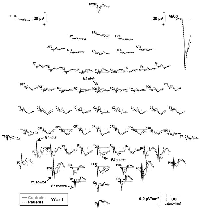
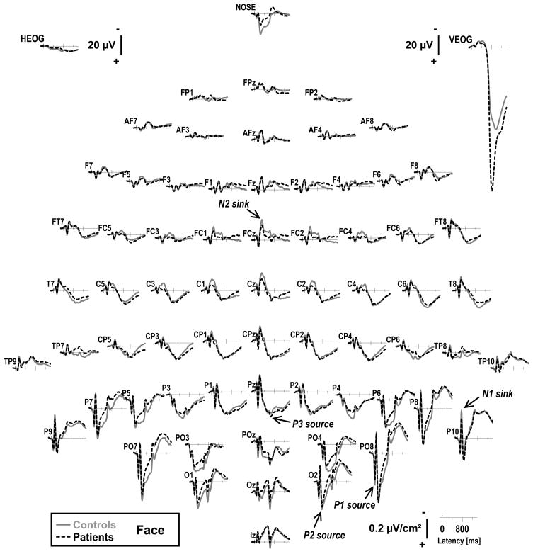
Figure 1 and 2 compare the grand mean CSD waveforms of patients and controls at all 67 recording sites for words and faces (averaged across condition), respectively.3 As one would expect for visual stimuli, the pattern of sink (negative) and source (positive) activity was most pronounced over posterior regions, and closely matched the stimulus-locked visual CSD component structure previously observed for words (Kayser et al., 2009). Distinct CSD components included inferior lateral-parietal P1 sources (approximate peak latency 80 ms at PO7 in controls) and left-lateralized N1 sinks (145 ms at P7), followed by an occipital P2 source (225 ms at O1), a central N2 sink (295 ms at FCz), and mid-parietal P3 sources (645 ms at Pz). As in our previous studies using visual stimuli (e.g., cf. Kayser et al., 1999, 2003, 2007, 2009), the P3 source was overlapped by a relative negativity at about 650 ms, which had a posterior topography, likely corresponding to a secondary N1 prompted by the stimulus offset (500 ms exposure time); however, this phenomenon did not differentially affect the experimental conditions.
Figure 1.
Reference-free current source density (CSD) [μV/cm2] waveforms (−100 to 1400 ms, 100 ms pre-stimulus baseline) for word stimuli (averaged across old and new items) comparing 44 controls (solid gray lines) and 57 patients (dashed black lines) at all 67 recording sites. Horizontal and vertical electrooculograms (EOG) [μV] are shown before blink correction. Distinct CSD components included inferior lateral-parietal P1 sources (approximate peak latency 80 ms at PO7) and N1 sinks (145 ms at P7), occipital P2 sources (225 ms at O1), a central N2 sink (295 ms at FCz), and mid-parietal P3 sources (645 ms at Pz).
Figure 2.
CSD waveforms as in Figure 1 for face stimuli. Distinct CSD components included inferior lateral-parietal P1 sources (approximate peak latency 90 ms at PO8) and N1 sinks (145 ms at P10), occipital P2 sources (220 ms at O2), a central N2 sink (295 ms at FCz), and mid-parietal P3 sources (645 ms at Pz).
The CSD component structure for faces (Figure 2) closely matched the one for words, including inferior lateral-parietal P1 sources (80 ms at PO8) and right-lateralized N1 sinks (145 ms at P10), followed by an occipital P2 source (225 ms at O2), a central N2 sink (295 ms at FCz), and mid-parietal P3 source (645 ms at Pz).
The overall CSD component structure was highly comparable in controls and patients for both tasks, despite notable reductions of prominent CSD components in patients (such as the mid-frontal N2 sink, which was markedly reduced for words, or the lateral occipitoparietal P3 source across tasks). Importantly, eye movements were largely comparable across groups and tasks (see bipolar eye activity traces included in Figure 1 and 2), and evidently did not affect the corresponding ERPs, suggesting that blink activity was effectively removed by the spatial blink filter applied to the continuous EEG data.
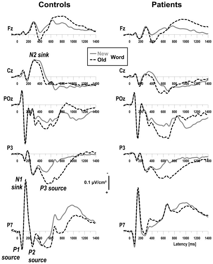
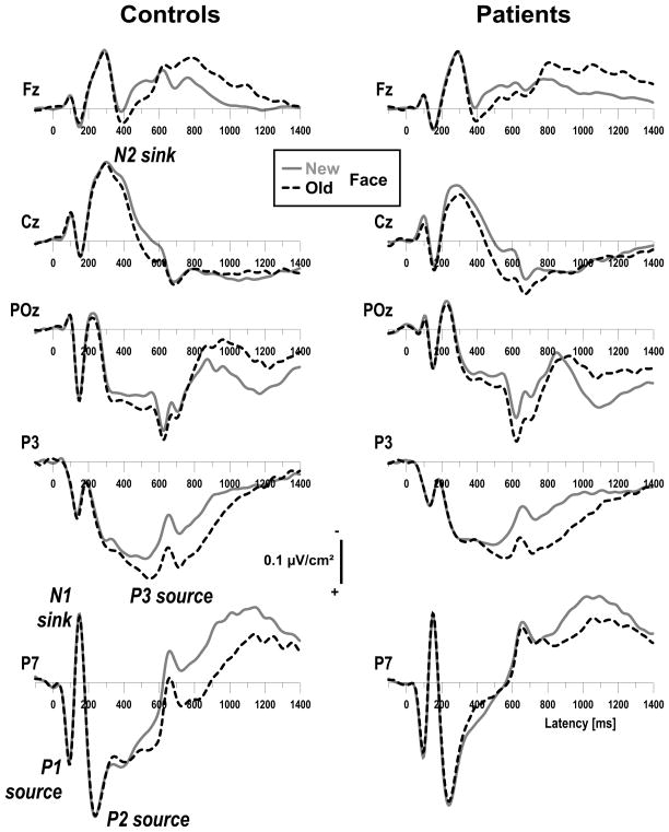
The corresponding old/new effects overlapping the stimulus-locked CSD component structure are depicted for each group at five representative sites (Fz, Cz, POz, P3, P7) in Figure 3 for words, and in Figure 4 for faces.4 Across groups and tasks, old items resulted in more positive-going current sources than new items, starting at about 300 ms and lasting for about 1 s. Early old/new effects were most prominent at mid-frontal sites and overlapped the falling phase of the N2 sink (Fz, Cz), whereas the later old/new effects were most prominent at left parietal sites, mostly overlapping P3 source but also an ensuing post-response negativity (cf. Kayser et al., 2003, 2007; Johansson and Mecklinger, 2003). Despite the presence of these old/new effects in each group, the late episodic memory effects seemed to be restricted to medial parietal sites in patients (POz, P3) and did not include more lateral sites as seen in controls (P7).
Figure 3.
CSD waveforms from 44 controls (left) and 57 patients (right) for words comparing old (dashed black lines) and new (solid gray lines) items at selected midline (Fz, Cz, POz) and left parietal (P3, P7) sites. Both groups showed more positive-going current sources for old than new words, overlapping the falling phase of the mid-frontal N2 sink (Fz, Cz) and the subsequent parietal P3 source (POz, P3, P7). The later old/new effects, however, were restricted to medial sites in patients (POz, P3).
Figure 4.
CSD waveforms as in Figure 3 for face stimuli. As for words, both groups had more positive-going current sources for old than new faces, overlapping the falling phase of the mid-frontal N2 sink (Fz, Cz) and the subsequent parietal P3 source (POz, P3, P7), with the later old/new effects confined to medial sites in patients (POz, P3).
3.2.2. PCA component waveforms and topographies
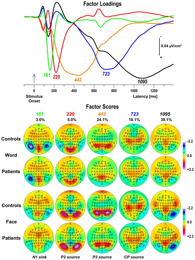
Figure 5 shows the time courses of factor loadings for the first five CSD factors extracted (89.3% explained variance after rotation) and the corresponding topographies of factor scores, separately plotted for tasks and groups. Labels were chosen to indicate the peak latency of the factor loadings relative to stimulus onset, and are supplemented by a brief functional interpretation if the factor had a signature topography. The mere purpose of these identifying labels is to ease referring to these CSD factors, which nevertheless consist of characteristic time courses and entire topographies.
Figure 5.
Unrestricted temporal PCA solution. Top: Time courses of Varimax-rotated covariance loadings for the first five CSD factors extracted (89.3% total variance explained). Labels indicate the peak latency of the factor loadings relative to stimulus onset. Bottom: Corresponding factor score topographies (nose at top) for both tasks and groups (pooled across old and new items) with percentage of explained variance. Factors 161, 220 and 442 clearly corresponded to lateral inferior-parietal N1 sinks (left-lateralized for words), task-specific occipital P2 sources, and parietal P3 sources, respectively. Factor 723 had a more complex topography, with a centroparietal (CP) source maximum accompanied by a mid-frontal and a right inferior occipitoparietal sink.
CSD factors corresponded to N1 sink (peak latency 161 ms; left lateral inferior-parietal maximum; 3.0% explained variance), P2 source (220 ms; occipital maximum with lateral-parietal sinks for words, but lateral occipital-parietal maximum with a mid-parietal sink for faces; 5.0%), and P3 source (442 ms; medial-parietal maximum; 24.1%). Two high-variance factors corresponded to late activity around the time subjects responded (723 ms; centroparietal source maximum with mid-frontal sink and right lateral inferior-parietal sink; 18.1%) and beyond (1095 ms; lateral occipital-parietal sink maximum with mid-parietal and temporal sources; 39.1%). Additional low-variance factors (not included in Figure 5) clearly corresponded to P1 source (87 ms; occipital-parietal maximum; 1.2%), early N1 sink (122 ms; left lateral inferior-parietal maximum for words, but mid-occipital maximum for faces; 1.5%), and N2 sink (294 ms; mid-frontocentral maximum; 1.0%). Because factors 442 and 723 explained most of the variance within the latency range of interest (i.e., old/new effects between 300 and 800 ms), the remainder of this report is focused on these two CSD-PCA components.5
3.2.3. Randomization tests of topographic old/new effects
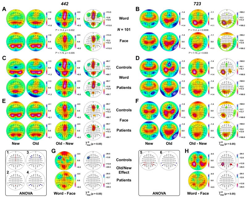
For the total sample (N = 101), both factors 442 and 723 yielded highly significant overall topographic old/new effects for each task (all multivariate T2 ≥ 7.74, all p ≤ 0.005; Figure 6AB). For the P3 source (factor 442), mid-frontal old/new effects were prominent in both tasks, as evidenced by univariate max(T2) statistics at each site (Figure 6A, column 4). However, these early old/new effects were somewhat more robust and broader for words (at FPz, AFz, Fz, FCz, Cz, F1, F2, FC1, FC2, AF4, all T2 > 30.9, all p < 0.0001) than faces (at AFz, Fz, FCz, Cz, F2, FC2, all T2 > 39.4, all p < 0.0001). These mid-frontal old/new effects were accompanied by inverted old/new effects (i.e., more negative-going sources for old than new items) at lateral anterior-frontal sites, particularly for words. There were also mid-occipitoparietal old/new effects for words (at POz, Oz, all T2 > 39.3, all p < 0.0001; at Pz, T2 = 14.6, p = 0.03) and faces (at POz, T2 = 14.2, p = 0.02; at Pz, T2 = 12.8, p = 0.04), which were accompanied by inverted old/new effects at lateral inferior temporoparietal sites, primarily for words and over the left hemisphere.
Figure 6.
Statistical evaluation of topographic old/new effects using randomization tests for paired samples for CSD factors 442 and 723, performed across groups for each task (A, B), and separately for 44 controls and 57 patients for words (C, D) and faces (E, F). Task-dependent differences of the old/new effect were also evaluated separately for each group (G, H). Shown are the mean factor score topographies for new and old items and their respective old-minus-new difference (A–F; multivariate Hotelling’s T2 statistics are reported for the total sample), or the old-minus-new difference for words minus the old-minus-new difference for faces (G, H), and squared univariate (channel-specific) paired samples T statistics thresholded at the 95th quantile (p = 0.05) of the corresponding randomization distribution (maximum of all 67-channel squared univariate paired samples T statistics). To facilitate comparisons of the max(T2) topographies with the underlying sink-source difference topographies, the sign of the difference at each site was applied to the respective T2 value, which is otherwise always positive. Please note that symmetric scales optimized for score ranges across new and old stimuli were used for the original topographies. To allow for better comparison of old/new effects across tasks and groups, the same symmetric scale range was used for all difference topographies for each factor, and within each set of max(T2) topographies. All topographies are two-dimensional representations of spherical spline interpolations (m = 2; λ = 0) derived from the mean factors scores or T2 statistics available for each recording site. Inset topographies show the sites selected for repeated measures ANOVA models performed on CSD factors 442 (1–4) and 723 (5–6), as indicated by colored locations (red: old/new effects; blue: inverted old/new effects).
In contrast, the later centroparietal source (factor 723) revealed robust left medial centrooccipitoparietal old/new effects (Figure 6B, column 4) for words (at CP3, CP5, P1, P3, P5, P7, PO3, PO7, O1, all T2 > 32.3, all p < 0.0001) and faces (at P1, P3, P5, P7, PO3, PO7, O1, Pz, POz, all T2 > 50.1, all p < 0.0001), with additional, but generally weaker old/new effects over corresponding right hemisphere regions and at midline sites (POz, Pz). These late old/new effects were also accompanied for both tasks by inverted old/new effects at lateral inferior temporoparietal sites.
Cell sizes were too small for the computation of multivariate T2 statistics (i.e., less than the 67 channels included in the EEG montage; cf. Maris, 2004) to evaluate overall topographic old/new effects for each group, but this constraint does not apply to the calculation of the corresponding univariate max(T2) statistics. As can be seen from Figure 6(C–F, columns 4), the observed old/new effects of both factors were also present for each group in both tasks, although to different degrees. For word stimuli, the early mid-frontal and mid-occipitoparietal old/new effects (factor 442) were highly comparable across groups (Figure 6C), whereas the late left parietal old/new effects (factor 723) appeared to be more robust in controls compared with patients (Figure 6D). For faces, however, early mid-frontal (Figure 6E) and late left parietal old/new effects (Figure 6F) were equally strong in patients and controls.
The univariate max(T2) statistics for evaluating the task-related differences of these old/new effects also suggested similar early mid-frontal and mid-parietal old/new effects (factor 442) across tasks, as there were no significant sites for controls or patients (Figure 6G). However, the early inverted old/new effects at left frontolateral sites were significantly larger for words than faces for controls only (at F5, F7, both T2 > 22.5, both p < 0.01; at FC5, T2 = 15.4, p = 0.03). In contrast, the late medial centrooccipitoparietal old/new effects (factor 723; Figure 6H) were significantly larger for words than faces for controls, particularly over the left hemisphere (at P5, T2 = 29.4, p = 0.0002; at P3, T2 = 19.2, p = 0.004; at P7, PO7, P6, P8, all T2 > 14.6, all p < 0.05). Thus, there were no task-related old/new differences for patients, nor did the inverted late old/new effects differ between tasks.
Based on these observations, recording sites were included in the conventional parametric statistics if they unambiguously contributed to old/new effects across tasks and groups. For the P3 source (factor 442), the ANOVA models were (Figure 6, left inset): 1) four mid-frontal sites (AFz, Fz, FCz, Cz); 2) one occipitoparietal site (POz); 3) four pairs of homologous lateral anterior-frontocentral sites (AF7/8, F7/8, F5/6, FC5/6); and 4) three pairs of homologous lateral inferior occipitoparietal sites (P7/8, P9/10, PO7/8). For the later centroparietal component (factor 723), the ANOVA models were (Figure 6, right inset): 5) six pairs of homologous medial centroparietal, parietal, and occipitoparietal sites (CP3/4, CP5/6, P3/4, P5/6, P7/8, PO7/8); and 6) two pairs of homologous lateral inferior temporoparietal sites (P9/10, TP9/10).
3.2.4. Repeated measures ANOVA
Table 3 summarizes the primary statistics obtained for the two CSD-PCA factors at regions representing old/new effects. As expected, all repeated measures ANOVA yielded highly significant “old-new” condition effects (all F[1, 97] > 31.0, all p < .0001), which validated the premise for selecting these sites for these analyses.
Table 3.
Summary of F ratios from repeated measures ANOVA performed on CSD-PCA factors at selected sites
| Factor (Sites) |
||||||
|---|---|---|---|---|---|---|
| 442 P3 source | 723 CP source | |||||
| (AFz, Fz, FCz, Cz) | (POz) | (AF7/8, F7/8, F5/6, FC5/6) | (P7/8, P9/10, PO7/8) | (CP3/4, CP5/6, P3/4, P5/6, P7/8, PO7/8) | (P9/10, TP9/10) | |
| C | 154.6 **** | 44.3 **** | 52.7 **** | 31.0 **** | 106.4 **** | 75.2 **** |
| G | 4.81 * | 7.80 ** | 7.24 ** | |||
| C × G | 3.04 | 8.84 ** | 16.3 **** | |||
| T | 32.8 **** | 20.2 **** | 25.7 **** | 18.0 **** | 7.27 ** | |
| T × G | 4.32 * | 7.59 ** | 4.80 * | |||
| T × C | 9.36 ** | 14.5 *** | 11.7 *** | 32.9 **** | 4.70 * | |
| T × C × G | 4.57 * | |||||
| H | - | - | 3.04 | 12.7 *** | 44.2 **** | 28.2 **** |
| H × C | - | - | 7.66 ** | 16.9 **** | 82.8 **** | 3.40 |
| H × C × G | - | - | 5.11 * | 3.29 | ||
| H × T | - | - | 34.8 **** | |||
| H × T × C | - | - | 20.1 **** | |||
Note. G = Group (patients, controls); T = task (word, face); C = condition (new, old); H = hemisphere (left, right). Only F ratios with p < .10 are reported for effects pooled over gender and site (subsets as indicated; for all tabled effects, df = 1, 97).
- Effect not applicable.
p ≤ .05.
p ≤ .01.
p ≤ .001,
p ≤ .0001.
The analysis for factor 442 at mid-frontal sites revealed a significant group main effect, with controls having greater sink activity than patients (M ± SD, −0.39 ± 0.86 vs. −0.18 ± 0.76). There was also a significant task main effect, resulting from less negative scores for words compared to faces (−0.17 ± 0.78 vs. −0.37 ± 0.83), and a significant task × condition interaction, stemming from a greater old/new effect for words (old vs. new, 0.04 ± 0.80 vs. −0.38 ± 0.70) when compared to faces (−0.22 ± 0.86 vs. −0.53 ± 0.78; Figure 6A, column 3). Task also interacted with group, with larger group differences observed for words (controls vs. patients, −0.33 ± 0.84 vs. −0.05 ± 0.72) than faces (−0.45 ± 0.89 vs. −0.31 ± 0.79). However, there were no interactions involving group and condition.
The analysis for factor 442 at the midline site POz revealed a significant task × group interaction, resulting from greater P3 sources in controls than patients for faces (1.00 ± 1.23 vs. 0.60 ± 0.94) but not words (0.74 ± 1.13 vs. 0.76 ± 1.08). There was also a task × condition interaction, stemming from greater old/new effects for words (0.93 ± 1.15 vs. 0.58 ± 1.02) than faces (0.85 ± 1.10 vs. 0.70 ± 1.08), but there were again no interactions involving group and condition. Thus, at anterior and parietal midline sites, patients showed the same early old/new effects as controls (cf. Figure 6CE, columns 3 and 4).
The analysis for factor 442 at lateral anterior-frontocentral sites revealed no significant effects involving group, although inverted old/new effects tended to be larger in controls (old vs. new, −0.56 ± 0.61 vs. −0.36 ± 0.56) than patients (−0.43 ± 0.66 vs. −0.29 ± 0.57). Thus, there was no evidence that task-related, inverted old/new effects differed between groups (cf. Figure 6G, column 4). At lateral inferior occipitoparietal sites, patients had an overall reduced P3 source compared to controls (0.17 ± 1.47 vs. 0.86 ± 1.69), and this reduction was greater for words (−0.17 ± 1.29 vs. 0.72 ± 1.43) than faces (0.52 ± 1.56 vs. 1.01 ± 1.90), yielding a significant task × group interaction. But again, there were no significant interactions involving group and condition, suggesting similar early inverted old/new effects in patients and controls (Figure 6CE, column 4).
The analysis for factor 723 at medial centroparietal, parietal, and occipitoparietal sites revealed a highly significant group main effect that originated from an overall larger source in controls compared to patients (0.22 ± 1.08 vs. −0.09 ± 1.17; cf. Figure 6DF, columns 1 and 2). More importantly, there was also a highly significant group × condition interaction, stemming from a greater old/new effect for controls (old vs. new, 0.51 ± 1.11 vs. −0.07 ± 0.96) than patients (0.06 ± 1.28 vs. −0.24 ± 1.03), and a significant group × condition × task interaction. Simple group × condition interactions for each task revealed that a greater old/new effect for controls than patients was significant only for words, F(1, 97) = 12.1, p = 0.0008 (old vs. new, for controls: 0.51 ± 1.02 vs. −0.26 ± 0.91; for patients: 0.02 ± 1.20 vs. −0.35 ± 0.92), but not for faces, F(1, 97) = 3.19, p = 0.08 (old vs. new, for controls: 0.51 ± 1.19 vs. 0.11 ± 0.98; for patients: 0.10 ± 1.35 vs. −0.12 ± 1.11). This three-way interaction qualified a significant task main effect, stemming from a greater source for faces than words, and a significant task × condition interaction, resulting from a greater old/new effects for words (old vs. new, 0.23 ± 1.15 vs. −0.31 ± 0.92) than faces (0.28 ± 1.30 vs. −0.02 ± 1.06) across groups. Thus, patients showed reduced late old/new effects, and these reductions were more pronounced for words than faces (cf. Figure 6H).
Overlapping a significant left-greater-than-right source asymmetry, a highly significant condition × hemisphere interaction confirmed that the late old/new effects were greater over the left (old vs. new, 0.49 ± 1.14 vs. −0.09 ± 0.94) than right hemisphere (0.02 ± 1.26 vs. −0.24 ± 1.06). There was also a three-way group × condition × hemisphere interaction. Simple group × condition interactions at each hemisphere revealed that a greater old/new effect for controls than patients was highly significant for the left parietal region, F(1, 97) = 11.1, p = 0.001 (old vs. new, for controls: 0.80 ± 0.98 vs. 0.02 ± 0.92; for patients: 0.25 ± 1.20 vs. −0.17 ± 0.94), but less robust for the right, F(1, 97) = 4.04, p = 0.05 (old vs. new, for controls: 0.21 ± 1.15 vs. −0.17 ± 0.99; for patients: −0.13 ± 1.33 vs. −0.30 ± 1.10). Thus, reductions of late parietal old/new effects in patients were also more prominent over the left than right hemisphere (Figure 6DF, column 4).
The analysis for the inverted old/new effects for factor 723 at lateral inferior temporoparietal sites revealed a highly significant group × condition interaction, stemming from greater inverted old/new effects in patients (old vs. new, −0.99 ± 1.02 vs. −0.44 ± 0.83) compared to controls (−0.70 ± 0.94 vs. −0.50 ± 0.80; cf. Figure 6DF, column 4). However, no other effect involving group attained a conventional level of significance.
To address the potential confound of group differences in age, identical repeated measures ANOVA were computed for participants falling within a restricted age range of 20–40 years, which formed sample subsets of 40 healthy adults (26.2 ± 5.0 years; 22 female) and 47 schizophrenic patients (27.7 ± 6.0 years; 20 female) with no significant difference in age (F[1, 83] = 2.49, n.s.). These additional analyses yielded almost identical results for both CSD factors (see supplementary Table A). Most importantly, the critical three-way group × condition × task interaction for factor 723 at medial centroparietal, parietal, and occipitoparietal sites was maintained at the same significance level, F(1, 83) = 5.63, p = 0.02.
Similarly, to address a potential confound for subjects whose native language was not English, identical repeated measures ANOVA were computed after excluding individuals learning English as a second language and bilingual participants, which formed sample subsets of 43 healthy adults (26 female) and 48 schizophrenic patients (23 female). These additional analyses also yielded highly comparable results for both CSD factors (see supplementary Table B). Again, the three-way group × condition × task interaction for factor 723 at medial centroparietal, parietal, and occipitoparietal sites was maintained at the same significance level, F(1, 87) = 5.67, p = 0.02.
In summary, the repeated measures ANOVA results were in complete agreement with the univariate max(T2) randomization statistics performed for each task and group as depicted in Figure 6.
4. Discussion
Compared to healthy adults, patients having schizophrenia or schizophrenia-spectrum disorders showed poorer recognition memory for common words and unfamiliar faces, which is in accordance with evidence of impaired episodic memory in schizophrenia (e.g., Barch, 2005; Pelletier et al., 2005). The behavioral data, however, did not support the notion of a selective deficit of verbal learning and memory in schizophrenia (e.g., Saykin et al., 1991; Gur et al., 1994) because lower accuracy and longer response latency were observed in patients for words and faces alike. This negative finding, however, should be interpreted with caution, not only for statistical reasons (i.e., null hypothesis), but also because participants were excluded if their task performance was at chance so as to obtain adequate processing for both tasks, thereby avoiding the pitfall of a possible task disengagement when exploring electrophysiologic abnormalities. Still, the extent of this performance deficit for each task was highly comparable to that previously reported for schizophrenia patients during visual and auditory implementations of the continuous recognition memory paradigm using words (Kayser et al., 1999, 2009).
In contrast to the behavioral data, the electrophysiologic findings showed evidence of a task-specific impairment of episodic memory processes in schizophrenia. Reference-free, high-density CSDs confirmed largely preserved old/new effects in patients over mid-parietal sites but marked old/new source reductions in patients over lateral parietal regions for words (cf. Kayser et al., 1999, 2009). These late old/new effects, however, were comparable in patients and healthy adults for unknown faces, which is in agreement with previous ERP findings (Guillem et al., 2001). This dissociation is even more surprising when considering that face recognition was clearly the more difficult task, yielding significantly lower recognition accuracy in both groups. This strongly suggests that reduced late old/new effects for words over lateral parietal sites cannot be attributed to a generalized cognitive dysfunction in schizophrenia (Blanchard and Neale, 1994), because this would predict greater deficits with increasing difficulty levels. Rather, this finding is reminiscent of the adequate performance of schizophrenic patients during difficult non-verbal working memory tests in contrast to their subpar performance on easier verbal memory tests (Wexler et al., 1998), and adds to the growing evidence that at least a subgroup of patients with schizophrenia has a selective deficit in verbal memory (e.g., Stevens et al., 2000; Wexler et al., 2002; Bruder et al., 2004).
In this regard, the new behavioral finding that manipulation of task difficulty (lag interval) was highly effective in the paradigm for faces but not words (cf. Kayser et al., 1999, 2003, 2007, 2009) offers an interesting interpretation. Encoding, storage and maintenance of verbal information in a phonological loop may be less affected by differences in lag, whereas this mechanism is largely unavailable for processing difficult-to-verbalize faces for which episodic memory decay is more critical. It seems then that healthy controls can benefit more than schizophrenic patients from accessing the phonological code, meaning that what is generally considered a deficit for patients is rather a selective advantage for controls. The similar late old/new effects for faces across groups, and for patients across tasks, are compatible with this post-hoc interpretation. It is also in line with recent ERP evidence suggesting that the phonological component of language processing may characterize a core deficit in schizophrenia (Angrilli et al., 2009).
4.1. Reduced left lateral parietal but preserved mid-frontal old/new effects in schizophrenia
Strong left parietal old/new effects for words and faces were present in both healthy adults and schizophrenic patients, likely reflecting conscious recollection processes associated with retrieval of contextual information (e.g., Friedman and Johnson, 2000; Rugg and Curran, 2007). The region for this old-greater-than-new source is entirely consistent with conventional ERP findings (e.g., Allan et al., 1998; Ally et al., 2008) as well as neuroimaging evidence implicating old/new effects for the lateral posterior parietal cortex (e.g., Wagner et al., 2005; Cabeza, 2008). Schizophrenia patients, however, revealed marked topographic abnormalities of old/new effects for words in that their lateral old/new sources were significantly reduced while their medial parietal old/new effects were largely preserved. This precisely replicates our previous CSD findings of abnormal late parietal old/new effects in schizophrenia for visually-presented words (Kayser et al., 2009). The current study adds that this lateral reduction is less evident for faces. Moreover, late parietal old/new effects were strongly left-lateralized in healthy adults across tasks, which is the typical topographical finding (e.g., Johnson, 1995; Allan et al., 1998; Friedman and Johnson, 2000), but were less asymmetric in patients (cf. Kayser et al., 1999, 2009).
The late parietal old/new source effects were preceded by prominent mid-frontal old/new effects across tasks and groups, which are likely the CSD equivalent of the mid-frontal ERP recognition memory effects (FN400) repeatedly observed in healthy adults (e.g., Curran, 1999; Curran and Cleary, 2003). Although the overall sink activity on which these early mid-frontal old/new effects were superimposed was greater in controls than patients, there were no significant group differences related to item repetition, which is consistent with our prior findings (Kayser et al., 2009) but in contrast to those reported by Guillem et al. (2001). If the mid-frontal old/new effect is indeed an electrophysiological correlate of item familiarity (e.g., Mecklinger, 2000; Rugg and Curran, 2007), our results suggest preserved implicit knowledge of previous word and face presentations in schizophrenia. However, this generalization should be viewed with caution, given the empirical and interpretational inconsistencies of ERP old/new effects for faces (cf. Curran and Hancock, 2007; Donaldson and Curran, 2007; MacKenzie and Donaldson, 2007, 2009).
As previously seen for visual items, the early mid-frontal old/new source effects were accompanied by mid-parietal old/new source effects (Kayser et al., 2003, 2007, 2009), but again, there were no group differences. However, the underlying P3 source was greater for controls than patients during face recognition, and consisted of bilateral medial-parietal maxima in controls across tasks, but had a mid-parietal maximum in patients for words. This also matches the reduced left inferior parietotemporal P3 source observed during visual word encoding in schizophrenia (Kayser et al., 2006). All of these findings add to the notion that schizophrenic patients are impaired during the encoding of linguistic information requiring left-lateralized resources subserving language functions (e.g., Price, 2000; cf. Crow, 1997; Condray, 2005).
Both early and late old/new effects were accompanied by inverted old/new effects over lateral anterior-frontal and/or lateral inferior occipitoparietal or temporoparietal sites. While we reported similar inverted old/new effects occurring at various information processing stages in our prior studies using this advanced CSD-PCA approach (Kayser et al., 2007, 2009), these observations are genuinely new to the literature. The inverted old/new effects consisted of reasonable local topographies and were clearly not a by-product of computational indeterminacies along the edge of the EEG montage when using a spherical spline CSD algorithm. While there were no group differences in the early inverted old/new effects, the late inverted old/new effects at lateral inferior temporoparietal sites were greater in patients than controls. The meaning of this finding is not immediately clear, but it seems to supplement the observation that the patients’ late old/new source effects (i.e., old-greater-than-new) were restricted to the medial parietal region. It also adds further evidence to the notion that the neuronal generator patterns underlying the late left parietal old/new effects are different between schizophrenic patients and healthy adults, a difference that will crucially affect ERP measures based on reference-dependent surface potentials.
With regard to the early mid-frontal old/new effects, the observed generator pattern is reminiscent of old/new effects overlapping a prominent response-related sink (cf. Kayser et al., 2007). It strongly suggests regional dipole activity oriented orthogonally to the cortical surface within the longitudinal fissure (anterior cingulate) with opposite orientations in the two hemispheres, which results in local field closure and typically manifests as focal activity at distinct midline recording locations (the error-related negativity is a prominent example; e.g., Gehring et al., 1993). Importantly, such local field cancellations are efficiently described by CSD methods but may not be appropriately resolved by popular inverse source localization algorithms, such as LORETA or BESA (Tenke and Kayser, 2008; see also Tenke at al., 2010).
As a final remarkable observation, the randomization tests of task-related differences in early and late old/new effects indicated for healthy adults a differential activation of left frontolateral regions (i.e., early inverted old/new effects) and left parietal regions (late old/new effects), each indicating greater activation when recognizing words than faces. It is intriguing that the CSD-PCA approach, in combination with unbiased randomization tests, implicated an involvement of regions that are entirely consistent with the classical language-related areas associated with speech production (Broca’s area) and speech comprehension (including Wernicke’s area; cf. Price, 2000).
4.2. Limitations and conclusions
As with many other ERP studies in schizophrenia, the current sample was heterogeneous with respect to meeting specific DSM-IV criteria. However, the unusually large sample of patients (n = 57) strongly suggests that the reported findings should generalize to (right-handed) individuals with schizophrenia spectrum disorders, although it will be of interest to follow-up the current report with more detailed analyses that take symptoms and subtypes of schizophrenia into account. As the sample also included a large number of unmedicated participants (n = 22), it is unlikely that the observed recognition memory deficits for words and faces, as well as the associated abnormalities of electrophysiologic old/new effects, were caused by antipsychotic medication (cf. Barch, 2005), but a moderating influence of drug treatment on cognition and brain function cannot be ruled out. Despite a larger proportion of individuals acquiring English as a second language among patients, this group difference failed to account for the observed task-related differences of left parietal ERP old/new effects between patients and controls, which is in accordance with other ERP and fMRI evidence indicating that age of language acquisition is of lesser importance than initially suggested (e.g., Friederici et al., 2002; Kotz, 2009). Although patients performed more poorly than healthy controls, their overall task performance was nevertheless adequate. Moreover, the ERP analysis was restricted to correct trials, which largely eliminates concerns that a generalized performance deficit in schizophrenia rather than a specific impairment in episodic memory was a major contributor to the current findings.
This report did not include analyses of earlier, mostly stimulus-driven visual components (P1, N1), which have been found to be reduced in schizophrenia and interpreted as indication for of an early perceptual processing deficit (e.g., Bruder et al., 1998; Doniger et al., 2002; Butler et al., 2007; Javitt et al., 2008; Javitt, 2009; Salisbury et al., 2009). However, given the patients’ adequate behavioral performance across tasks, combined with an intact and robust ERP/CSD component structure, we would argue that any group differences in early perceptual processing were of subordinate importance to their later, task-specific electrophysiologic abnormalities of verbal episodic memory. A different case can be made for N2, a component linked to stimulus categorization, for which we and others have repeatedly found robust reductions in schizophrenia across stimulus modalities (e.g., O’Donnell et al., 1993; Bruder et al., 1998, 1999; Kayser et al., 1999, 2001, 2009; Alain et al., 2001, 2002; Umbricht et al., 2006). A cursory inspection of the CSD waveforms suggested that the previously reported reductions in patients were also present in the current data, and we will address in a different report to what extent these reductions may be task-specific and how this may relate to impairments of later old/new effects. Likewise, our prior study (Kayser et al., 2009) also reported marked reductions of a mid-frontal, response-related sink in schizophrenia indicative of a prominent performance monitoring deficit. Additional response-locked analyses are planned to investigate whether the current data can replicate this interesting finding.
The reference-free CSD-PCA approach eliminates many of the pitfalls of volume-conducted scalp potentials (e.g., redundancy, reference-dependence), allowing one to more fully exploit the available temporal resolution, and to more efficiently characterize the neuroanatomical origins of brain activity associated with successive stages of information processing. This is an important advantage over other neuroimaging methods that rely on slow measures only indirectly related to neuronal activity. CSD is not merely a conservative bridge without the additional assumptions imposed by standard inverse solutions, it often provides a more appropriate description of neuroanatomical current generators underlying the potentials recorded from the scalp surface (Tenke et al., 2010).
Merging CSD and PCA methods into a generic ERP strategy is a powerful approach for advancing knowledge of cognitive dysfunction in schizophrenia. The present CSD-PCA findings replicate robust reductions of late old/new effects over left lateral temporoparietal, but not mid-parietal, sites in schizophrenia. In addition, they suggest that these abnormalities are specific to word recognition memory, implicating impaired phonological stimulus representation and/or encoding involving left parietal-temporal regions typically associated with language-related processing.
Supplementary Material
Figure A (Supplement to Figure 1). Nose-referenced, grand mean event-related surface potential (ERP) [μV] waveforms (−100 to 1400 ms, 100 ms pre-stimulus baseline) for word stimuli (averaged across old and new items) comparing 44 controls (solid gray lines) and 57 patients (dashed black lines) at all 67 recording sites. Horizontal and vertical electrooculograms (EOG) are shown at a smaller scale before blink correction. Distinct ERP components are labeled at PO7 (P1; approximate peak latency 85 ms), P7 (N1; 150 ms), O1 (P2; 235 ms), FCz (N2; 295 ms), and Pz (P3; 580 ms).
Figure B (Supplement to Figure 2). ERP waveforms as in Figure A for face stimuli. Distinct ERP components are labeled at PO8 (P1; approximate peak latency 90 ms), P10 (N1; 150 ms), O2 (P2; 225 ms), FCz (N2; 270 ms), and Pz (P3; 700 ms).
Figure C (Supplement to Figure 3). Reference-free current source density (CSD) [μV/cm2] waveforms (−100 to 1400 ms, 100 ms pre-stimulus baseline) for words in 44 controls comparing new (solid gray lines) and old items (dashed black lines) at all 67 recording sites. Horizontal and vertical electrooculograms (EOG) [μV] are shown before blink correction. Distinct CSD components are labeled at sites where they are most prominent (PO7, P7, O1, FCz, Pz).
Figure D (Supplement to Figure 3). CSD waveforms as in Figure C for 57 patients.
Figure E (Supplement to Figure 4). Reference-free current source density (CSD) [μV/cm2] waveforms (−100 to 1400 ms, 100 ms pre-stimulus baseline) for faces in 44 controls comparing new (solid gray lines) and old items (dashed black lines) at all 67 recording sites. Horizontal and vertical electrooculograms (EOG) [μV] are shown before blink correction. Distinct CSD components are labeled at sites where they are most prominent (PO7, P7, O1, FCz, Pz).
Figure F (Supplement to Figure 4). CSD waveforms as in Figure E for 57 patients.
Acknowledgments
This research was supported by grants MH50715 and MH066597 from the National Institute of Mental Health (NIMH). We thank Charles L. Brown, III, for developing, providing and improving excellent waveform plotting software (Disaver, Pan). Preliminary analyses of these data were presented at the 49th Annual Meeting of the Society for Psychophysiological Research (SPR), Berlin, Germany, October 21–24, 2009. We are grateful for several helpful comments received during the review process by two anonymous referees and the editors of this special issue, Stuart Steinhauer and Ruth Condray.
Footnotes
Although our previous work in schizophrenia indicated marked reductions of early negative, stimulus-locked components (N1, N2; e.g., Bruder et al., 1998; Kayser et al., 1999, 2001, 2009), as well as for negativities associated with response monitoring (FRN; Kayser et al., 2009), the focus of the current report is on old/new effects typically observed between 300 and 800 ms post stimulus. A detailed analysis of early stimulus-locked and response-locked effects will be presented elsewhere.
Although core psychotic symptoms, such as thought disorder, disorganized thinking, paranoid thoughts, and hallucinations, dominated the clinical profile of four male patients, their final consensus diagnosis was psychosis not otherwise specified. As these individuals clearly fall in the range of schizophrenia spectrum disorders, they were included in the study.
Corresponding figures showing the original, nose-referenced ERP waveforms are included as supplementary material (Figures A and B). For both controls and patients, the ERP for words were highly comparable to those of our prior studies (e.g., see Figure 1 in Kayser et al., 1999; Figure 1B in Kayser et al., 2009).
Corresponding figures for the full EEG montage are included as supplementary material (Figures C to F). Animated topographies of CSD waveforms for old and new words and faces can be obtained at URL http://psychophysiology.cpmc.columbia.edu/crm2009csd.html.
Separate PCA solutions derived from the CSD data for patients only (n = 57) or controls only (n = 44) revealed highly comparable factor structures, thereby validating the use of a common extraction with both groups.
Publisher's Disclaimer: This is a PDF file of an unedited manuscript that has been accepted for publication. As a service to our customers we are providing this early version of the manuscript. The manuscript will undergo copyediting, typesetting, and review of the resulting proof before it is published in its final citable form. Please note that during the production process errors may be discovered which could affect the content, and all legal disclaimers that apply to the journal pertain.
References
- Alain C, Cortese F, Bernstein LJ, He Y, Zipursky RB. Auditory feature conjunction in patients with schizophrenia. Schizophr Res. 2001;49(1–2):179–191. doi: 10.1016/s0920-9964(00)00138-9. [DOI] [PubMed] [Google Scholar]
- Alain C, Bernstein LJ, He Y, Cortese F, Zipursky RB. Visual feature conjunction in patients with schizophrenia: an event-related brain potential study. Schizophr Res. 2002;57(1):69–79. doi: 10.1016/s0920-9964(01)00303-6. [DOI] [PubMed] [Google Scholar]
- Albus M, Hubmann W, Mohr F, Hecht S, Hinterberger Weber P, Seitz NN, Kuchenhoff H. Neurocognitive functioning in patients with first-episode schizophrenia: results of a prospective 5-year follow-up study. Eur Arch Psychiatry Clin Neurosci. 2006;256(7):442–451. doi: 10.1007/s00406-006-0667-1. [DOI] [PubMed] [Google Scholar]
- Allan K, Wilding EL, Rugg MD. Electrophysiological evidence for dissociable processes contributing to recollection. Acta Psychol (Amst) 1998;98(2–3):231–252. doi: 10.1016/s0001-6918(97)00044-9. [DOI] [PubMed] [Google Scholar]
- Ally BA, Simons JS, McKeever JD, Peers PV, Budson AE. Parietal contributions to recollection: Electrophysiological evidence from aging and patients with parietal lesions. Neuropsychologia. 2008;46(7):1800–1812. doi: 10.1016/j.neuropsychologia.2008.02.026. [DOI] [PMC free article] [PubMed] [Google Scholar]
- American Psychiatric Association. Diagnostic and Statistical Manual of the Mental Disorders. 4. American Psychiatric Association; Washington, DC: 1994. (DSM-IV) [Google Scholar]
- Andreasen NC. The Scale for the Assessment of Negative Symptoms (SANS) The University of Iowa; Iowa City, IA: 1983. [Google Scholar]
- Andreasen NC. The Scale for the Assessment of Positive Symptoms (SAPS) The University of Iowa; Iowa City, IA: 1984. [Google Scholar]
- Angrilli A, Spironelli C, Elbert T, Crow TJ, Marano G, Stegagno L. Schizophrenia as failure of left hemispheric dominance for the phonological component of language. PLoS One. 2009;4(2):4507. doi: 10.1371/journal.pone.0004507. [DOI] [PMC free article] [PubMed] [Google Scholar]
- Arnold SE, Hyman BT, Van Hoesen GW, Damasio AR. Some cytoarchitectural abnormalities of the entorhinal cortex in schizophrenia. Arch Gen Psychiatry. 1991;48(7):625–632. doi: 10.1001/archpsyc.1991.01810310043008. [DOI] [PubMed] [Google Scholar]
- Baddeley AD. Working Memory. Philos Trans R Soc Lond, B, Biol Sci. 1983;302(1110):311–324. [Google Scholar]
- Barch DM. The cognitive neuroscience of schizophrenia. Annu Rev Clin Psychol. 2005;1:321–353. doi: 10.1146/annurev.clinpsy.1.102803.143959. [DOI] [PubMed] [Google Scholar]
- Barta PE, Pearlson GD, Powers RE, Richards SS, Tune LE. Auditory hallucinations and smaller superior temporal gyral volume in schizophrenia. Am J Psychiatry. 1990;147(11):1457–1462. doi: 10.1176/ajp.147.11.1457. [DOI] [PubMed] [Google Scholar]
- Baving L, Rockstroh B, Rossner P, Cohen R, Elbert T, Roth WT. Event-related potential correlates of acquisition and retrieval of verbal associations in schizophrenics and controls. J Psychophysiol. 2000;14(2):87–96. [Google Scholar]
- BioSemi, Inc. ActiveTwo - Multichannel, DC amplifier, 24-bit resolution, biopotential measurement system with active electrodes [ Amsterdam, NL: Author; 2001. http://www.biosemi.com. [Google Scholar]
- Blanchard JJ, Neale JM. The neuropsychological signature of schizophrenia: generalized or differential deficit? Am J Psychiatry. 1994;151(1):40–48. doi: 10.1176/ajp.151.1.40. [DOI] [PubMed] [Google Scholar]
- Bogerts B, Ashtari M, Degreef G, Alvir JM, Bilder RM, Lieberman JA. Reduced temporal limbic structure volumes on magnetic resonance images in first episode schizophrenia. Psychiatry Res. 1990;35(1):1–13. doi: 10.1016/0925-4927(90)90004-p. [DOI] [PubMed] [Google Scholar]
- Bogerts B, Lieberman JA, Ashtari M, Bilder RM, Degreef G, Lerner G, Johns C, Masiar S. Hippocampus-amygdala volumes and psychopathology in chronic schizophrenia. Biol Psychiatry. 1993;33(4):236–246. doi: 10.1016/0006-3223(93)90289-p. [DOI] [PubMed] [Google Scholar]
- Bruder G, Kayser J, Tenke C, Amador X, Friedman M, Sharif Z, Gorman J. Left temporal lobe dysfunction in schizophrenia: event-related potential and behavioral evidence from phonetic and tonal dichotic listening tasks. Arch Gen Psychiatry. 1999;56(3):267–276. doi: 10.1001/archpsyc.56.3.267. [DOI] [PubMed] [Google Scholar]
- Bruder G, Kayser J, Tenke C, Rabinowicz E, Friedman M, Amador X, Sharif Z, Gorman J. The time course of visuospatial processing deficits in schizophrenia: an event-related brain potential study. J Abnorm Psychol. 1998;107(3):399–411. doi: 10.1037//0021-843x.107.3.399. [DOI] [PubMed] [Google Scholar]
- Bruder GE, Wexler BE, Sage MM, Gil RB, Gorman JM. Verbal memory in schizophrenia: additional evidence of subtypes having different cognitive deficits. Schizophr Res. 2004;68(2–3):137–147. doi: 10.1016/S0920-9964(03)00156-7. [DOI] [PubMed] [Google Scholar]
- Butler PD, Martinez A, Foxe JJ, Kim D, Zemon V, Silipo G, Mahoney J, Shpaner M, Jalbrzikowski M, Javitt DC. Subcortical visual dysfunction in schizophrenia drives secondary cortical impairments. Brain. 2007;130(2):417–430. doi: 10.1093/brain/awl233. [DOI] [PMC free article] [PubMed] [Google Scholar]
- Cabeza R. Role of parietal regions in episodic memory retrieval: The dual attentional processes hypothesis. Neuropsychologia. 2008;46(7):1813–1827. doi: 10.1016/j.neuropsychologia.2008.03.019. [DOI] [PMC free article] [PubMed] [Google Scholar]
- Coltheart M. The MRC psycholinguistic database. Q J Exp Psychol A. 1981;33:497–505. [Google Scholar]
- Condray R. Language disorder in schizophrenia as a developmental learning disorder. Schizophr Res. 2005;73(1):5–20. doi: 10.1016/j.schres.2004.05.022. [DOI] [PubMed] [Google Scholar]
- Crow TJ. Temporal lobe asymmetries as the key to the etiology of schizophrenia. Schizophr Bull. 1990;16(3):433–443. doi: 10.1093/schbul/16.3.433. [DOI] [PubMed] [Google Scholar]
- Crow TJ. Schizophrenia as failure of hemispheric dominance for language. Trends Neurosci. 1997;20(8):339–343. doi: 10.1016/s0166-2236(97)01071-0. [DOI] [PubMed] [Google Scholar]
- Crow TJ. Cerebral asymmetry and the lateralization of language: core deficits in schizophrenia as pointers to the gene. Curr Opin Psychiatry. 2004;17(2):97–106. [Google Scholar]
- Curran T. The electrophysiology of incidental and intentional retrieval: ERP old/new effects in lexical decision and recognition memory. Neuropsychologia. 1999;37(7):771–785. doi: 10.1016/s0028-3932(98)00133-x. [DOI] [PubMed] [Google Scholar]
- Curran T, Cleary AM. Using ERPs to dissociate recollection from familiarity in picture recognition. Cogn Brain Res. 2003;15(2):191–205. doi: 10.1016/s0926-6410(02)00192-1. [DOI] [PubMed] [Google Scholar]
- Curran T, Hancock J. The FN400 indexes familiarity-based recognition of faces. Neuroimage. 2007;36(2):464–471. doi: 10.1016/j.neuroimage.2006.12.016. [DOI] [PMC free article] [PubMed] [Google Scholar]
- Damasio AR. Time-locked multiregional retroactivation: a systems-level proposal for the neural substrates of recall and recognition. Cognition. 1989;33(1–2):25–62. doi: 10.1016/0010-0277(89)90005-x. [DOI] [PubMed] [Google Scholar]
- DeLisi LE, Sakuma M, Kushner M, Finer DL, Hoff AL, Crow TJ. Anomalous cerebral asymmetry and language processing in schizophrenia. Schizophr Bull. 1997;23(2):255–271. doi: 10.1093/schbul/23.2.255. [DOI] [PubMed] [Google Scholar]
- Dixon WJ, editor. BMDP Statistical Software Manual: To Accompany the 7.0 Software Release. University of California Press; Berkeley, CA: 1992. [Google Scholar]
- Donaldson DI, Curran T. Potential (ERP) studies of recognition memory for faces. Neuroimage. 2007;36(2):488–489. doi: 10.1016/j.neuroimage.2006.12.049. [DOI] [PMC free article] [PubMed] [Google Scholar]
- Doniger GM, Foxe JJ, Murray MM, Higgins BA, Javitt DC. Impaired visual object recognition and dorsal/ventral stream interaction in schizophrenia. Arch Gen Psychiatry. 2002;59(11):1011–1020. doi: 10.1001/archpsyc.59.11.1011. [DOI] [PubMed] [Google Scholar]
- Duncan CC. Event-related brain potentials: a window on information processing in schizophrenia. Schizophr Bull. 1988;14(2):199–203. doi: 10.1093/schbul/14.2.199. [DOI] [PubMed] [Google Scholar]
- Egan MF, Duncan CC, Suddath RL, Kirch DG, Mirsky AF, Wyatt RJ. Event-related potential abnormalities correlate with structural brain alterations and clinical features in patients with chronic schizophrenia. Schizophr Res. 1994;11(3):259–271. doi: 10.1016/0920-9964(94)90020-5. [DOI] [PubMed] [Google Scholar]
- Falkai P, Bogerts B, Schneider T, Greve B, Pfeiffer U, Pilz K, Gonsiorzcyk C, Majtenyi C, Ovary I. Disturbed planum temporale asymmetry in schizophrenia. A quantitative postmortem study. Schizophr Res. 1995;14(2):161–176. doi: 10.1016/0920-9964(94)00035-7. [DOI] [PubMed] [Google Scholar]
- First MB, Spitzer RL, Gibbon M, Williams JBW. Structured Clinical Interview for DSM-IV Axis-I Disorders - Non-patient Edition (SCID-NP) Biometrics Research Department, New York State Psychiatric Institute; New York, NY: 1996. [Google Scholar]
- Flor-Henry P. Psychosis and temporal lobe epilepsy. A controlled investigation. Epilepsia. 1969;10(3):363–395. doi: 10.1111/j.1528-1157.1969.tb03853.x. [DOI] [PubMed] [Google Scholar]
- Flor-Henry P. Lateralized temporal-limbic dysfunction and psychopathology. Ann N Y Acad Sci. 1976;280:777–797. doi: 10.1111/j.1749-6632.1976.tb25541.x. [DOI] [PubMed] [Google Scholar]
- Ford JM. Schizophrenia: the broken P300 and beyond. Psychophysiology. 1999;36(6):667–682. [PubMed] [Google Scholar]
- Ford JM, White PM, Csernansky JG, Faustman WO, Roth WT, Pfefferbaum A. ERPs in schizophrenia: effects of antipsychotic medication. Biol Psychiatry. 1994;36(3):153–170. doi: 10.1016/0006-3223(94)91221-1. [DOI] [PubMed] [Google Scholar]
- Friederici AD, Steinhauer K, Pfeifer E. Brain signatures of artificial language processing: evidence challenging the critical period hypothesis. Proc Natl Acad Sci USA. 2002;99(1):529–534. doi: 10.1073/pnas.012611199. [DOI] [PMC free article] [PubMed] [Google Scholar]
- Friedman D. Event-related brain potential investigations of memory and aging. Biol Psychol. 2000;54(1–3):175–206. doi: 10.1016/s0301-0511(00)00056-9. [DOI] [PubMed] [Google Scholar]
- Friedman D, Johnson R., Jr Event-related potential (ERP) studies of memory encoding and retrieval: a selective review. Microsc Res Tech. 2000;51(1):6–28. doi: 10.1002/1097-0029(20001001)51:1<6::AID-JEMT2>3.0.CO;2-R. [DOI] [PubMed] [Google Scholar]
- Gehring WJ, Goss B, Coles MGH, Meyer DE, Donchin E. A neural system for error-detection and compensation. Psychol Sci. 1993;4(6):385–390. [Google Scholar]
- Goldberg TE, Torrey EF, Gold JM, Ragland JD, Bigelow LB, Weinberger DR. Learning and memory in monozygotic twins discordant for schizophrenia. Psychol Med. 1993;23(1):71–85. doi: 10.1017/s0033291700038861. [DOI] [PubMed] [Google Scholar]
- Guillem F, N’Kaoua B, Rougier A, Claverie B. Intracranial topography of event-related potentials (N400/P600) elicited during a continuous recognition memory task. Psychophysiology. 1995;32(4):382–392. doi: 10.1111/j.1469-8986.1995.tb01221.x. [DOI] [PubMed] [Google Scholar]
- Guillem F, Bicu M, Hooper R, Bloom D, Wolf MA, Messier J, Desautels R, Debruille JB. Memory impairment in schizophrenia: a study using event-related potentials in implicit and explicit tasks. Psychiatry Res. 2001;104(2):157–173. doi: 10.1016/s0165-1781(01)00305-5. [DOI] [PubMed] [Google Scholar]
- Gur RE, Jaggi JL, Shtasel DL, Ragland JD, Gur RC. Cerebral blood flow in schizophrenia: effects of memory processing on regional activation. Biol Psychiatry. 1994;35(1):3–15. doi: 10.1016/0006-3223(94)91160-6. [DOI] [PubMed] [Google Scholar]
- Hill SK, Beers SR, Kmiec JA, Keshavan MS, Sweeney JA. Impairment of verbal memory and learning in antipsychotic-naive patients with first-episode schizophrenia. Schizophr Res. 2004;68(2–3):127–136. doi: 10.1016/S0920-9964(03)00125-7. [DOI] [PubMed] [Google Scholar]
- Iidaka T, Matsumoto A, Nogawa J, Yamamoto Y, Sadato N. Frontoparietal network involved in successful retrieval from episodic memory. Spatial and temporal analyses using fMRI and ERP. Cereb Cortex. 2006;16(9):1349–1360. doi: 10.1093/cercor/bhl040. [DOI] [PubMed] [Google Scholar]
- Javitt DC. When doors of perception close: bottom-up models of disrupted cognition in schizophrenia. Annu Rev Clin Psychol. 2009;5:249–275. doi: 10.1146/annurev.clinpsy.032408.153502. [DOI] [PMC free article] [PubMed] [Google Scholar]
- Javitt DC, Spencer KM, Thaker GK, Winterer G, Hajos M. Neurophysiological biomarkers for drug development in schizophrenia. Nat Rev Drug Discov. 2008;7(1):68–83. doi: 10.1038/nrd2463. [DOI] [PMC free article] [PubMed] [Google Scholar]
- Jeon YW, Polich J. Meta-analysis of P300 and schizophrenia: patients, paradigms, and practical implications. Psychophysiology. 2003;40(5):684–701. doi: 10.1111/1469-8986.00070. [DOI] [PubMed] [Google Scholar]
- Johansson M, Mecklinger A. The late posterior negativity in ERP studies of episodic memory: action monitoring and retrieval of attribute conjunctions. Biol Psychol. 2003;64(1–2):91–117. doi: 10.1016/s0301-0511(03)00104-2. [DOI] [PubMed] [Google Scholar]
- Johnson R., Jr . Event-related potential insights into the neurobiology of memory systems. In: Boller F, Grafman J, editors. Handbook of Neuropsychology. Vol. 10. Elsevier; Amsterdam: 1995. pp. 135–163. [Google Scholar]
- Kawasaki Y, Maeda Y, Higashima M, Nagasawa T, Koshino Y, Suzuki M, Ide Y. Reduced auditory P300 amplitude, medial temporal volume reduction and psychopathology in schizophrenia. Schizophr Res. 1997;26(2–3):107–115. doi: 10.1016/s0920-9964(97)00055-8. [DOI] [PubMed] [Google Scholar]
- Kawasaki Y, Suzuki M, Takahashi T, Nohara S, McGuire PK, Seto H, Kurachi M. Anomalous cerebral asymmetry in patients with schizophrenia demonstrated by voxel-based morphometry. Biol Psychiatry. 2008;63(8):793–800. doi: 10.1016/j.biopsych.2007.08.008. [DOI] [PubMed] [Google Scholar]
- Kay SR, Opler LA, Fishbein A. Positive and negative syndrome scale (PANSS) rating manual. Multihealth System Inc; Toronto, Canada: 1992. [Google Scholar]
- Kayser J. Polygraphic Recording Data Exchange - PolyRex. New York State Psychiatric Institute: Department of Biopsychology; 2003. http://psychophysiology.cpmc.columbia.edu/PolyRex.htm. [Google Scholar]
- Kayser J, Tenke CE. Optimizing PCA methodology for ERP component identification and measurement: theoretical rationale and empirical evaluation. Clin Neurophysiol. 2003;114(12):2307–2325. doi: 10.1016/s1388-2457(03)00241-4. [DOI] [PubMed] [Google Scholar]
- Kayser J, Tenke CE. Trusting in or breaking with convention: towards a renaissance of principal components analysis in electrophysiology. Clin Neurophysiol. 2005;116(8):1747–1753. doi: 10.1016/j.clinph.2005.03.020. [DOI] [PubMed] [Google Scholar]
- Kayser J, Tenke CE. Principal components analysis of Laplacian waveforms as a generic method for identifying ERP generator patterns: I. Evaluation with auditory oddball tasks. Clin Neurophysiol. 2006a;117(2):348–368. doi: 10.1016/j.clinph.2005.08.034. [DOI] [PubMed] [Google Scholar]
- Kayser J, Tenke CE. Principal components analysis of Laplacian waveforms as a generic method for identifying ERP generator patterns: II. Adequacy of low-density estimates. Clin Neurophysiol. 2006b;117(2):369–380. doi: 10.1016/j.clinph.2005.08.033. [DOI] [PubMed] [Google Scholar]
- Kayser J, Tenke CE. Consensus on PCA for ERP data, and sensibility of unrestricted solutions. Clin Neurophysiol. 2006c;117(3):703–707. [Google Scholar]
- Kayser J, Tenke CE. Electrical distance as a reference-free measure for identifying artifacts in multichannel electroencephalogram (EEG) recordings. Psychophysiology. 2006d;43:S51. [Google Scholar]
- Kayser J, Bruder GE, Friedman D, Tenke CE, Amador XF, Clark SC, Malaspina D, Gorman JM. Brain event-related potentials (ERPs) in schizophrenia during a word recognition memory task. Int J Psychophysiol. 1999;34(3):249–265. doi: 10.1016/s0167-8760(99)00082-3. [DOI] [PubMed] [Google Scholar]
- Kayser J, Bruder GE, Tenke CE, Stuart BK, Amador XF, Gorman JM. Event-related brain potentials (ERPs) in schizophrenia for tonal and phonetic oddball tasks. Biol Psychiatry. 2001;49(10):832–847. doi: 10.1016/s0006-3223(00)01090-8. [DOI] [PubMed] [Google Scholar]
- Kayser J, Fong R, Tenke CE, Bruder GE. Event-related brain potentials during auditory and visual word recognition memory tasks. Cogn Brain Res. 2003;16(1):11–25. doi: 10.1016/s0926-6410(02)00205-7. [DOI] [PubMed] [Google Scholar]
- Kayser J, Tenke CE, Gates NA, Bruder GE. Reference-independent ERP old/new effects of auditory and visual word recognition memory: joint extraction of stimulus- and response-locked neuronal generator patterns. Psychophysiology. 2007;44(6):949–967. doi: 10.1111/j.1469-8986.2007.00562.x. [DOI] [PubMed] [Google Scholar]
- Kayser J, Tenke CE, Gates NA, Kroppmann CJ, Gil RB, Bruder GE. ERP/CSD indices of impaired verbal working memory subprocesses in schizophrenia. Psychophysiology. 2006;43(3):237–252. doi: 10.1111/j.1469-8986.2006.00398.x. [DOI] [PubMed] [Google Scholar]
- Kayser J, Tenke CE, Gil RB, Bruder GE. Stimulus- and response-locked neuronal generator patterns of auditory and visual word recognition memory in schizophrenia. Int J Psychophysiol. 2009;73(3):186–206. doi: 10.1016/j.ijpsycho.2009.02.003. [DOI] [PMC free article] [PubMed] [Google Scholar]
- Kim JJ, Kwon JS, Park HJ, Youn T, Kang do H, Kim MS, Lee DS, Lee MC. Functional disconnection between the prefrontal and parietal cortices during working memory processing in schizophrenia: a[15(O)]H2O PET study. Am J Psychiatry. 2003;160(5):919–923. doi: 10.1176/appi.ajp.160.5.919. [DOI] [PubMed] [Google Scholar]
- Kim MS, Kwon JS, Kang SS, Youn T, Kang KW. Impairment of recognition memory in schizophrenia: event-related potential study using a continuous recognition task. Psychiatry Clin Neurosci. 2004;58(5):465–472. doi: 10.1111/j.1440-1819.2004.01287.x. [DOI] [PubMed] [Google Scholar]
- Kotz SA. A critical review of ERP and fMRI evidence on L2 syntactic processing. Brain Lang. 2009;109(2–3):68–74. doi: 10.1016/j.bandl.2008.06.002. [DOI] [PubMed] [Google Scholar]
- Kucera N, Francis WN. Computational analysis of present-day American English. Brown University Press; Providence, RI: 1967. [Google Scholar]
- MacKenzie G, Donaldson DI. Dissociating recollection from familiarity: electrophysiological evidence that familiarity for faces is associated with a posterior old/new effect. Neuroimage. 2007;36(2):454–463. doi: 10.1016/j.neuroimage.2006.12.005. [DOI] [PubMed] [Google Scholar]
- MacKenzie G, Donaldson DI. Examining the neural basis of episodic memory: ERP evidence that faces are recollected differently from names. Neuropsychologia. 2009;2010;47(13):2756–2765. doi: 10.1016/j.neuropsychologia.2009.05.025. [DOI] [PubMed] [Google Scholar]
- Maris E. Randomization tests for ERP topographies and whole spatiotemporal data matrices. Psychophysiology. 2004;41(1):142–151. doi: 10.1111/j.1469-8986.2003.00139.x. [DOI] [PubMed] [Google Scholar]
- Matsumoto K, Matsuoka H, Yamazaki H, Sakai H, Kato T, Miura N, Nakamura M, Osakabe K, Saito H, Ueno T, Sato M. Impairment of an event-related potential correlate of memory in schizophrenia: effects of immediate and delayed word repetition. Clin Neurophysiol. 2001;112(4):662–673. doi: 10.1016/s1388-2457(01)00475-8. [DOI] [PubMed] [Google Scholar]
- Matsumoto K, Yamazaki H, Nakamura M, Sakai H, Miura N, Kato T, Miwa S, Ueno T, Saito H, Matsuoka H. Reduced word-repetition effect in the event-related potentials of thought-disordered patients with schizophrenia. Psychiatry Res. 2005;134(3):225–231. doi: 10.1016/j.psychres.2004.03.013. [DOI] [PubMed] [Google Scholar]
- Matsuoka H, Matsumoto K, Yamazaki H, Sakai H, Miwa S, Yoshida S, Numachi Y, Saito H, Ueno T, Sato M. Lack of repetition priming effect on visual event-related potentials in schizophrenia. Biol Psychiatry. 1999;46(1):137–140. doi: 10.1016/s0006-3223(98)00330-8. [DOI] [PubMed] [Google Scholar]
- McCarley RW, Faux SF, Shenton ME, Nestor PG, Adams J. Event-related potentials in schizophrenia: their biological and clinical correlates and a new model of schizophrenic pathophysiology. Schizophr Res. 1991;4(2):209–231. doi: 10.1016/0920-9964(91)90034-o. [DOI] [PubMed] [Google Scholar]
- McCarley RW, Shenton ME, O’Donnell BF, Faux SF, Kikinis R, Nestor PG, Jolesz FA. Auditory P300 abnormalities and left posterior superior temporal gyrus volume reduction in schizophrenia. Arch Gen Psychiatry. 1993;50(3):190–197. doi: 10.1001/archpsyc.1993.01820150036003. [DOI] [PubMed] [Google Scholar]
- McCarley RW, Salisbury DF, Hirayasu Y, Yurgelun Todd DA, Tohen M, Zarate C, Kikinis R, Jolesz FA, Shenton ME. Association between smaller left posterior superior temporal gyrus volume on magnetic resonance imaging and smaller left temporal P300 amplitude in first-episode schizophrenia. Arch Gen Psychiatry. 2002;59(4):321–331. doi: 10.1001/archpsyc.59.4.321. [DOI] [PubMed] [Google Scholar]
- McCarley RW, Nakamura M, Shenton ME, Salisbury DF. Combining ERP and structural MRI information in first episode schizophrenia and bipolar disorder. Clin EEG Neurosci. 2008;39(2):57–60. doi: 10.1177/155005940803900206. [DOI] [PMC free article] [PubMed] [Google Scholar]
- Mecklinger A. Interfacing mind and brain: a neurocognitive model of recognition memory. Psychophysiology. 2000;37(5):565–582. [PubMed] [Google Scholar]
- Menon RR, Barta PE, Aylward EH, Richards SS, Vaughn DD, Tien AY, Harris GJ, Pearlson GD. Posterior superior temporal gyrus in schizophrenia: Grey matter changes and clinical correlates. Schizophr Res. 1995;16(2):127–135. doi: 10.1016/0920-9964(94)00067-i. [DOI] [PubMed] [Google Scholar]
- Mozley LH, Gur RC, Gur RE, Mozley PD, Alavi A. Relationships between verbal memory performance and the cerebral distribution of fluorodeoxyglucose in patients with schizophrenia. Biol Psychiatry. 1996;40(6):443–451. doi: 10.1016/0006-3223(95)00421-1. [DOI] [PubMed] [Google Scholar]
- Nestor PG, Kubicki M, Kuroki N, Gurrera RJ, Niznikiewicz M, Shenton ME, McCarley RW. Episodic memory and neuroimaging of hippocampus and fornix in chronic schizophrenia. Psychiatry Res. 2007;155(1):21–28. doi: 10.1016/j.pscychresns.2006.12.020. [DOI] [PubMed] [Google Scholar]
- NeuroScan, Inc. STIM software manual. Author; Herndon, VA: 1994. [Google Scholar]
- NeuroScan, Inc. SCAN 4.3 - Vol II EDIT 4.3 - Offline analysis of acquired data (Document number 2203, Revision D) Compumedics Neuroscan; El Paso, TX: 2003. [Google Scholar]
- Nunez PL, Srinivasan R. Electric fields of the brain: the neurophysics of EEG. Oxford University Press; New York: 2006. [Google Scholar]
- Nunez PL, Westdorp AF. The surface Laplacian, high resolution EEG and controversies. Brain Topogr. 1994;6(3):221–226. doi: 10.1007/BF01187712. [DOI] [PubMed] [Google Scholar]
- Nurnberger JI, Jr, Blehar MC, Kaufmann CA, York Cooler C, Simpson SG, Harkavy Friedman J, Severe JB, Malaspina D, Reich T. Diagnostic interview for genetic studies. Rationale, unique features, and training. NIMH Genetics Initiative. Arch Gen Psychiatry. 1994;51(11):849–859. doi: 10.1001/archpsyc.1994.03950110009002. [DOI] [PubMed] [Google Scholar]
- O’Donnell BF, Shenton ME, McCarley RW, Faux SF, Smith RS, Salisbury DF, Nestor PG, Pollak SD, Kikinis R, Jolesz FA. The auditory N2 component in schizophrenia: relationship to MRI temporal lobe gray matter and to other ERP abnormalities. Biol Psychiatry. 1993;34(1–2):26–40. doi: 10.1016/0006-3223(93)90253-a. [DOI] [PubMed] [Google Scholar]
- O’Donnell BF, McCarley RW, Potts GF, Salisbury DF, Nestor PG, Hirayasu Y, Niznikiewicz MA, Barnard J, Shen ZJ, Weinstein DM, Bookstein FL, Shenton ME. Identification of neural circuits underlying P300 abnormalities in schizophrenia. Psychophysiology. 1999;36(3):388–398. doi: 10.1017/s0048577299971688. [DOI] [PMC free article] [PubMed] [Google Scholar]
- Oldfield RC. The assessment and analysis of handedness: the Edinburgh inventory. Neuropsychologia. 1971;9(1):97–113. doi: 10.1016/0028-3932(71)90067-4. [DOI] [PubMed] [Google Scholar]
- Paivio A, Yuille JC, Madigan SA. Concreteness, imagery, and meaningfulness values for 925 nouns. J Exp Psychol. 1968;76(1):1–25. doi: 10.1037/h0025327. [DOI] [PubMed] [Google Scholar]
- Pearlson GD, Barta PE, Powers RE, Menon RR, Richards SS, Aylward EH, Federman EB, Chase GA, Petty RG, Tien AY. Medial and superior temporal gyral volumes and cerebral asymmetry in schizophrenia versus bipolar disorder. Biol Psychiatry. 1997;41(1):1–14. doi: 10.1016/s0006-3223(96)00373-3. [DOI] [PubMed] [Google Scholar]
- Pelletier M, Achim AM, Montoya A, Lal S, Lepage M. Cognitive and clinical moderators of recognition memory in schizophrenia: a meta-analysis. Schizophr Res. 2005;74(2–3):233–252. doi: 10.1016/j.schres.2004.08.017. [DOI] [PubMed] [Google Scholar]
- Penney CG. Modality effects and the structure of short-term verbal memory. Mem Cognit. 1989;17(4):398–422. doi: 10.3758/bf03202613. [DOI] [PubMed] [Google Scholar]
- Perrin F, Pernier J, Bertrand O, Echallier JF. Spherical splines for scalp potential and current density mapping [Corrigenda EEG 02274, EEG Clin. Neurophysiol 1990. 1989;76:565. doi: 10.1016/0013-4694(89)90180-6. [DOI] [PubMed] [Google Scholar]; Electroenceph Clin Neurophysiol. 72(2):184–187. doi: 10.1016/0013-4694(89)90180-6. [DOI] [PubMed] [Google Scholar]
- Pfefferbaum A, Ford JM, White PM, Roth WT. P3 in schizophrenia is affected by stimulus modality, response requirements, medication status, and negative symptoms. Arch Gen Psychiatry. 1989;46(11):1035–1044. doi: 10.1001/archpsyc.1989.01810110077011. [DOI] [PubMed] [Google Scholar]
- Pivik RT, Broughton RJ, Coppola R, Davidson RJ, Fox N, Nuwer MR. Guidelines for the recording and quantitative analysis of electroencephalographic activity in research contexts. Psychophysiology. 1993;30(6):547–558. doi: 10.1111/j.1469-8986.1993.tb02081.x. [DOI] [PubMed] [Google Scholar]
- Price CJ. The anatomy of language: contributions from functional neuroimaging. J Anat. 2000;197(3):335–359. doi: 10.1046/j.1469-7580.2000.19730335.x. [DOI] [PMC free article] [PubMed] [Google Scholar]
- Rugg MD, Curran T. Event-related potentials and recognition memory. Trends Cogn Sci. 2007;11(6):251–257. doi: 10.1016/j.tics.2007.04.004. [DOI] [PubMed] [Google Scholar]
- Rugg MD, Roberts RC, Potter DD, Pickles CD, Nagy ME. Event-related potentials related to recognition memory. Effects of unilateral temporal lobectomy and temporal lobe epilepsy. Brain. 1991;114(5):2313–2332. doi: 10.1093/brain/114.5.2313. [DOI] [PubMed] [Google Scholar]
- Salisbury DF, Collins KC, McCarley RW. Reductions in the N1 and P2 Auditory Event-Related Potentials in First-Hospitalized and Chronic Schizophrenia. Schizophr Bull. 2009 doi: 10.1093/schbul/sbp003. Advance Access published online on March 12, 2009 http://dx.doi.org/10.1093/schbul/sbp003. [DOI] [PMC free article] [PubMed]
- Salisbury DF, Shenton ME, Sherwood AR, Fischer IA, Yurgelun-Todd DA, Tohen M, McCarley RW. First-episode schizophrenic psychosis differs from first-episode affective psychosis and controls in P300 amplitude over left temporal lobe. Arch Gen Psychiatry. 1998;55(2):173–180. doi: 10.1001/archpsyc.55.2.173. [DOI] [PMC free article] [PubMed] [Google Scholar]
- Saykin AJ, Gur RC, Gur RE, Mozley PD, Mozley LH, Resnick SM, Kester DB, Stafiniak P. Neuropsychological function in schizophrenia. Selective impairment in memory and learning. Arch Gen Psychiatry. 1991;48(7):618–624. doi: 10.1001/archpsyc.1991.01810310036007. [DOI] [PubMed] [Google Scholar]
- Saykin AJ, Shtasel DL, Gur RE, Kester DB, Mozley LH, Stafiniak P, Gur RC. Neuropsychological deficits in neuroleptic naive patients with first-episode schizophrenia. Arch Gen Psychiatry. 1994;51(2):124–131. doi: 10.1001/archpsyc.1994.03950020048005. [DOI] [PubMed] [Google Scholar]
- Shenton ME, Kikinis R, Jolesz FA, Pollak SD, LeMay M, Wible CG, Hokama H, Martin J, Metcalf D, Coleman M, et al. Abnormalities of the left temporal lobe and thought disorder in schizophrenia: A quantitative magnetic resonance imaging study. N Engl J Med. 1992;327(9):604–612. doi: 10.1056/NEJM199208273270905. [DOI] [PubMed] [Google Scholar]
- Smith ME, Halgren E. Dissociation of recognition memory components following temporal lobe lesions. J Exp Psychol Learn Mem Cogn. 1989;15(1):50–60. doi: 10.1037//0278-7393.15.1.50. [DOI] [PubMed] [Google Scholar]
- Snodgrass JG, Corwin J. Pragmatics of measuring recognition memory: applications to dementia and amnesia. J Exp Psychol Gen. 1988;117(1):34–50. doi: 10.1037//0096-3445.117.1.34. [DOI] [PubMed] [Google Scholar]
- Spitzer RL, Williams JBW, Gibbon M, First MB. Structured Clinical Interview for DSM-III-R: Patient Edition. American Psychiatric Press; Washington, DC: 1990. [Google Scholar]
- Stevens AA, Donegan NH, Anderson M, Goldman-Rakic PS, Wexler BE. Verbal processing deficits in schizophrenia. J Abnorm Psychol. 2000;109(3):461–471. [PubMed] [Google Scholar]
- Strik WK, Dierks T, Franzek E, Stober G, Maurer K. P300 asymmetries in schizophrenia revisited with reference-independent methods. Psychiatry Res. 1994;55(3):153–166. doi: 10.1016/0925-4927(94)90023-x. [DOI] [PubMed] [Google Scholar]
- Tendolkar I, Ruhrmann S, Brockhaus A, Pukrop R, Klosterkotter J. Remembering or knowing: electrophysiological evidence for an episodic memory deficit in schizophrenia. Psychol Med. 2002;32(7):1261–1271. doi: 10.1017/s0033291702006335. [DOI] [PubMed] [Google Scholar]
- Tenke CE, Kayser J. A convenient method for detecting electrolyte bridges in multichannel electroencephalogram and event-related potential recordings. Clin Neurophysiol. 2001;112(3):545–550. doi: 10.1016/s1388-2457(00)00553-8. [DOI] [PubMed] [Google Scholar]
- Tenke CE, Kayser J. Reference-free quantification of EEG spectra: combining current source density (CSD) and frequency principal components analysis (fPCA) Clin Neurophysiol. 2005;116(12):2826–2846. doi: 10.1016/j.clinph.2005.08.007. [DOI] [PubMed] [Google Scholar]
- Tenke CE, Kayser J. Neuroscience Meeting Planner. Society for Neuroscience; Washington, DC: 2008. ERP generators within the longitudinal fissure: are putative inverses flawed? Program No. 872.28.2008. Online. [Google Scholar]
- Tenke CE, Kayser J, Shankman SA, Griggs CB, Leite P, Stewart JW, Bruder GE. Hemispatial PCA dissociates temporal from parietal ERP generator patterns: CSD components in healthy adults and depressed patients during a dichotic oddball task. Int J Psychophysiol. 2008;67(1):1–16. doi: 10.1016/j.ijpsycho.2007.09.001. [DOI] [PMC free article] [PubMed] [Google Scholar]
- Tenke CE, Kayser J, Stewart JW, Bruder GE. Novelty P3 reductions in depression: characterization using principal components analysis (PCA) of current source density (CSD) waveforms. Psychophysiology. 2010;47(1):133–146. doi: 10.1111/j.1469-8986.2009.00880.x. [DOI] [PMC free article] [PubMed] [Google Scholar]
- Umbricht DS, Bates JA, Lieberman JA, Kane JM, Javitt DC. Electrophysiological indices of automatic and controlled auditory information processing in first-episode, recent-onset and chronic schizophrenia. Biol Psychiatry. 2006;59(8):762–772. doi: 10.1016/j.biopsych.2005.08.030. [DOI] [PubMed] [Google Scholar]
- van der Stelt O, Frye J, Lieberman JA, Belger A. Impaired P3 generation reflects high-level and progressive neurocognitive dysfunction in schizophrenia. Arch Gen Psychiatry. 2004;61(3):237–248. doi: 10.1001/archpsyc.61.3.237. [DOI] [PubMed] [Google Scholar]
- Vita A, Dieci M, Giobbio GM, Caputo A, Ghiringhelli L, Comazzi M, Garbarini M, Mendini AP, Morganti C, Tenconi F, et al. Language and thought disorder in schizophrenia: brain morphological correlates. Schizophr Res. 1995;15(3):243–251. doi: 10.1016/0920-9964(94)00050-i. [DOI] [PubMed] [Google Scholar]
- Wagner AD, Shannon BJ, Kahn I, Buckner RL. Parietal lobe contributions to episodic memory retrieval. Trends Cogn Sci. 2005;9(9):445–453. doi: 10.1016/j.tics.2005.07.001. [DOI] [PubMed] [Google Scholar]
- Wegesin DJ, Nelson CA. Effects of inter-item lag on recognition memory in seizure patients preceding temporal lobe resection: evidence from event-related potentials. Int J Psychophysiol. 2000;37(3):243–255. doi: 10.1016/s0167-8760(00)00101-x. [DOI] [PubMed] [Google Scholar]
- Wexler BE, Donegan N, Stevens AA, Jacob SA. Deficits in language-mediated mental operations in patients with schizophrenia. Schizophr Res. 2002;53(3):171–179. doi: 10.1016/s0920-9964(01)00194-3. [DOI] [PubMed] [Google Scholar]
- Wexler BE, Stevens AA, Bowers AA, Sernyak MJ, Goldman-Rakic PS. Word and tone working memory deficits in schizophrenia. Arch Gen Psychiatry. 1998;55(12):1093–1096. doi: 10.1001/archpsyc.55.12.1093. [DOI] [PubMed] [Google Scholar]
- Winterer G, Coppola R, Egan MF, Goldberg TE, Weinberger DR. Functional and effective frontotemporal connectivity and genetic risk for schizophrenia. Biol Psychiatry. 2003;54(11):1181–1192. doi: 10.1016/s0006-3223(03)00532-8. [DOI] [PubMed] [Google Scholar]
- Woods SW. Chlorpromazine equivalent doses for the newer atypical antipsychotics. J Clin Psychiatry. 2003;64(6):663–667. doi: 10.4088/jcp.v64n0607. [DOI] [PubMed] [Google Scholar]
- Yonelinas AP. Components of episodic memory: the contribution of recollection and familiarity. P. 2001;356(1413):1363–1374. doi: 10.1098/rstb.2001.0939. [DOI] [PMC free article] [PubMed] [Google Scholar]
- Yonelinas AP, Otten LJ, Shaw KN, Rugg MD. Separating the brain regions involved in recollection and familiarity in recognition memory. J Neurosci. 2005;25(11):3002–3008. doi: 10.1523/JNEUROSCI.5295-04.2005. [DOI] [PMC free article] [PubMed] [Google Scholar]
Associated Data
This section collects any data citations, data availability statements, or supplementary materials included in this article.
Supplementary Materials
Figure A (Supplement to Figure 1). Nose-referenced, grand mean event-related surface potential (ERP) [μV] waveforms (−100 to 1400 ms, 100 ms pre-stimulus baseline) for word stimuli (averaged across old and new items) comparing 44 controls (solid gray lines) and 57 patients (dashed black lines) at all 67 recording sites. Horizontal and vertical electrooculograms (EOG) are shown at a smaller scale before blink correction. Distinct ERP components are labeled at PO7 (P1; approximate peak latency 85 ms), P7 (N1; 150 ms), O1 (P2; 235 ms), FCz (N2; 295 ms), and Pz (P3; 580 ms).
Figure B (Supplement to Figure 2). ERP waveforms as in Figure A for face stimuli. Distinct ERP components are labeled at PO8 (P1; approximate peak latency 90 ms), P10 (N1; 150 ms), O2 (P2; 225 ms), FCz (N2; 270 ms), and Pz (P3; 700 ms).
Figure C (Supplement to Figure 3). Reference-free current source density (CSD) [μV/cm2] waveforms (−100 to 1400 ms, 100 ms pre-stimulus baseline) for words in 44 controls comparing new (solid gray lines) and old items (dashed black lines) at all 67 recording sites. Horizontal and vertical electrooculograms (EOG) [μV] are shown before blink correction. Distinct CSD components are labeled at sites where they are most prominent (PO7, P7, O1, FCz, Pz).
Figure D (Supplement to Figure 3). CSD waveforms as in Figure C for 57 patients.
Figure E (Supplement to Figure 4). Reference-free current source density (CSD) [μV/cm2] waveforms (−100 to 1400 ms, 100 ms pre-stimulus baseline) for faces in 44 controls comparing new (solid gray lines) and old items (dashed black lines) at all 67 recording sites. Horizontal and vertical electrooculograms (EOG) [μV] are shown before blink correction. Distinct CSD components are labeled at sites where they are most prominent (PO7, P7, O1, FCz, Pz).
Figure F (Supplement to Figure 4). CSD waveforms as in Figure E for 57 patients.


