Abstract
Rationale
The mitochondrial ATP sensitive potassium channel (mKATP) is implicated in cardioprotection by ischemic preconditioning (IPC), but the molecular identity of the channel remains controversial. The validity of current methods to assay mKATP activity is disputed.
Objective
We sought to develop novel methods to assay mKATP activity and its regulation.
Methods & Results
Using a thallium (Tl+) sensitive fluorophore, we developed a novel Tl+ flux based assay for mKATP activity, and used this assay probe several aspects of mKATP function. The following key observations were made: (i) Time-dependent run-down of mKATP activity was reversed by phosphatidylinositol-4,5-bisphosphate (PIP2). (ii) Dose responses of mKATP to nucleotides revealed a UDP EC50 of ~20 μmol/L and an ATP IC50 of ~5 μmol/L. (iii) The antidepressant fluoxetine (Prozac™) inhibited mKATP (IC50 2.4 μmol/L). Fluoxetine also blocked cardioprotection triggered by IPC, but did not block protection triggered by a mKATP independent stimulus. The related antidepressant zimelidine was without effect on either mKATP or IPC.
Conclusions
The Tl+ flux mKATP assay was validated by correlation with a classical mKATP channel osmotic swelling assay (R2 0.855). The pharmacologic profile of mKATP (response to ATP, UDP, PIP2, and fluoxetine) is consistent with that of an inward rectifying K+ channel (KIR) and is somewhat closer to that of the KIR6.2 than the KIR6.1 isoform. The effect of fluoxetine on mKATP-dependent cardioprotection has implications for the growing use of antidepressants in patients who may benefit from preconditioning.
Keywords: mitochondrial ATP sensitive potassium channel, ischemia, reperfusion, ischemic preconditioning, fluoxetine
INTRODUCTION
The mitochondrial ATP sensitive potassium channel (mKATP) is thought to be essential for cardioprotection recruited by ischemic preconditioning (IPC),1, 2 but despite intense research the molecular identity of this channel remains unclear. The simplest thesis is that mKATP channels are derivative of surface KATP channels, and thus composed of inward rectifying K+ channels (KIR) and sulfonylurea receptors (SUR). The cardiomyocyte surface KATP channel is comprised of KIR6.2 and SUR2A isoforms,3 but efforts to conclusively assign these proteins to the cardiac mKATP have been unsuccessful to date.
Neither Kir6 nor SUR genes contain mitochondrial target sequences, and Kir6/SUR proteins are not found in mitochondrial proteome databases or prediction engines.4, 5 Furthermore, immune-based methods to detect KIR/SUR subunits in mitochondria are plagued by issues of antibody specificity6 and mitochondrial purity/contamination. Several of the key pharmacologic reagents used to study mKATP channels (e.g. the agonist diazoxide (DZX) and antagonist 5-hydroxydecanoate (5-HD)) are also known to exhibit off-target effects.7, 8 Targeted gene deletion in mice to identify the mKATP channel involved in IPC has proven futile, due to the confounding cardiovascular effects of knocking out KIR6 and SUR genes (Kcnj8, Kcnj11, Abcc8, and Abcc9) on surface KATP channel function. In general, KIR and SUR knockouts exhibit profound defects in glucose/insulin handling,9–12 which impacts the response to IPC.13
A recent study using custom-made antibodies and SUR knockout mice identified shortform splice variants of SUR2 in mitochondria.14 Furthermore, recent pharmacological evidence suggests that complex II of the respiratory chain (succinate dehydrogenase) may be a regulatory component of the mKATP channel.15, 16 However, both these findings leave the identity of the K+ channel-forming subunit of mKATP unknown. In this regard, mKATP is similar to other mitochondrial ion channels which exist at a phenomenological level but have not been molecularly identified (e.g., the mitochondrial Ca2+ uniporter).
A major obstacle in studying the mKATP channel has been the availability of a reliable assay. Most studies to date have utilized an isolated mitochondrial rapid swelling assay, in which K+ uptake into mitochondria is followed by osmotically-obliged water, leading to mild swelling that is assayed as light scattering in a spectrophotometer.17, 18 This assay has been criticized as irreproducible by some laboratories,19 with the precise timing of mitochondrial isolation appearing to be a critical factor.20
Studying the literature on surface KATP channels, two key biochemical properties that appeared to have been overlooked in the mKATP channel field were the permeability of surface KATP channels for the heavy metal thallium (Tl+),21 and the modulation of channel run-down by phospholipids such as phosphatidylinositol-4,5-bisphosphate (PIP2).22, 23 Herein, we developed a novel Tl+ fluorescence based assay for mKATP channel activity, and used this assay to show that the channel is subject to run-down that is reversed by PIP2. It is anticipated that both these discoveries will advance the study of this channel. Furthermore, the antidepressant fluoxetine (FLX), which is known to modulate KIR channels,24, 25 was found herein to block the mKATP channel and to block IPC, but FLX did not block mKATP-independent cardioprotection. The implications of these data for clinical use of FLX in cardiovascular disease patients are discussed.
METHODS
Full experimental details are in the online supplement. Cardiac mitochondria were rapidly isolated from male Sprague-Dawley rat hearts by differential centrifugation in sucrose-based buffer as previously described.20 Protein was determined by the Folin-phenol method.26 Within 1.5 hr of mitochondrial isolation the activity of mKATP was monitored by the osmotic swelling assay as previously described.20
A novel fluorescence-based Tl+ flux assay for mKATP activity was also developed. The ionic radii of Tl+ (0.154 nm) and K+ (0.144 nm) are similar,27 and thus Tl+ is widely used as an analog to study membrane K+ transport.21, 27–30 The assay made use of the fluorescent indicator BTC-AM, which is better known as a ratiometric Ca2+ sensor, but is also sensitive to Tl+ with a distinct spectral response preventing signal overlap between these sensitivities. Mitochondria were loaded with BTC-AM during the isolation procedure and stored on ice until use. In the assay, 0.3 mg BTC-AM loaded mitochondria were added to a rapidly stirred cuvet containing 2 ml of chloride-free Tl+ assay buffer at 37°C. Tested compounds were present from the beginning of the assay, and baseline fluorescence was recorded for 10 s. prior to addition of TlSO4 (2 mmol/L final) via a syringe port. Fluorescence was monitored in a Varian Cary Eclipse spectrofluorometer (λex=488nm, λem525nm) and normalized to baseline. Full details including the concentrations and preparation methods for all reagents used in the assay, are in the online supplement.
Isolated rat heart perfusions (Langendorff) were performed as previously described.16 Following 20 min. equilibration, hearts were divided into 7 groups: (i) IR alone, comprising 20 min. vehicle (water or DMSO) infusion, 30 s. wash-out, 25 min. global ischemia, 120 min. reperfusion; (ii) FLX + IR, comprising 20 min. FLX infusion (5 μmol/L), 30 s. wash-out, then IR; (iii) IPC + IR, comprising 3 × 5 min. ischemia interspersed with 5 min. reperfusion, then IR; (iv) FLX + IPC + IR, comprising 5 min. FLX infusion (5 μmol/L), plus FLX infused throughout the 3 reperfusion phases of IPC (i.e. 20 min. total FLX delivery), 30 s. wash-out, then IR; (v) Zimelidine + IPC + IR. As above, replacing FLX with zimelidine (5 μmol/L). (vi) FCCP + IR, comprising 20 min. FCCP infusion (30 nmol/L 31, 32), then IR; vii) FCCP + FLX + IR, comprising 20 min. infusion of both FCCP (30 nmol/L) and FLX (5 μmol/L), then IR. Following reperfusion hearts were stained with tetrazolium chloride (TTC), imaged, and infarct size measured as previously described.16
In all experiments, each “N” was an independent heart perfusion or mitochondrial isolation from a single animal on one day. Statistical differences between groups were determined using ANOVA, with significance defined as p<0.05.
RESULTS
In seeking to develop an assay for mKATP channel activity that does not measure secondary effects such as water uptake (as is the case for the osmotic swelling assay), we discerned that the heavy metal thallium (Tl+) is widely used as a surrogate substrate to study K+ channel function.21, 27–30 A fluorescent probe that responds to [Tl+] is commercially available (FluxOR™, Invitrogen, Carlsbad CA), but careful analysis of the literature underlying this reagent revealed that the active component was BTC-AM, a more economical reagent.28–30 Thus, isolated mitochondria were loaded with BTC-AM as the basis for a Tl+ uptake assay of K+ channel activity.
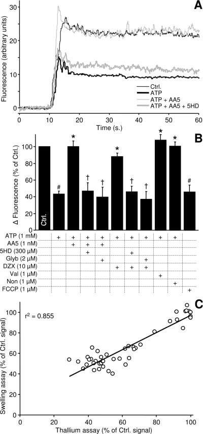
Figure 1A shows the addition of Tl+ to BTC-AM loaded mitochondria resulted in increased fluorescence due to rapid Tl+ influx and the establishment of a new steady-state. The fluorescence increase was largely inhibited by ATP, consistent with Tl+ transport by a KATP channel. Furthermore, the effect of ATP could be overridden by the mKATP channel opener AA5, and this effect was in-turn blocked by the mKATP antagonist 5-HD. These data are quantified in Figure 1B, which also shows the effects of mKATP reagents DZX (agonist) and glyburide (antagonist). The ionophores valinomycin and nonactin, both of which transport Tl+,33 resulted in maximal Tl+ flux into mitochondria, and the mitochondrial uncoupler FCCP inhibited Tl+ uptake indicating a requirement for membrane potential. It was hypothesized that the steady state is likely due to balancing of Tl+ influx by its efflux through the K+/H+ exchanger (KHE). However, attempts to modulate KHE activity with the inhibitors DCCD and quinine were inconclusive (data not shown). Validation of the Tl+ assay for mKATP channel activity was also performed by a direct comparison with results from mKATP osmotic swelling assays run in parallel under a variety of open/closed conditions. Figure 1C shows that the 2 assays correlated well (r2 = 0.855).
Figure 1. BTC-AM Tl+ fluorescence assay for mKATP activity in isolated heart mitochondria.
(A): Representative traces of BTC-AM Tl+ fluorescence in isolated mitochondria. Fluorescence (λex 488 nm, λem 525 nm) was normalized to the 10 s. of baseline prior to the injection of TlSO4 (arrow). Where indicated, 1 mmol/L ATP (thick, black), 1 nmol/L AA5 plus ATP (thin, grey), or 300 μmol/L 5HD plus AA5 plus ATP (thick grey) were present from the beginning of incubations. (B): Magnitude of the change in BTC-AM fluorescence following Tl+ addition, relative to control. Change in fluorescence was determined by subtracting the average baseline fluorescence from the stabilized average fluorescence at 30–60 s. Delta fluorescence in controls was 26.4 ± 3.3 arbitrary units. Experimental conditions are listed below the x-axis. Data are mean ± SEM, N≥4. # P<0.05 versus control, * P<0.05 versus ATP, † P<0.05 versus the effect of ATP+AA5 or ATP+DZX. (C): Correlation between two assays for mKATP channel activity. Experiments using a variety of conditions that modulate the mKATP were performed using either the osmotic swelling assay and the BTC-AM-Tl+ assay. Both sets of results were expressed as a percent of their respective controls. Linear regression curve fit revealed an r2 of 0.855.
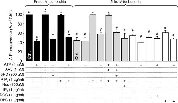
A general property of KIR channels is their tendency to “run-down” over time, a phenomenon attributed to loss of the phospholipid PIP2 from a binding site on the channel.34 The mKATP channel (which is constitutively open in isolated mitochondria) also loses activity following mitochondrial isolation, which may underlie the reported poor reproducibility of mKATP channel activity measurements.19 Upon investigating the relationship between these phenomena, it was found that incubation of mitochondria on ice for 5 hrs. resulted in complete loss of mKATP channel activity, and that PIP2 addition restored channel activity (Figure 2). Furthermore, the full pharmacologic profile of mKATP channel activity (i.e. inhibition by ATP, activation by AA5, and re-inhibition by 5HD) was recovered in PIP2-treated aged mitochondria. The same concentrations of the PIP2 breakdown products inositol triphosphate (IP3), 1,2-dioctanoyl glycerol (DOG), or 1,2-dipalmitoyl glycerol (DPG) did not affect mKATP activity. The polyvalent cation neomycin, which is known to inhibit KIR channel activity by sequestering PIP2,35 was able to reverse the mKATP channel-restorative effects of PIP2. Identical results were obtained with the osmotic swelling mKATP channel assay (Figure S1). Overall these data suggest that the mKATP channel contains a PIP2 sensitive subunit, possibly a KIR channel. Consistent with this, both the Tl+ and swelling assays revealed that mKATP sensitivity to the nucleotides UDP and ATP (Figure S2) was closer to that of the KIR6.2 than the KIR6.1 isoform.36–39
Figure 2. PIP2 modulation of mKATP channel activity using the swelling and BTC-AM-Tl+ assays.
mKATP activity was monitored using the BTC-AM-Tl+ assay in fresh mitochondria (black bars), or mitochondria 5 hrs. post isolation (gray and white bars). Fresh mitochondria data were normalized to control (Δ fluorescence 26.4±3.3) while 5 hr. mitochondria were normalized to control + PIP2 (3rd gray bar, Δ fluorescence 29.3±6.4). Experimental conditions are listed below the x-axis. Data in both panels are means ± SEM, N≥4. Fresh mitochondria: # P<0.05 versus control, * P<0.05 versus ATP, † P<0.05 versus ATP+AA5. 5 hr. mitochondria: # P<0.05 versus control+PIP2, * P<0.05 versus ATP, † P<0.05 versus ATP+AA5, like symbols are not significantly different.
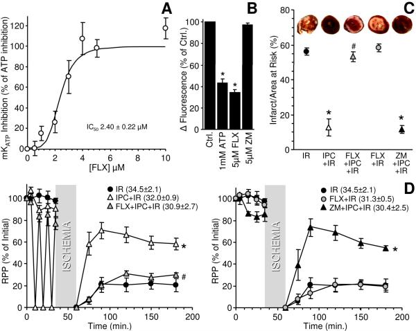
Several classes of KIR channel are known to be inhibited by fluoxetine (Prozac™), an antidepressant of the selective serotonin reuptake inhibitor (SSRI) class.24, 25 As shown in Figure S3, KIR6 channels (components of KATP channels) are an order of magnitude more sensitive to FLX than KIR4 channels, a well-known FLX target.40 Thus, we investigated the possibility that FLX might block mKATP channels. As shown in Figure 3, FLX blocked mKATP channel activity with an IC50 of 2.3 μmol/L, while a related SSRI zimelidine (ZM) did not. Identical results were obtained with the osmotic swelling mKATP assay (Figure S3). Furthermore, FLX blocked AA5- or DZX-mediated opening of mKATP (Figure S3).
Figure 3. Modulation of mKATP activity and IPC-mediated cardioprotection by fluoxetine.
(A): FLX dose-response of mKATP activity. Activity was measured using the BTC-AM-Tl+ assay. Data were plotted as % mKATP inhibition, with 100% inhibition defined as the condition in the presence of 1 mmol/L ATP, and 0% closed (i.e. open) being the baseline (Ctrl.) condition without ATP. FLX experiments were measured in the absence of ATP. Curve fit using the Hill equation revealed the FLX IC50 to be 2.39 ± 0.22 μmol/L. Data are means ± SEM, N≥4. (B): mKATP activity was measured by the BTC-AM-Tl+ assay. Where indicated, FLX or ZM were present. Data are means ± SEM, N≥4. *P<0.05 versus control. (C): Effect of FLX or ZM on IR injury and IPC. Hearts were subjected to Langendorff perfusion as detailed in the methods. Infarct size / area at risk was quantified from TTC staining, with representative stained hearts shown above each condition. Data are means ± SEM, N≥4, *P<0.05 vs. IR. (D): Rate pressure product (RPP, expressed as % of initial) in hearts subjected to each protocol. Data are split across two panels for clarity (IR data shown in both panels), and are means ± SEM, N≥4. *P<0.05 vs. IR, #P<0.05 vs. IPC+IR. The initial RPP (mmHg·min−1, ×103) for each group is listed in the legend.
Given the importance of the mKATP channel in IPC, we hypothesized that FLX may block IPC. Figure 3 shows that 5 μmol/L FLX completely blocked IPC-mediated cardioprotection in a rat perfused heart model of IR injury, while ZM was without effect. Notably FLX did not enhance baseline IR injury in this model, indicating that blockage of IPC was not due to an equal-but-opposite injurious effect, canceling out cardioprotection. Furthermore, FLX had no effect on cardioprotection mediated by FCCP (Figure S4), which occurs independent of the mKATP channel.31, 32
DISCUSSION
The major findings of this study are as follows: (i) Development of a novel Tl+ flux based assay for the mKATP channel; (ii) Time-dependent loss of mKATP channel activity is a genuine run-down phenomenon and is reversed by PIP2; (iii) FLX blocks both mKATP channel activity and IPC-mediated cardioprotection. This is the first demonstration of the modulation of a mitochondrial ion channel by PIP2, and the first identification of a mitochondrial ion channel target for FLX. Collectively, the data support the concept that mKATP contains a bona fide KIR channel. The effects of FLX on IPC may elucidate some of the reported negative impact of SSRI use on the outcome of cardiac surgery in humans.41
Work on the mKATP channel to date has relied on a variety of assays, many of which measure downstream effects of mitochondrial K+ uptake such as changes in respiration,42 matrix alkalinization,42 flavoprotein fluorescence,43 and swelling induced light scatter.42 Such methods are limited by the ability of other mitochondrial phenomena (e.g. electron transport chain activity, volume changes, membrane potential) to interfere with the measured parameters. Direct measurement of mitochondrial K+ fluxes using the potassium-binding fluorescent indicator (PBFI) is difficult because its Kd for K+ of ~8 mmol/L42 would result in saturation at typical intramitochondrial K+ levels (~150 mmol/L).44 Thus herein we chose to exploit another property of K+ channels, their ability to transport Tl+ as a surrogate for K+.21, 27 While Tl+ acetate has previously been used in swelling-based studies on mKATP45, this study is the first application of a Tl+ sensitive probe, BTC-AM,28–30 to study mKATP.
The kinetics of the Tl+ based mKATP channel assay are superior to those of the swelling based assay.16 Following Tl+ addition maximal fluorescence is attained within 2–4 s., compared to a time-lag of 20–30 s. for maximal signal intensity in the osmotic swelling assay. Unfortunately the high flux rate of Tl+ through K+ channels (~2x K+ flux 21), coupled with the relatively slow mixing time in the fluorescence cuvet, does not permit precise channel kinetics to be determined in this apparatus. Current best estimates for mKATP channel conductivity range from 10 to 300 pS.15, 46, 47
Another barrier to investigating the mKATP channel has been the rapid loss of channel activity over time in isolated mitochondrial preparations.20 Previous work showed that the purified mKATP channel runs-down in an electrophysiology setting and can be re-activated by very high concentrations of UDP.48 However, the cause of channel activity loss in intact mitochondria was unknown, and could easily be due to proteolytic degradation. The finding herein that time-dependent mKATP channel inactivation in intact mitochondria can be reversed by PIP2 indicates this is a genuine run-down phenomenon, which is a common property of KIR channels.49
KATP channels were the first channels identified to depend on phosphoinositides such as PIP2,22, 23 and this is the first study to identify a mitochondrial ion channel that responds to PIP2. Such regulation of mKATP channel activity by PIP2 may have implications for the function of this channel in IPC. PIP2 has been found in mitochondrial membranes, 50 but its endogenous source in mitochondria is unknown. Notably, the run-down of a planar lipid bilayer reconstituted mKATP was reversed by ATP/Mg2+,51 suggesting the mitochondrial high energy phosphate pool may be important in maintaining membrane lipid phosphorylation status. However, this phenomenon required very specific experimental conditions (i.e. addition and removal of ATP/Mg2+ to and from different sides of the membrane in a particular order), and attempts to reproduce this in isolated mitochondria were unsuccessful (data not shown). The role of lipid kinases (e.g., PI3K), phospholipases (e.g., PLC), and other components of the IP3/DAG signaling pathway in regulating mKATP is also unknown, but the involvement of such signaling components in IPC52, 53 suggests a potential novel pharmacological target (i.e., mitochondrial PIP2 turnover) to modulate preconditioning.
The Tl+ assay was also used to probe the response of mKATP to nucleotides (Figure S2). The mKATP EC50 for UDP (~20 μmol/L) was closer to that of KIR6.2 (~200 μmol/L) than KIR6.1 (~4 mmol/L), and the mKATP IC50 for ATP (~4.5 μmol/L) was also closer to that of KIR6.2 (~15 μmol/L) than KIR 6.1 (~350 μmol/L). While these data agree with previous studies on mKATP,48 a variety of labeling, electrophysiological and genetic studies across multiple species and tissues have suggested the presence of either KIR6.1, KIR6.2, both, or neither in mitochondria. 6, 10, 54–62 An overall consensus is that mKATP likely contains a KIR channel, but the definitive assignment of a particular KIR isoform is not yet possible.
The discovery that mKATP activity is blocked by FLX is also consistent with the consensus that mKATP contains a KIR. Fluoxetine has previously been shown to inhibit KIR channels, while related SSRIs (e.g. zimelidine) had no effect.24, 25 Our data (Figure S3) suggest that KIR6 channels may be the most sensitive to FLX of all KIR isoforms,24, 25, 40, 63 and in agreement with this the mKATP exhibits a strikingly low FLX IC50 of 2.4 μmol/L (Figure 3). Physiological concentrations of FLX are in the range of 1 – 20 μmol/L.63 The fact that FLX is a lipophilic cation (LogP 4.8),64 coupled with the highly membranous nature of mitochondria, may serve to concentrate FLX in the organelle. In a mitochondria-rich tissue such as myocardium, the mitochondrion may be a primary target for FLX.
The discovery that FLX can block IPC-mediated cardioprotection is both consistent with its effect on mKATP activity, and consistent with a critical role for mKATP in IPC signaling.1, 16, 65 The lack of effect of another SSRI, zimelidine, on either IPC or mKATP activity suggests that this effect is not mediated via the SSRI mode of action. The observation that FLX also blocks mKATP channel opening by the highly specific agonist AA5 also suggests a direct mKATP effect. Furthermore, the lack of effect of FLX on FCCP-mediated cardioprotection, which is completely independent of mKATP channels,31, 32 suggests that the protection-blocking effect of FLX is specific to mKATP channel-mediated protection, and does not extend to all modes of protection. The current lack of a molecular identity for the mKATP does not permit decisive knock-out experiments to verify whether the effects of FLX observed in the intact heart are mediated by mKATP.
In the US, antidepressants are the most commonly prescribed class of medication,66 with FLX alone prescribed >23 million times in 2008.67 Although SSRIs are known to negatively impact the outcome of cardiac surgery,41 they are widely prescribed to patients with acute coronary syndrome.68 Notably, while IPC elicits solid protection in animal models of IR injury, its application in humans is limited by confounding effects such as age,69 gender,70 diabetes,13 and other medications.71 To this list of medications FLX must now be added, with the implication that successful cardioprotection in humans may require FLX withdrawal. Furthermore, the mood enhancer lithium is also known to both increase cardiac PIP2 lelvels72 and to induce cardioprotection,73 suggesting that some of the protective effects of lithium previously attributed to GSK-3β inhibition73 may be mediated via the mKATP channel.
In summary, we have developed herein a novel assay for the mKATP channel, and used this assay to reveal novel sensitivities of the channel to phosphoinositides and antidepressants. It is anticipated that this assay may find widespread use in the mKATP field, leading ultimately to the identification of this important channel.
Novelty and Significance (Wojtovich et al.).
What is known?
The mitochondrial ATP-sensitive potassium channel (mKATP) mediates protection from cardiac ischemia reperfusion injury by ischemic preconditioning.
The molecular identity of the mKATP remains controversial and the validity of current methods to assay mKATP activity is disputed.
Another limitation to the investigation of this channel is the rapid (~ 1 hr.) loss of channel activity following mitochondrial isolation.
What new information does this article contribute?
We describe a novel thallium flux assay for measurement of mKATP activity.
The rapid loss of mKATP activity after isolation is shown to be due to classical channel run-down, and is recovered by the phospholipid PIP2.
Both the mKATP channel and ischemic preconditioning are inhibited by fluoxetine (Prozac™).
Summary of Novelty and Significance
Cardiac ischemia/reperfusion (IR) injury is an important worldwide morbidity factor. Strategies to protect the heart from IR injury (such as during heart attack) are limited, but one promising avenue is ischemic preconditioning (IPC). The mitochondrial ATPsensitive K+ channel (mKATP) has been suggested to mediate the protection afforded by IPC; however, the molecular identity of this channel is unknown, and its assay is also technically challenging, thus hindering drug-development efforts. Using a Tl+-sensitive fluorophore, a novel assay was developed herein to measure mKATP activity. Using this assay, we show that loss of mKATP channel activity over time is reversed by the lipid PIP2. These findings should greatly facilitate mKATP research, hopefully leading to a molecular identity. Furthermore, this is the first report of a PIP2 sensitive phenomenon in mitochondria; it may possibly relate to the mechanism of channel regulation in IPC itself. Finally, we found that the antidepressant fluoxetine (Prozac™) inhibited mKATP and also blocked the protective effects of IPC. Given the widespread use of fluoxetine in cardiac patients, this may have important implications for the potential application ofIPC in humans.
Supplementary Material
ACKNOWLEDGEMENTS
We thank Teresa A. Sherman (URMC) for technical assistance.
SOURCES OF FUNDING This work was funded by a pre-doctoral fellowship award to APW from the American Heart Association, Founders Affiliate (0815770D), and by grants from the US National Institutes of Health to PSB (RO1-HL071158), and PSB & KWN (RO1-GM-087483).
ABBREVIATIONS
- FLX
Fluoxetine
- ZM
Zimelidine
- BTC-AM
Benzothiazole coumarin acetyoxymethyl ester
- PIP2
phosphatidylinositol-4,5-bisphosphate
- 5-HD
5-hydroxydecanoate
- AA5
Atpenin A5
- Glyb
Glyburide
- DCCD
N,N′-Dicyclohexylcarbodiimide
- DZX
Diazoxide
- IPC
Ischemic preconditioning
- Kir
Inward rectifying potassium channel
- SUR
sulfonylurea receptor
- Neo
Neomycin
- DOG
1,2-dioctanoyl glycerol
- IP3
Inositol triphosphate
- DPG
1,2-dipalmitoyl glycerol
- FCCP
carbonyl cyanide-p-trifluoromethoxyphenylhydrazone
Footnotes
DISCLOSURES None
Publisher's Disclaimer: This is a PDF file of an unedited manuscript that has been accepted for publication. As a service to our customers we are providing this early version of the manuscript. The manuscript will undergo copyediting, typesetting, and review of the resulting proof before it is published in its final citable form. Please note that during the production process errors may be discovered which could affect the content, and all legal disclaimers that apply to the journal pertain.
REFERENCES
- (1).Auchampach JA, Grover GJ, Gross GJ. Blockade of ischaemic preconditioning in dogs by the novel ATP dependent potassium channel antagonist sodium 5-hydroxydecanoate. Cardiovasc Res. 1992;26:1054–62. doi: 10.1093/cvr/26.11.1054. [DOI] [PubMed] [Google Scholar]
- (2).Schultz JE, Qian YZ, Gross GJ, Kukreja RC. The ischemia-selective KATP channel antagonist, 5-hydroxydecanoate, blocks ischemic preconditioning in the rat heart. J Mol Cell Cardiol. 1997;29:1055–60. doi: 10.1006/jmcc.1996.0358. [DOI] [PubMed] [Google Scholar]
- (3).Nichols CG. KATP channels as molecular sensors of cellular metabolism. Nature. 2006;440:470–6. doi: 10.1038/nature04711. [DOI] [PubMed] [Google Scholar]
- (4).Pagliarini DJ, Calvo SE, Chang B, Sheth SA, Vafai SB, Ong SE, Walford GA, Sugiana C, Boneh A, Chen WK, Hill DE, Vidal M, Evans JG, Thorburn DR, Carr SA, Mootha VK. A mitochondrial protein compendium elucidates complex I disease biology. Cell. 2008;134:112–23. doi: 10.1016/j.cell.2008.06.016. [DOI] [PMC free article] [PubMed] [Google Scholar]
- (5).Guda C, Guda P, Fahy E, Subramaniam S. MITOPRED: a web server for the prediction of mitochondrial proteins. Nucleic Acids Res. 2004;32:W372–W374. doi: 10.1093/nar/gkh374. [DOI] [PMC free article] [PubMed] [Google Scholar]
- (6).Foster DB, Rucker JJ, Marban E. Is Kir6.1 a subunit of mitoKATP? Biochem Biophys Res Commun. 2008;366:649–56. doi: 10.1016/j.bbrc.2007.11.154. [DOI] [PMC free article] [PubMed] [Google Scholar]
- (7).Schafer G, Wegener C, Portenhauser R, Bojanovski D. Diazoxide, an inhibitor of succinate oxidation. Biochem Pharmacol. 1969;18:2678–81. [PubMed] [Google Scholar]
- (8).Hanley PJ, Gopalan KV, Lareau RA, Srivastava DK, von MM, Daut J. Beta-oxidation of 5-hydroxydecanoate, a putative blocker of mitochondrial ATP-sensitive potassium channels. J Physiol. 2003;547:387–93. doi: 10.1113/jphysiol.2002.037044. [DOI] [PMC free article] [PubMed] [Google Scholar]
- (9).Chutkow WA, Samuel V, Hansen PA, Pu J, Valdivia CR, Makielski JC, Burant CF. Disruption of Sur2-containing KATP channels enhances insulin-stimulated glucose uptake in skeletal muscle. Proc Natl Acad Sci USA. 2001;98:11760–4. doi: 10.1073/pnas.201390398. [DOI] [PMC free article] [PubMed] [Google Scholar]
- (10).Miki T, Suzuki M, Shibasaki T, Uemura H, Sato T, Yamaguchi K, Koseki H, Iwanaga T, Nakaya H, Seino S. Mouse model of Prinzmetal angina by disruption of the inward rectifier Kir6.1. Nat Med. 2002;8:466–72. doi: 10.1038/nm0502-466. [DOI] [PubMed] [Google Scholar]
- (11).Stoller D, Kakkar R, Smelley M, Chalupsky K, Earley JU, Shi NQ, Makielski JC, McNally EM. Mice lacking sulfonylurea receptor 2 (SUR2) ATP-sensitive potassium channels are resistant to acute cardiovascular stress. J Mol Cell Cardiol. 2007;43:445–54. doi: 10.1016/j.yjmcc.2007.07.058. [DOI] [PMC free article] [PubMed] [Google Scholar]
- (12).Elrod JW, Harrell M, Flagg TP, Gundewar S, Magnuson MA, Nichols CG, Coetzee WA, Lefer DJ. Role of sulfonylurea receptor type 1 subunits of ATP-sensitive potassium channels in myocardial ischemia/reperfusion injury. Circulation. 2008;117:1405–13. doi: 10.1161/CIRCULATIONAHA.107.745539. [DOI] [PubMed] [Google Scholar]
- (13).Kersten JR, Toller WG, Gross ER, Pagel PS, Warltier DC. Diabetes abolishes ischemic preconditioning: role of glucose, insulin, and osmolality. Am J Physiol. 2000;278:H1218–H1224. doi: 10.1152/ajpheart.2000.278.4.H1218. [DOI] [PubMed] [Google Scholar]
- (14).Ye B, Kroboth S, Pu JL, Sims J, Aggarwal N, McNally E, Makielski J, Shi NQ. Molecular Identification and Functional Characterization of a Mitochondrial Sulfonylurea Receptor 2 Splice Variant Generated by Intraexonic Splicing. Circ Res. 2009;105:1083–93. doi: 10.1161/CIRCRESAHA.109.195040. [DOI] [PMC free article] [PubMed] [Google Scholar]
- (15).Ardehali H, Chen Z, Ko Y, Mejia-Alvarez R, Marban E. Multiprotein complex containing succinate dehydrogenase confers mitochondrial ATP-sensitive K+ channel activity. Proc Natl Acad Sci USA. 2004;101:11880–5. doi: 10.1073/pnas.0401703101. [DOI] [PMC free article] [PubMed] [Google Scholar]
- (16).Wojtovich AP, Brookes PS. The complex II inhibitor atpenin A5 protects against cardiac ischemia-reperfusion injury via activation of mitochondrial KATP channels. Basic Res Cardiol. 2009;104:121–9. doi: 10.1007/s00395-009-0001-y. [DOI] [PMC free article] [PubMed] [Google Scholar]
- (17).Beavis AD, Brannan RD, Garlid KD. Swelling and contraction of the mitochondrial matrix. I. A structural interpretation of the relationship between light scattering and matrix volume. J Biol Chem. 1985;260:13424–33. [PubMed] [Google Scholar]
- (18).Garlid KD, Beavis AD. Swelling and contraction of the mitochondrial matrix. II. Quantitative application of the light scattering technique to solute transport across the inner membrane. J Biol Chem. 1985;260:13434–41. [PubMed] [Google Scholar]
- (19).Das M, Parker JE, Halestrap AP. Matrix volume measurements challenge the existence of diazoxide/glibencamide-sensitive KATP channels in rat mitochondria. J Physiol. 2003;547:893–902. doi: 10.1113/jphysiol.2002.035006. [DOI] [PMC free article] [PubMed] [Google Scholar]
- (20).Wojtovich AP, Brookes PS. The endogenous mitochondrial complex II inhibitor malonate regulates mitochondrial ATP-sensitive potassium channels: Implications for ischemic preconditioning. Biochim Biophys Acta. 2008;1777:882–9. doi: 10.1016/j.bbabio.2008.03.025. [DOI] [PMC free article] [PubMed] [Google Scholar]
- (21).Hille B. Potassium channels in myelinated nerve. Selective permeability to small cations. J Gen Physiol. 1973;61:669–86. doi: 10.1085/jgp.61.6.669. [DOI] [PMC free article] [PubMed] [Google Scholar]
- (22).Fan Z, Makielski JC. Anionic phospholipids activate ATP-sensitive potassium channels. J Biol Chem. 1997;272:5388–95. doi: 10.1074/jbc.272.9.5388. [DOI] [PubMed] [Google Scholar]
- (23).Hilgemann DW, Ball R. Regulation of cardiac Na+,Ca2+ exchange and KATP potassium channels by PIP2. Science. 1996;273:956–9. doi: 10.1126/science.273.5277.956. [DOI] [PubMed] [Google Scholar]
- (24).Kobayashi T, Washiyama K, Ikeda K. Inhibition of G protein-activated inwardly rectifying K+ channels by fluoxetine (Prozac) Br J Pharmacol. 2003;138:1119–28. doi: 10.1038/sj.bjp.0705172. [DOI] [PMC free article] [PubMed] [Google Scholar]
- (25).Kobayashi T, Washiyama K, Ikeda K. Modulators of G protein-activated inwardly rectifying K+ channels: potentially therapeutic agents for addictive drug users. Ann N Y Acad Sci. 2004;1025:590–4. doi: 10.1196/annals.1316.073. [DOI] [PubMed] [Google Scholar]
- (26).Lowry OH, Rosenbrough NJ, Farr AL, Randall RJ. Protein measurement with the Folin phenol reagent. J Biol Chem. 1951;193:265–75. [PubMed] [Google Scholar]
- (27).Lee AG. Coordination Chemistry of Thallium(I) Coordination Chemistry Reviews. 1972;8:289–349. [Google Scholar]
- (28).Weaver CD, Harden D, Dworetzky SI, Robertson B, Knox RJ. A thallium-sensitive, fluorescence-based assay for detecting and characterizing potassium channel modulators in mammalian cells. J Biomol Screen. 2004;9:671–7. doi: 10.1177/1087057104268749. [DOI] [PubMed] [Google Scholar]
- (29).Hougaard C, Eriksen BL, Jorgensen S, Johansen TH, Dyhring T, Madsen LS, Strobaek D, Christophersen P. Selective positive modulation of the SK3 and SK2 subtypes of small conductance Ca2+-activated K+ channels. Br J Pharmacol. 2007;151:655–65. doi: 10.1038/sj.bjp.0707281. [DOI] [PMC free article] [PubMed] [Google Scholar]
- (30).Diwan JJ, Paliwal R, Kaftan E, Bawa R. A mitochondrial protein fraction catalyzing transport of the K+ analog Tl+ FEBS Lett. 1990;273:215–8. doi: 10.1016/0014-5793(90)81088-6. [DOI] [PubMed] [Google Scholar]
- (31).Brennan JP, Berry RG, Baghai M, Duchen MR, Shattock MJ. FCCP is cardioprotective at concentrations that cause mitochondrial oxidation without detectable depolarisation. Cardiovasc Res. 2006;72:322–30. doi: 10.1016/j.cardiores.2006.08.006. [DOI] [PubMed] [Google Scholar]
- (32).Brennan JP, Southworth R, Medina RA, Davidson SM, Duchen MR, Shattock MJ. Mitochondrial uncoupling, with low concentration FCCP, induces ROS-dependent cardioprotection independent of KATP channel activation. Cardiovasc Res. 2006;72:313–21. doi: 10.1016/j.cardiores.2006.07.019. [DOI] [PubMed] [Google Scholar]
- (33).Cornelius G, Gartner W, Haynes DH. Cation complexation by valinomycin- and nigericin-type inophores registered by the fluorescence signal of Tl+ Biochemistry. 1974;13:3052–7. doi: 10.1021/bi00712a009. [DOI] [PubMed] [Google Scholar]
- (34).Logothetis DE, Jin T, Lupyan D, Rosenhouse-Dantsker A. Phosphoinositide-mediated gating of inwardly rectifying K+ channels. Pflugers Arch. 2007;455:83–95. doi: 10.1007/s00424-007-0276-5. [DOI] [PubMed] [Google Scholar]
- (35).Winkler M, Lutz R, Russ U, Quast U, Bryan J. Analysis of two KCNJ11 neonatal diabetes mutations, V59G and V59A, and the analogous KCNJ8 I60G substitution: differences between the channel subtypes formed with SUR1. J Biol Chem. 2009;284:6752–62. doi: 10.1074/jbc.M805435200. [DOI] [PMC free article] [PubMed] [Google Scholar]
- (36).Takano M, Xie LH, Otani H, Horie M. Cytoplasmic terminus domains of Kir6.x confer different nucleotide-dependent gating on the ATP-sensitive K+ channel. J Physiol. 1998;512:395–406. doi: 10.1111/j.1469-7793.1998.395be.x. [DOI] [PMC free article] [PubMed] [Google Scholar]
- (37).Kono Y, Horie M, Takano M, Otani H, Xie LH, Akao M, Tsuji K, Sasayama S. The properties of the Kir6.1–6.2 tandem channel co-expressed with SUR2A. Pflugers Arch. 2000;440:692–8. doi: 10.1007/s004240000315. [DOI] [PubMed] [Google Scholar]
- (38).Alekseev AE, Hodgson DM, Karger AB, Park S, Zingman LV, Terzic A. ATP-sensitive K+ channel channel/enzyme multimer: metabolic gating in the heart. J Mol Cell Cardiol. 2005;38:895–905. doi: 10.1016/j.yjmcc.2005.02.022. [DOI] [PMC free article] [PubMed] [Google Scholar]
- (39).Tucker SJ, Gribble FM, Zhao C, Trapp S, Ashcroft FM. Truncation of Kir6.2 produces ATP-sensitive K+ channels in the absence of the sulphonylurea receptor. Nature. 1997;387:179–83. doi: 10.1038/387179a0. [DOI] [PubMed] [Google Scholar]
- (40).Furutani K, Ohno Y, Inanobe A, Hibino H, Kurachi Y. Mutational and in silico analyses for antidepressant block of astroglial inward-rectifier Kir4.1 channel. Mol Pharmacol. 2009;75:1287–95. doi: 10.1124/mol.108.052936. [DOI] [PubMed] [Google Scholar]
- (41).Xiong GL, Jiang W, Clare R, Shaw LK, Smith PK, Mahaffey KW, O'Connor CM, Krishnan KR, Newby LK. Prognosis of patients taking selective serotonin reuptake inhibitors before coronary artery bypass grafting. Am J Cardiol. 2006;98:42–7. doi: 10.1016/j.amjcard.2006.01.051. [DOI] [PubMed] [Google Scholar]
- (42).Costa AD, Quinlan CL, Andrukhiv A, West IC, Jaburek M, Garlid KD. The direct physiological effects of mitoKATP opening on heart mitochondria. Am J Physiol. 2006;290:H406–H415. doi: 10.1152/ajpheart.00794.2005. [DOI] [PubMed] [Google Scholar]
- (43).Liu Y, Sato T, O'Rourke B, Marban E. Mitochondrial ATP-dependent potassium channels: novel effectors of cardioprotection? Circulation. 1998;97:2463–9. doi: 10.1161/01.cir.97.24.2463. [DOI] [PubMed] [Google Scholar]
- (44).Kowaltowski AJ, Cosso RG, Campos CB, Fiskum G. Effect of Bcl-2 overexpression on mitochondrial structure and function. J Biol Chem. 2002;277:42802–7. doi: 10.1074/jbc.M207765200. [DOI] [PubMed] [Google Scholar]
- (45).Nikitina ER, Glazunov VV. Involvement of K+-ATP-dependent channel in transport of monovalent thallium (Tl+) across the inner membrane of rat liver mitochondria. Dokl Biochem Biophys. 2003;392:244–6. doi: 10.1023/a:1026130527827. [DOI] [PubMed] [Google Scholar]
- (46).Inoue I, Nagase H, Kishi K, Higuti T. ATP-sensitive K+ channel in the mitochondrial inner membrane. Nature. 1991;352:244–7. doi: 10.1038/352244a0. [DOI] [PubMed] [Google Scholar]
- (47).Paucek P, Mironova G, Mahdi F, Beavis AD, Woldegiorgis G, Garlid KD. Reconstitution and partial purification of the glibenclamide-sensitive, ATP-dependent K+ channel from rat liver and beef heart mitochondria. J Biol Chem. 1992;267:26062–9. [PubMed] [Google Scholar]
- (48).Mironova GD, Negoda AE, Marinov BS, Paucek P, Costa AD, Grigoriev SM, Skarga YY, Garlid KD. Functional distinctions between the mitochondrial ATP-dependent K+ channel (mitoKATP) and its inward rectifier subunit (mitoKIR) J Biol Chem. 2004;279:32562–8. doi: 10.1074/jbc.M401115200. [DOI] [PubMed] [Google Scholar]
- (49).Reimann F, Ashcroft FM. Inwardly rectifying potassium channels. Curr Opin Cell Biol. 1999;11:503–8. doi: 10.1016/S0955-0674(99)80073-8. [DOI] [PubMed] [Google Scholar]
- (50).Watt SA, Kular G, Fleming IN, Downes CP, Lucocq JM. Subcellular localization of phosphatidylinositol 4,5-bisphosphate using the pleckstrin homology domain of phospholipase C delta1. Biochem J. 2002;363:657–66. doi: 10.1042/0264-6021:3630657. [DOI] [PMC free article] [PubMed] [Google Scholar]
- (51).Bednarczyk P, Dolowy K, Szewczyk A. New properties of mitochondrial ATP-regulated potassium channels. J Bioenerg Biomembr. 2008;40:325–35. doi: 10.1007/s10863-008-9153-y. [DOI] [PubMed] [Google Scholar]
- (52).Ban K, Cooper AJ, Samuel S, Bhatti A, Patel M, Izumo S, Penninger JM, Backx PH, Oudit GY, Tsushima RG. Phosphatidylinositol 3-kinase gamma is a critical mediator of myocardial ischemic and adenosine-mediated preconditioning. Circ Res. 2008;103:643–53. doi: 10.1161/CIRCRESAHA.108.175018. [DOI] [PubMed] [Google Scholar]
- (53).Solenkova NV, Solodushko V, Cohen MV, Downey JM. Endogenous adenosine protects preconditioned heart during early minutes of reperfusion by activating Akt. Am J Physiol. 2006;290:H441–H449. doi: 10.1152/ajpheart.00589.2005. [DOI] [PubMed] [Google Scholar]
- (54).Suzuki M, Kotake K, Fujikura K, Inagaki N, Suzuki T, Gonoi T, Seino S, Takata K. Kir6.1: a possible subunit of ATP-sensitive K+ channels in mitochondria. Biochem Biophys Res Commun. 1997;241:693–7. doi: 10.1006/bbrc.1997.7891. [DOI] [PubMed] [Google Scholar]
- (55).Zhou M, Tanaka O, Sekiguchi M, Sakabe K, Anzai M, Izumida I, Inoue T, Kawahara K, Abe H. Localization of the ATP-sensitive potassium channel subunit (Kir6.1/uK(ATP)-1) in rat brain. Brain Res Mol Brain Res. 1999;74:15–25. doi: 10.1016/s0169-328x(99)00232-6. [DOI] [PubMed] [Google Scholar]
- (56).Zhou M, He HJ, Suzuki R, Tanaka O, Sekiguchi M, Yasuoka Y, Kawahara K, Itoh H, Abe H. Expression of ATP sensitive K+ channel subunit Kir6.1 in rat kidney. Eur J Histochem. 2007;51:43–51. [PubMed] [Google Scholar]
- (57).Lacza Z, Snipes JA, Miller AW, Szabo C, Grover G, Busija DW. Heart mitochondria contain functional ATP-dependent K+ channels. J Mol Cell Cardiol. 2003;35:1339–47. doi: 10.1016/s0022-2828(03)00249-9. [DOI] [PubMed] [Google Scholar]
- (58).Lacza Z, Snipes JA, Kis B, Szabo C, Grover G, Busija DW. Investigation of the subunit composition and the pharmacology of the mitochondrial ATP-dependent K+ channel in the brain. Brain Res. 2003;994:27–36. doi: 10.1016/j.brainres.2003.09.046. [DOI] [PubMed] [Google Scholar]
- (59).Singh H, Hudman D, Lawrence CL, Rainbow RD, Lodwick D, Norman RI. Distribution of Kir6.0 and SUR2 ATP-sensitive potassium channel subunits in isolated ventricular myocytes. J Mol Cell Cardiol. 2003;35:445–59. doi: 10.1016/s0022-2828(03)00041-5. [DOI] [PubMed] [Google Scholar]
- (60).Jiang MT, Ljubkovic M, Nakae Y, Shi Y, Kwok WM, Stowe DF, Bosnjak ZJ. Characterization of human cardiac mitochondrial ATP-sensitive potassium channel and its regulation by phorbol ester in vitro. Am J Physiol. 2006;290:H1770–H1776. doi: 10.1152/ajpheart.01084.2005. [DOI] [PubMed] [Google Scholar]
- (61).Kuniyasu A, Kaneko K, Kawahara K, Nakayama H. Molecular assembly and subcellular distribution of ATP-sensitive potassium channel proteins in rat hearts. FEBS Lett. 2003;552:259–63. doi: 10.1016/s0014-5793(03)00936-0. [DOI] [PubMed] [Google Scholar]
- (62).Suzuki M, Sasaki N, Miki T, Sakamoto N, Ohmoto-Sekine Y, Tamagawa M, Seino S, Marban E, Nakaya H. Role of sarcolemmal KATP channels in cardioprotection against ischemia/reperfusion injury in mice. J Clin Invest. 2002;109:509–16. doi: 10.1172/JCI14270. [DOI] [PMC free article] [PubMed] [Google Scholar]
- (63).Ohno Y, Hibino H, Lossin C, Inanobe A, Kurachi Y. Inhibition of astroglial Kir4.1 channels by selective serotonin reuptake inhibitors. Brain Res. 2007;1178:44–51. doi: 10.1016/j.brainres.2007.08.018. [DOI] [PubMed] [Google Scholar]
- (64).Elfving B, Bjornholm B, Ebert B, Knudsen GM. Binding characteristics of selective serotonin reuptake inhibitors with relation to emission tomography studies. Synapse. 2001;41:203–11. doi: 10.1002/syn.1076. [DOI] [PubMed] [Google Scholar]
- (65).Jaburek M, Yarov-Yarovoy V, Paucek P, Garlid KD. State-dependent inhibition of the mitochondrial KATP channel by glyburide and 5-hydroxydecanoate. J Biol Chem. 1998;273:13578–82. [PubMed] [Google Scholar]
- (66).Olfson M, Marcus SC. National patterns in antidepressant medication treatment. Arch Gen Psychiatry. 2009;66:848–56. doi: 10.1001/archgenpsychiatry.2009.81. [DOI] [PubMed] [Google Scholar]
- (67).2008 top 200 generic drugs by total prescriptions. Drug Topics E-News. 2009:4–6. 2009. [Google Scholar]
- (68).Regan KL. Depression treatment with selective serotonin reuptake inhibitors for the postacute coronary syndrome population: a literature review. J Cardiovasc Nurs. 2008;23:489–96. doi: 10.1097/01.JCN.0000338929.89210.af. [DOI] [PubMed] [Google Scholar]
- (69).Boengler K, Schulz R, Heusch G. Loss of cardioprotection with ageing. Cardiovasc Res. 2009;83:247–61. doi: 10.1093/cvr/cvp033. [DOI] [PubMed] [Google Scholar]
- (70).Murphy E, Steenbergen C. Gender-based differences in mechanisms of protection in myocardial ischemia-reperfusion injury. Cardiovasc Res. 2007;75:478–86. doi: 10.1016/j.cardiores.2007.03.025. [DOI] [PubMed] [Google Scholar]
- (71).Shim YH, Kersten JR. Preconditioning, anesthetics, and perioperative medication. Best Pract Res Clin Anaesthesiol. 2008;22:151–65. doi: 10.1016/j.bpa.2007.08.003. [DOI] [PubMed] [Google Scholar]
- (72).Scholz J, Schaefer B, Schmitz W, Scholz H, Steinfath M, Lohse M, Schwabe U, Puurunen J. Alpha-1 adrenoceptor-mediated positive inotropic effect and inositol trisphosphate increase in mammalian heart. J Pharmacol Exp Ther. 1988;245:327–35. [PubMed] [Google Scholar]
- (73).Juhaszova M, Zorov DB, Kim SH, Pepe S, Fu Q, Fishbein KW, Ziman BD, Wang S, Ytrehus K, Antos CL, Olson EN, Sollott SJ. Glycogen synthase kinase-3beta mediates convergence of protection signaling to inhibit the mitochondrial permeability transition pore. J Clin Invest. 2004;113:1535–49. doi: 10.1172/JCI19906. [DOI] [PMC free article] [PubMed] [Google Scholar]
Associated Data
This section collects any data citations, data availability statements, or supplementary materials included in this article.


