Abstract
Several groups have recently reported that people with autism may suffer from a deficit in visual motion processing and proposed that these deficits may be related to a general dorsal stream dysfunction. In order to test the dorsal stream deficit hypothesis, we investigated coherent and biological motion perception as well as coherent form perception in a group of adolescents with autism and a group of age-matched typically developing controls. If the dorsal stream hypothesis were true, we would expect to document deficits in both coherent and biological motion processing in this group but find no deficit in coherent form perception. Using the method of constant stimuli and standard psychophysical analysis techniques, we measured thresholds for coherent motion, biological motion and coherent form. We found that adolescents with autism showed reduced sensitivity to both coherent and biological motion but performed as well as age-matched controls during coherent form perception. Correlations between intelligence quotient and task performance, however, appear to drive much of the group difference in coherent motion perception. Differences between groups on coherent motion perception did not remain significant when intelligence quotient was controlled for, but group differences in biological motion perception were more robust, remaining significant even when intelligence quotient differences were accounted for. Additionally, aspects of task performance on the biological motion perception task were related to autism symptomatology. These results do not support a general dorsal stream dysfunction in adolescents with autism but provide evidence of a more complex impairment in higher-level dynamic attentional processes.
Keywords: autism, visual motion, biological motion, coherent motion, dorsal stream
Introduction
Reports of putative visual motion perception impairments in people with autism spectrum disorders have recently renewed interest in possible perceptual deficits in autism. Autism itself is defined as a neurodevelopmental disorder and characterized by deficits in social understanding and behaviour, delayed and/or impoverished verbal and non-verbal language skills as well as restricted and stereotyped interests and repetitive actions. A wide range of other difficulties, including differences in perceptual processing, have been reported in autism but are not included in the diagnostic criteria. Visual motion perception has been investigated across different ages and across different levels of the visual system. Deficits have not been reported for one test of magnocellular function—flicker contrast sensitivity (Bertone et al., 2005; Pellicano et al., 2005; Pellicano and Gibson, 2008) nor for simple first-order (luminance-defined) motion discrimination, a task thought to be accomplished in early visual cortex (Bertone et al., 2005). However, second-order motion perception impairments have been reported in autism (Bertone et al., 2003). Reports on coherent motion perception, a more complex task requiring integration across space and thought to be accomplished in extrastriate cortex, have been mixed. While several groups have reported impairments for both adults and children assessed on a global dot motion task (Spencer et al., 2000; Milne et al., 2002, 2006; Pellicano et al., 2005; Tsermentseli et al., 2008; Atkinson, 2009), at least two other groups have found no such impairment (Del Viva et al., 2006; White et al., 2006). Additionally, two groups using quite different tests of coherent motion (plaid motion and motion signal detection in Gaussian noise) have also found no impairments in autism (Sanchez-Marin and Padilla-Medina, 2007; Vandenbroucke et al., 2008). Contradictory results may be due in part to perceptual differences in the stimuli, but may also result from differences in the attentional requirements of the tasks. In particular, shorter presentation times, faster dot speeds and greater dot densities may require greater spatiotemporal attention and produce different between-group results. Additionally, studies likely differed in the particular cross-section of autism participants recruited. Genotypic and phenotypic subtypes of autism could vary in the severity of visual motion deficits, making replication more difficult even when tasks are identical.
Biological motion perception has also been repeatedly reported as impaired in people with autism. Biological motion is one very important source of information for understanding and predicting the movements and reactions of others in social settings and thus is sometimes termed ‘social perception’ (Zilbovicius et al., 2006). It also appears to be the system through which we process those subtle, transitory cues used for understanding others’ emotions and intentions (eye movements and facial expressions) (Campbell et al., 2001; Blakemore and Frith, 2004). One common symptom of autism [and an important part of the Autism Diagnostic Observation Schedule (ADOS)] is a deficit in perceiving and understanding gaze shifts as well as difficulty in coordinating eye-gaze, facial expressions and verbal communication. As such, it is easy to see how impairments in biological motion perception could have an impact on those with autism.
People with autism are not blind to biological motion and can usually identify simple point of light displays of biological motion (Moore et al., 1997). They have been shown, however, to be less accurate than controls when identifying biological motion both in a forced-choice paradigm (Atkinson, 2009) and when compared with scrambled motion (Blake et al., 2003; Freitag et al., 2008). Even in cases where recognition is unimpaired, both children and adults with autism have been shown to be less sensitive to higher order information in these displays such as emotional content (Hubert et al., 2007; Parron et al., 2008; Atkinson, 2009). Impairment in biological motion perception has been reported in children as young as 15 months (Klin and Jones, 2008; Klin et al., 2009) and so could contribute to the development of abnormal social cognition. Importantly, in at least one study, autism severity was correlated with performance on a biological motion perception task (Blake et al., 2003). While biological motion perception is generally thought of as a task performed primarily in the dorsal stream, it is a ‘form-from-motion’ task and activates areas in both the dorsal and the ventral stream in fMRI experiments (Cowey and Vaina, 2000; Grossman et al., 2000; Vaina et al., 2001). Biological motion is thought to be processed principally in the superior temporal sulcus, a multi-modal region where dorsal and ventral information combine (Grossman et al., 2000; Vaina et al., 2001; Pelphrey et al., 2005). While the superior temporal sulcus has been shown to be particularly involved in the perception of biological motion, it has also been suggested to be involved in the understanding of any dynamic social signal (Calder and Young, 2005).
In broad terms, the human visual system functionally divides into at least two pathways (for a review see Goodale and Milner, 1992; Ungerleider and Haxby, 1994). The ventral pathway is generally specialized for fine detail, static form and colour perception. The dorsal pathway is predominantly responsible for processing and perceiving moving stimuli, localizing objects and directing visually guided action (Trevarthen, 1968; Schneider, 1969; Jeannerod, 1988). Several groups have proposed that a specific visual motion perception deficit in people with autism could be reflective of disruptions in the dorsal stream of the visual system (Spencer et al., 2000; Braddick et al., 2003; Milne et al., 2005). While a brain imaging study designed to explore dorsal stream function has, to our knowledge, not yet been published, several studies in those with autism have suggested that the superior temporal sulcus may be affected in those with autism both structurally (Boddaert et al., 2004) and functionally (Gervais et al., 2004; Pelphrey et al., 2005, 2007; Pelphrey and Carter, 2008). While the superior temporal sulcus integrates information from both the ventral and dorsal streams, it appears to be primarily involved in the understanding of dynamic stimuli (Puce and Perrett, 2003; Calder and Young, 2005) rather than the detailed perception of static form. This, paired with its close proximity and connections with hMT+ and motion-sensitive parietal areas (Zeki, 1974; Allison et al., 2000; Ochiai et al., 2004), has often caused researchers to place the biological motion-sensitive areas of the superior temporal sulcus in the dorsal stream.
The possibility of a dorsal stream deficit in autism has been framed in two different ways. Braddick and colleagues (Atkinson et al., 1997, 2001; Spencer et al., 2000; Gunn et al., 2002; Braddick et al., 2003) have suggested that the dorsal stream is more ‘vulnerable’ during development and that deficits in the dorsal stream are common among those with developmental disabilities. On the other hand, Milne and colleagues (2002, 2004, 2005, 2006; White et al., 2006) suggest that deficits on dorsal stream tasks may be fundamental to autism and that the pattern of these deficits may have explanatory power in the neurogenesis of some autism symptoms. Despite these differences, deficits in global motion perception that are not accompanied by corresponding deficits in global form perception have led both of these groups to conclude that the dorsal stream is selectively impaired in people with autism.
An alternative explanation for the range of motion perception impairments in autism is an increased sensitivity to local information in a scene rather than the global or Gestalt information. It has been noted, both formally and anecdotally, that individuals with autism have a bias towards focusing attention on fine details and local features in images. Global information in scenes (i.e. textures, groups and Gestalt information), on the other hand, is ignored, lost or blunted. For example, individuals with autism are thought to have superior visual search skills (Plaisted et al., 1998; O’Riordan et al., 2001), superior embedded figure test performance (Shah and Frith, 1983, 1993; Jolliffe and Baron-Cohen, 1997; Mottron et al., 2003) and superior perception of the features that make up Navon stimuli (Pomerantz, 1983; Plaisted et al., 1999). On the other hand, there is also evidence that contradicts the hypothesis of a global deficit. Several studies, for example, have reported unimpaired perception of global form in those with autism (Spencer et al., 2000; Milne et al., 2002, 2006; Blake et al., 2003), although not all reports agree (see Spencer and O’Brien, 2006; Tsermentseli et al., 2008). Others have more recently suggested that there may be a bias towards local processing in those with autism, but that global perception can be invoked when the task requires it and when task parameters and expectations are clearly explained (Mottron et al., 2003; Happe and Frith, 2006; Happe and Booth, 2008).
In the present study, we investigated global motion, global form and biological motion tasks in order to analyse more fully if visual impairments seen in people with autism are consistent with the theory of dorsal stream dysfunction, if they fit better with the theory of global integration deficits or if they lend support to neither theory. If there is a generalized dorsal stream deficit in those with autism, coherent motion and biological motion perception should be similarly impacted, with global form being spared. If deficits in visual processing seen in autism are due instead to a global integration deficit, we would expect to find impairments in the performance on all three tasks.
It was also our intention to examine psychophysical data generated from these tasks closely. As participants with autism may differ from typical participants not only on measures of sensitivity but also on lapse rates, reaction time, response biases and attentional capabilities, there may be important information hidden at a deeper level than that explored via outcome measures typically reported in psychophysical studies. We chose to employ a method of constant stimuli rather than an adaptive psychophysical procedure to assess more fully any influence of generalized inattention or random error.
Lastly, we investigated whether any aspect of performance on the tasks might relate to or predict the degree of autism symptomatology. If we are to understand how visual motion perception deficits fit within the phenotype of autism, or if they can be used as part of the definition of a subtype of autism, it is important to understand how they may relate to the already well-defined behavioural symptoms used to define and diagnose autism.
Methods
Participants
Participants included 32 typically developing adolescents (two female) and 30 adolescents (two female) with autism spectrum disorder (three participants had a diagnosis of Asperger syndrome, the remaining 26 were diagnosed with Autistic Disorder). Autism diagnosis was confirmed by completing the ADOS (Lord et al., 2000) for all participants with autism. An additional 14 participants with autism diagnoses were initially recruited but excluded either because their non-verbal IQ was <75 (n = 8), they could not complete the protocol (n = 2), or they did not meet autism criteria on the ADOS (n = 4). An additional 10 typically developing adolescents served as pilot participants during stimulus and paradigm testing and their data are not included here. Because of the visual–spatial nature of the tasks, we used non-verbal IQ as our primary IQ measure. When time allowed, verbal IQ scores were also obtained. Six participants with autism and two typically developing participants were missing verbal IQ data. The two groups were matched on age and gender but differed on IQ as measured by the Wechsler Abbreviated Scale of Intelligence (Table 1). All participants had normal or corrected to normal vision. All participants received modest monetary compensation for their participation.
Table 1.
Participant information
| Measure | Control (n = 32) |
Autism (n = 30) |
t | P-value | ||
|---|---|---|---|---|---|---|
| Mean (SD) | Range | Mean (SD) | Range | |||
| Performance IQ (WASI) | 114.20(10.39) | 94–143 | 104.74 (13.31) | 82–133 | 3.08 | 0.003 |
| Verbal IQa (WASI) | 123.60 (13.88) | 87–143 | 110.82 (19.85) | 77–149 | 2.59 | 0.014 |
| Full-Score IQa (WASI) | 121.30 (12.17) | 101–144 | 107.80 (16.00) | 82–142 | 3.47 | 0.001 |
| Age | 15.78 (2.41) | 11.90–19.72 | 15.12 (2.64) | 11.41–19.53 | 1.55 | 0.126 |
| SCQ | 3.19 (2.57) | 0–8 | 24.83 (6.03) | 14–33 | −12 | <0.001 |
| ADOS (social) | – | – | 8.90 (2.55) | 5–15 | – | – |
| ADOS (communication) | – | – | 5.07 (1.51) | 2–8 | – | – |
| ADOS (total) | – | – | 13.97 (3.76) | 8–22 | – | – |
a Missing data on six participants with autism and two control participants WASI = Wechsler Abbreviated Scale of Intelligence; SCQ = Social Communication Questionnaire.
Adolescents with autism were recruited through the Medical Investigation of Neurodevelopmental Disorders Institute. Typically developing children were recruited from the local community through phone calls and advertisements. Potential participants were excluded on the basis of any history of birth or brain trauma, or the presence of any non-corrected visual deficit and all participants were required to have a non-verbal IQ of at least 75. Autism participants were further excluded if they were diagnosed with any associated disorder (e.g. fragile X) while control participants were further excluded on the basis of any developmental disorder or an immediate family history of autism spectrum disorders. Parents of all participants filled out the Social Communication Questionnaire (Berument et al., 1999), which was used to screen typical controls for any significant concern of autism spectrum disorders. Due to the prevalence of attention-deficit hyperactivity disorder (ADHD) symptomatology in those with autism, control participants were not excluded on the basis of a diagnosis of ADHD. Six participants in the control group reported a diagnosis of ADHD, whereas nine participants with autism reported significant symptoms of attention deficit. Every participant signed an assent form and a parent or guardian of each participant signed an informed consent approved by the University of California at Davis Institutional Review Board.
Standardized measures
Autism Diagnostic Observation Schedule
The ADOS is a structured observational assessment that provides opportunities for interaction and play while measuring social and communicative behaviours that are diagnostic of autism (Lord et al., 2000). Items are scored from 0 (typical for age or not autistic in quality) to 3 (unquestionably abnormal and autistic in quality). In this study, 24 participants completed Module 3 and six completed Module 4. ADOS items are summed into three scores: a social score, a communication score and a social–communication total score. The range for autism spectrum disorder on Modules 3 and 4 is 2–3 points for the communication scale, 4–6 points for the social scale and 7–9 points for the social–communication total score. The range for full autism is ≤3 for the communication scale, ≥6 for the social scale and ≥10 points for the total score.
Wechsler's Abbreviated Scales of Intelligence
Wechsler Abbreviated Scales of Intelligence provides a short and reliable means of assessing intelligence in individuals aged 6–89 years (Wechsler, 1999). The Wechsler Abbreviated Scale of Intelligence produces three separate scores: verbal, performance and full-scale IQ. It consists of four subtests: vocabulary, block design, similarities and matrix reasoning. These scales provide standard scores with a mean of 100 and a standard deviation of 15.
Social Communication Questionnaire
The Social Communication Questionnaire is a brief 40-item parent-report screening questionnaire to evaluate communication and social skills in people aged 4 years or older (Rutter et al., 2003). For this study we used the lifetime form, which asks parents to report on the social and communications skills of their child throughout their entire developmental history. A conservative cut-off score of 11 was used as an exclusion criterion for the typically developing group.
Apparatus and stimuli
All stimuli were presented on a 17 inch screen with a resolution of 1280 × 1024 pixels and a 50 Hz refresh rate. PresentationTM was used to present trials and collect participant responses. Biological and coherent motion stimuli were presented as 2 s video clips with a frame rate of 30 Hz. Participants were seated 60 cm from the screen and asked to keep their heads still while performing the task but given no explicit instructions on where to fixate. All psychophysical testing was completed in a single testing session (typically ∼1.5 h of testing), with breaks taken as needed by participants. Each task was broken up into two blocks for a total of six blocks. The block order varied between individuals and was counterbalanced both within and between participant groups.
Coherent motion stimuli
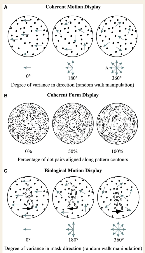
Coherent motion was assessed through a Global Dot Motion task (Newsome and Pare, 1988), where dots were presented in a rectangle 10.67° by 8.5° of visual angle centred on the screen (Fig. 1A). We manipulated the coherence of the display by the use of a standard ‘random walk’ paradigm (e.g. Williams and Sekuler, 1984). Every dot in the display was given the same intended direction (left or right). Between frames, each dot was independently given a new direction chosen from a uniform probability distribution centred on the intended direction. We manipulated the probability of perceiving coherent motion in the intended direction by varying the range of the distribution of direction vectors. In the current study, these ranges will be expressed in degrees. At 360°, no coherent direction can be perceived; only local, random movement (Brownian motion) can be seen. At 0°, no variance is allowed in any direction—all dots move in a straight line in the intended direction. The direction of each individual dot was independent from any other dot and independent of the direction it had moved in the last frame. All dots were assigned movement directions from the same distribution. Every dot conveys the same amount of information so that no single dot gives more directional information than another. As the coherence level drops, it becomes virtually impossible to track individual dot trajectories. To facilitate the use of the coherent motion display as a mask in the biological motion task, dots varied in their speed (moving between 4.5 and 9° of visual angle per second) and in the length of time between choosing new path directions (between 33.3 and 166.67 ms). These values for speed and step size were assigned to each dot independently and randomly, and were changed each time a dot was assigned a new direction. These settings were the same on all trials of both the coherent motion and the biological motion paradigms. Black dots were displayed on a white screen in an outlined rectangular box centred on the screen. The global direction (mean direction) was leftward on 50% of the trials and rightward on 50%. The method of constant stimuli was used so that the same six coherence levels (0°, 252°, 288°, 324°, 342° and 360°) were presented as a two-alternative forced choice to all participants. The coherence levels between 252° and 360° were chosen to assess sensitivity in the dynamic range of participants while the 0° condition allowed us to measure lapse rates directly. Participants were presented with 20 practice trials before the beginning of the experimental block. In each trial, participants were asked to indicate the direction of global motion and encouraged to guess when they did not know. No feedback was provided. Each stimulus was shown for 2 s and participants could answer at any time.
Figure 1.
Stimuli examples. (A) Depiction of the coherent motion stimuli across coherence levels where global motion is to the left. (B) Depiction of circular glass patterns at different coherence levels. (C) Depiction of the biological motion task where the global motion of the mask is to the left. Note that arrows and red dots/lines shown in either (A) or (C) were not visible during testing.
Coherent form stimuli
Concentric or expanding glass patterns (Glass, 1969; Glass and Perez, 1973) were presented one at a time in the centre of the screen (Fig. 1B). These patterns are an array of dot pairs where the coherence of the pattern is manipulated by the percentage of dot pairs aligned along a pattern. All dot pairs had the same spacing and each pattern at every coherence level was unique. Concentric patterns have dot pairs aligned along concentric circles while expanding patterns have pairs aligned along lines radiating from the centre. All patterns were presented in circular apertures with a diameter of 8° visual angle centred in the middle of the screen. A small fixation cross was placed at the centre of each pattern to encourage participants not to move their eyes while viewing the patterns. The average dot density was roughly consistent across each pattern as well as between different patterns. Six coherence levels (0%, 5%, 10%, 15%, 20% and 100%) were presented as a two-alternative forced choice to all participants. Concentric and expanding patterns occurred with equal frequency and were presented for 500 ms. Prior to testing, participants were shown patterns at 100% coherence and told that it would be harder to see the pattern on some trials than others. The concentric patterns were explained as circles inside of each other and were called ‘circle’ patterns. Expanding patterns were explained as lines coming out from the middle ‘like fireworks’ and called ‘burst’ patterns. Participants were first presented with 20 practice trials at 100% coherence to be sure they both understood the task and were able to map the correct response onto the correct button. In each trial, participants indicated whether patterns were concentric or expanding.
Biological motion stimuli
Thresholds for biological motion perception were tested by introducing noise into a standard point-light biological motion display of a walking human (Johansson, 1973) (Fig. 1C). Each walker consisted of 13 points of ‘light’ (black dots on a white background) placed on the major joints and head of a walking figure. The original stimuli were obtained from a standard stimulus set (Vanrie and Verfaillie, 2004) and then manipulated in MATLAB. The ‘walker’ stayed in one location as though walking on a treadmill. To introduce noise into these walker stimuli, we used exactly the same stimuli utilized in the coherent motion task described above and superimposed them on the point-light-walker displays. When the coherence of this noise ‘mask’ is high, the biological motion figure tends to ‘pop-out’ from the background. As the coherence of the mask is reduced, it becomes much more difficult to pick out the figure and determine in which direction it is walking. The ‘walker’ remained constant across trials, except for the direction of movement, while the coherence of the mask was manipulated at six difference coherence levels (0°, 72°, 144°, 216°, 288° and 360°). It is important to note here that the 0° coherence condition does not represent a condition without noise but is simply the condition at which the movement of the masking dots is completely coherent and the walking figure is most easily seen. The walker was presented at a slightly different position for each trial, moving around within the middle 1/16th of the stimulus rectangle to reduce the likelihood of participants forming a specific ‘template’ of the walker. Each stimulus was presented for 2 s and participants were asked to report in which direction the figure was walking. Prior to experimental blocks, participants were presented with 10 trials without noise to familiarize them with the walking figure. They then also completed 10 practice trials at the lowest noise level (360°).
Threshold estimation
To obtain detection thresholds for each task, a logistic function was fitted to each individual participant's data as a function of coherence level using the psignifit toolbox (version 2.5.6) for MATLAB. This program implements the maximum-likelihood procedure described by Wichmann and Hill (2001a, b). To obtain the best fit for most participants on all tasks, lambda was allowed to vary but was constrained to values between 0 and 0.05 and delta was set as a constant at 0.5. To estimate thresholds, slope, ‘lapse rate’ and error, a bootstrapping technique was used which included 5000 replications for each fitted function. Lapse rate is simply a measure that describes the rate of errors made at the tail end of the psychometric function, when there is little noise in the display and participants should be at close to 100% performance. The distribution of these fits was used to generate 95% confidence intervals for the estimates of each parameter. Error was described most succinctly by a deviance value, which is reflective of a combination of the residuals at each tested coherence level.
Participants were excluded from the data set for any particular task if their data would not fit adequately to the logistic function (Wichmann and Hill, 2001a). Fits were considered to be inadequate if either residual scores or deviance scores were larger than two standard deviations above the mean of the entire group of participants. Five participants with autism were dropped from the biological motion task analysis leaving 25 controls and 25 participants with autism. Four participants with autism and four control participants were dropped from the coherent motion task analysis, leaving 28 typical controls and 26 participants with autism. One participant with autism was dropped from the coherent form task, leaving 32 control participants and 29 participants with autism. Although we focused primarily on the threshold data obtained for each participant, lapse rate, response bias, reaction times and slope were also gathered. Groups did not differ on any of these measures (Table 2).
Table 2.
Details of psychophysical data
| Measure | Control group |
Autism group |
t | P-value | ||
|---|---|---|---|---|---|---|
| Mean (SD) | Range | Mean (SD) | Range | |||
| Coherent form | ||||||
| Lapse rate | 0.03 (0.02) | 0.00–0.05 | 0.02 (0.02) | 0.00–0.05 | 0.79 | 0.43 |
| Slope | 0.03 (0.02) | 0.01–0.08 | 0.04 (0.04) | 0.01–0.23 | −1.11 | 0.27 |
| Response bias | 0.05 (0.03) | 0.00–0.12 | 0.12 (0.08) | 0.00–0.29 | −0.15 | 0.88 |
| Reaction time (ms) | 1647.7 (577.1) | 598.7–3001.8 | 1128.2 (499) | 316.6–2237.5 | −1.26 | 0.21 |
| Coherent motion | ||||||
| Lapse rate | 0.01 (0.02) | 0.00–0.05 | 0.03 (0.02) | 0.00–0.05 | −1.13 | 0.27 |
| Slope | 0.05 (0.04) | 0.01–0.21 | 0.04 (0.02) | 0.01–0.07 | 0.82 | 0.42 |
| Response bias | 0.10 (0.08) | 0.02–0.35 | 0.06 (0.05) | 0.01–0.26 | −0.56 | 0.58 |
| Reaction time (ms) | 990.5 (323.1) | 460.5–1937.5 | 1695.8 (530.1) | 1080.1–2920.7 | −0.32 | 0.75 |
| Biological motion | ||||||
| Lapse rate | 0.01 (0.02) | 0.00–0.05 | 0.01 (0.02) | 0.00–0.05 | −0.37 | 0.71 |
| Slope | 0.01 (0.01) | 0.00–0.04 | 0.02 (0.02) | 0.01–0.09 | −1.49 | 0.15 |
| Response bias | 0.05 (0.03) | 0.01–0.12 | 0.05 (0.04) | 0.01–0.17 | −0.86 | 0.39 |
| Reaction time (ms) | 2097.3 (0.03) | 1198.3–3295.3 | 2042.7 (644.7) | 1096.9–3648.2 | 0.36 | 0.72 |
More lapsing is represented by higher values in the lapse rate column. Likewise, higher values for response bias represent greater bias towards one particular response in the two alternative forced choice tasks.
Results
Coherent motion
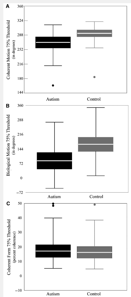
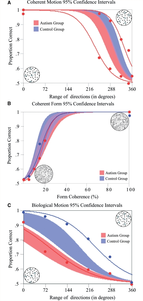
There was a significant difference between groups on 75% threshold [control mean = 309.82°, autism mean = 285.56°; t(52) = −2.586, P = 0.01] but there was also a strong relationship between performance IQ and 75% threshold for the autism group (r = 0.627, P = 0.001). This same relationship is seen with both verbal IQ (r = 0.515, P = 0.02) and full-score IQ (r = −0.607, P = 0.005) but because of missing data on these two measures and the non-verbal nature of the tasks, we will report principally on the effects of performance IQ. While the relationship between performance IQ and 75% threshold was significant for the autism group it was both non-significant and in the opposite direction for the control group (r = −0.218, P = 0.284). There is additionally a large degree of variability in the autism group such that some individual participants with autism performed better than the 95% confidence interval of the control group and others performed worse than the 95% confidence interval of controls (Figs 2A and 3A). Differences in the relationship between intellectual ability and visual motion perception between groups at least partially drive the threshold sensitivity difference between groups on this task. A one-way ANOVA was conducted with 75% threshold on the coherent motion task as a dependent variable, group as a fixed factor and performance IQ entered as a covariate. The results revealed IQ to be a significant factor [F(1,51) = 11.4, P = 0.001] but group was no longer a significant factor in between-group differences [F(1,55) = 1.1, P = 0.297]. To be sure that the ANCOVA analysis was sufficient to account for the influence of IQ on our dependent measures, we additionally created sub-samples matched on performance IQ, verbal IQ, full-score IQ and age (n = 23 in each group for performance IQ analysis, 19 in the autism group for verbal IQ and full-score IQ analyses). Similar to the results seen in the ANCOVA analysis, in this smaller sub-sample the difference between groups on 75% threshold dropped in significance to trend levels [control mean = 311.98, autism mean = 295.67, t(40) = −2.023, P = 0.061].
Figure 2.
Thresholds of 75% for (A) coherent motion, (B) coherent form and (C) biological motion perception for autism and control groups. Boxes represent plus and minus twice the standard error of the mean. The white line represents the mean of the group. Bars above and below the boxes include the entire data set except for outliers (defined as anyone >2 SDs from the mean of their group) whose data are depicted as individual dots. Higher 75% thresholds indicate better performance in (A) and (C) while <75% thresholds indicate better performance in (B).
Figure 3.
Individual subject data fitted with a psychometric function overlaid over each groups’ 95% confidence intervals for (A) coherent motion perception, (B) coherent form perception and (C) biological motion perception. Areas shaded in blue represent the control group's 95% confidence interval, and areas shaded red, the confidence interval for the autism group. Purple shading denotes overlap between the two groups’ confidence intervals.
Coherent form
No significant differences were revealed between groups for sensitivity during glass pattern discrimination trials [control mean = 16.2; autism mean = 17.1; t(59) = −0.313, P = 0.755]. In contrast to the coherent motion task, there was no clear influence of IQ on sensitivity for this task for either the control group (r = 0.307, P = 0.1) or the autism group (r = −0.085, P = 0.667; Figs 2B and 3B).
Biological motion
There was not only a significant difference between groups on 75% threshold [control mean = 153.94°, autism mean = 68.69°, t(55) = −3.287, P = 0.002] but, again, also a significant correlation between performance IQ and 75% threshold for the autism group (r = 0.412, P = 0.041) but not for the control group (r = 0.188, P = 0.321). This same relationship was seen with both verbal IQ (r = −0.462, P = 0.05) and full-score IQ (r = −0.479, P = 0.04) but because of missing data on these two measures and the non-verbal nature of the tasks, we again will focus principally on the effects of performance IQ. When the data were analysed using a one-way ANOVA with group entered as a fixed factor and performance IQ entered as a covariate, performance IQ emerged as a significant factor [F(1,55) = 8.2, P = 0.006] but, despite this influence, the main effect of group remained significant [F(1,55) = 5.23, P = 0.026; Figs 2C and 3C]. In our smaller IQ and age-matched sub-sample, the two groups remained significantly different on 75% threshold [control mean = 178.45°, autism mean = 78.30°, t(41) = −3.33, P = 0.002].
Correlation analyses
Relationships between tasks
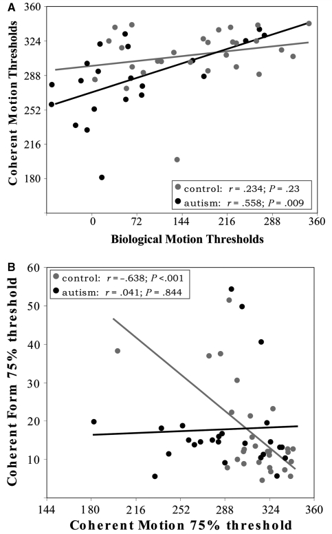
Performance on biological motion and coherent motion was correlated in the autism group (r = 0.558, P = 0.009) but not in the control group (r = 0.234, P = 0.23; Fig. 4A). Performing a partial correlation to account for the possible influence of performance IQ on this relationship reduced but did not eliminate the correlation between dynamic tasks in the autism group (r = 0.456, P = 0.04). Performance on the coherent form task was correlated with performance on the coherent motion task in the control group (r = −0.638, P < 0.001) but not in the autism group (r = 0.041, P = 0.844; Fig. 4B).
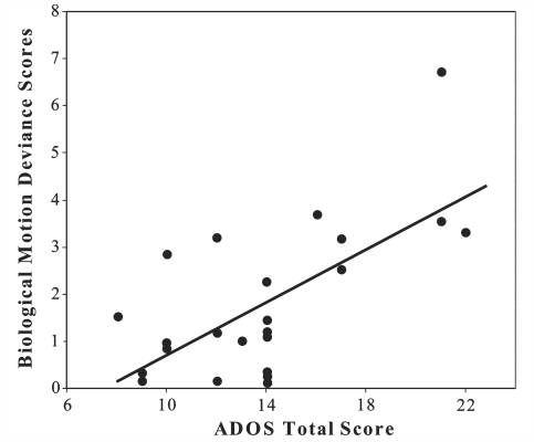
Figure 5.
The correlation between individual participant deviance scores (a measure of goodness-of-fit of the psychometric function) during the biological motion perception task with the degree of autism symptomatology as measured by the ADOS total score.
Figure 4.
Correlations between participant's performance on various tasks. In (A) the correlation between performance on the coherent and biological motion tasks. In (B) the correlation between performance on the coherent form and coherent motion tasks.
Relationships with autism and ADHD symptomatology
To be sure that ADHD symptomatology was not driving differences between our groups, we re-ran our statistics after removing the 15 participants with either an ADHD diagnosis or significant ADHD symptomatology. These groups showed almost identical results to our complete sample. Performance remained significantly different on both the coherent motion [control mean = 312.37°, autism mean = 276.48°, t(40) = −3.53, P = 0.001] and the biological motion [control mean = 153.14°, autism mean = 42.12°, t(43) = −4.05, P < 0.001] while performance on the coherent form task remained similar [control mean = 15.87, autism mean = 17.06, t(46) = −0.37, P = 0.71]. Similar to the results in the whole group, when the data were analysed using a one-way ANOVA with performance IQ and age entered as covariates, the main effect of group remained significant for the biological motion task [F(1,35) = 11.54, P = 0.002] but did not for the coherent motion task [F(1,35) = 3.44, P = 0.078].
In contrast to the correlation with IQ, there are no easily interpretable relationships between autism symptomatology as measured by the ADOS (or its sub-scores) and sensitivity measured on any of the tasks. The goodness-of-fit, one measure not typically reported in psychophysical experiments, did correlate strongly with ADOS measures. While all participants retained in these analyses had data that were well fitted by a logistic psychometric function, measures of goodness-of-fit (deviance scores) differed between individuals. After the exclusion of participants whose fits were not adequate, deviance scores for the biological motion task did not differ significantly between groups. As deviance values were not normally distributed, we tested possible differences between groups using the Mann–Whitney test (control mean = 2.91, SD = 2.25; autism mean = 1.74, SD = 1.56; U = 282, Z = −1.9, P = 0.06). Deviance scores were strongly related to the ADOS total score [roh = 0.513, P = 0.04 (Bonferroni corrected)] within the autism group. Neither deviance values nor ADOS scores were significantly correlated with performance IQ, verbal IQ or full-score IQ within the autism group. This relationship also could not be explained by possible influences from lambda values (performance at the highest tested coherence level) or by response biases, as none of these measures were different between groups (Table 2) or correlated with either ADOS scores, task performance or deviance values.
Discussion
Adolescents with autism performed as well as typical participants on a coherent form task that required discrimination between two static glass patterns but showed significant deficits on both a coherent motion and a biological motion perception task. In contrast with previous findings (Pellicano et al., 2005; Milne et al., 2006; Pellicano and Gibson, 2008), IQ scores correlated with task performance on both motion tasks in the autism group. Both factoring IQ into the between-group comparison and matching smaller groups on IQ removed much of the between-group differences on the coherent motion task but not on the biological motion task. This suggests that differences between groups on biological motion perception were not primarily due to differences in intellectual ability. IQ scores were not correlated with performance on the coherent form task, suggesting that the influence of IQ was specific to the dynamic tasks. Additionally, IQ did not influence performance on any task in the control group despite a similar range of IQ scores in the two groups, demonstrating that whatever influence IQ was exerting was specific to those with autism. IQ, even divided into verbal and performance subcomponents, is a large construct, reflective not only of intellectual ability but also of social and cultural competence. As a consequence, IQ measures may be assessing something different in those with autism than in the typically developing population and we can only speculate on the neural basis for such correlations. In our data, we observe a greater influence of IQ during dynamic tasks than during static tasks, which might lead us to the conjecture that IQ is acting as a proxy for temporal integration ability in the autism group. Such speculation, however, would have to be explicitly tested in future research. Although challenging to interpret, our IQ data are intriguing and warrant further study to ascertain explicity what influence IQ is exerting, as well as to determine whether IQ is measuring the same construct in those with autism as it is in typically developing populations.
The autism group was able to perform the coherent form task as well as typical participants and this demonstrates that deficits seen on the dynamic tasks cannot be the result of differences between groups in general attentiveness, task understanding or motivation. Including or excluding a group of participants with ADHD symptomatology also did not substantially affect our findings, implying that general attentional differences between groups were not driving our results. Additionally, the lack of difference between groups in lapse rates, response bias and average reaction time on all tasks argues against straightforward differences in general attentional engagement (Table 2).
Results from the coherent motion task do not support the theory of a simple sensory deficit specific to the processing of moving stimuli within the dorsal stream. Both the substantial influence of IQ and the wide range of sensitivity between individuals suggest that there is no uniform ‘perceptual’ deficit in those with autism. If a general dorsal stream deficit is present in some of those with autism, it is neither pervasive nor systematic. Additionally, in agreement with previous studies (Pellicano et al., 2005; Pellicano and Gibson, 2008), there is no indication that coherent motion deficits are connected with the degree of autism symptoms.
Our results from the coherent form task also argue against the possibility that coherent motion deficits seen in previous studies could be due only to a global integration deficit. The current study used glass patterns to ensure that there is no spatial frequency confound that could have allowed the pattern to be identified locally rather than through integration across the display (Dakin and Frith, 2005). Importantly, performance on the coherent motion and biological motion task was not correlated with performance on the coherent form task in our autism group. This also argues against a global integration deficit as the complete explanation for visual perception deficits in autism.
Our results from the biological motion task are more complex. Performance on this task was highly correlated with performance on the coherent motion task within the autism group but not highly correlated with performance on the form task. Both this correlation and its specificity to the autism group is in agreement with the findings of a recent paper investigating the relationship between coherent motion processing and the recognition of emotional states in biological motion displays (Atkinson, 2009). This relationship suggests that the deficit seen in the biological motion task was specific to dynamic stimuli and not to the integration of separate items into a coherent form. It is also important to point out that, unlike our other tasks, coherence levels in the biological motion task reflect changes in the masking noise rather than alterations of the biological motion ‘signal’ itself. There were no experimental trials without noise and the task required not only the dynamic integration of moving dots into a coherent form but also the segmentation of the biological motion figure from the masking noise. Thus, from our data set, we cannot determine if the impairment we saw was specific to biological motion perception itself or a result of difficulty with segmentation in noisy dynamic displays.
That we see a deficit, which is specific to dynamic stimuli and more severe in the biological motion condition, where both dynamic segmentation and dynamic form detection are required, is suggestive of a deficit in dynamic or ‘spatiotemporal’ attention. The integration of moving dots into a coherent percept of motion, although not attention intensive, is modulated by spatiotemporal attention (Bulakowski et al., 2007). The relatively simple integration of the biological motion cues into a coherent form also requires an allocation of dynamic attentional resources (Cavanagh et al., 2001; Battelli et al., 2003). Additionally, segmenting a moving form from a moving background requires significant spatiotemporal attention (Cavanagh et al., 2001; Thornton et al., 2002). This sort of dynamic attention is not the same as generalized attention and would have a selective impact upon dynamic stimuli. It would also have a larger effect on the biological motion task, where dynamic integration and dynamic segmentation are both required to complete the task accurately. While a deficit in spatiotemporal attention could more parsimoniously explain the mix of visual motion perception results from both this and earlier studies than either a specific dorsal stream deficit or a bias towards local processing, our data cannot directly address the question of such a deficit, as this study was not designed to assess spatiotemporal attention.
It is also important to remember that biological motion perception is not just a form-from-motion task, but it is also a social perception task. As such, those with autism may show impairment on a task requiring biological motion perception in noise not because of simple motion processing deficits or the dynamic attention necessary to perform the task but because of the social perception necessary to complete it. Indeed, several theories of autism posit that many of its symptoms develop directly from early deficits in social orientation and social reciprocity (Schultz et al., 2000, 2005; Dawson et al., 2005). Our data set cannot directly address the extent to which social cognition deficits might have an impact upon biological motion perception (or vice versa) as we did not manipulate the ‘socialness’ of our stimuli. Further studies including both biological and non-biological form-from-motion perception tasks that are matched on difficulty as well as assessments that better measure social cognition will be necessary to assess the true relationship between social perception deficits and biological motion perception.
The correlation between ADOS scores and deviance values on the biological motion task is intriguing. At the first level of analysis, it suggests that those participants who are most affected by autism (as measured by the ADOS) struggle, across coherence levels, to perceive the biological motion figure in the background noise consistently. In support of this idea, it is also important to note that while five participants with autism had to be removed from the biological motion analysis because their data would not be fit adequately, this was not the case for any of the control participants. Usually, researchers discard, discount or attempt to factor out information related to whether participants are able to do a task consistently (Klein, 2001). Motivation, generalized attention, lapsing and deviance from a perfect fit are all considered confounds. The relationship between ADOS scores and deviance scores on the biological motion task suggests that psychophysical measures beyond sensitivity should be examined more thoroughly. Measures of goodness-of-fit, lapsing and motivation may actually be important for understanding possible perceptual deficits in autism. In future studies it may not only be beneficial to take advantage of existing measures that naturally fall out of psychophysical testing, like the results reported here, but also to develop tools that can better help us determine which aspects of perception or task performance are reflected in these measures.
There is some indication in the literature that motion perception deficits may differentiate those with Asperger syndrome from those with other autism spectrum disorder diagnoses (Gepner and Mestre, 2002; Spencer and O’Brien, 2006; Tsermentseli et al., 2008). Our data cannot comment substantially on this question as only three participants with autism, who did not meet full criteria for autism on the ADOS, were included in our sample (all met autism spectrum criteria on the communication sub-score, full autism criteria on the social sub-score and were one or two points below full-autism diagnosis on the total score). Removing them from the data set made no significant difference to our findings and they were not outliers on any dependent measure. These data may, however, contribute to the characterization of subtypes within the greater autism spectrum. While our sample was too small to reveal the possibility of a bimodal distribution in scores, not all participants with autism showed impairment on either the coherent or the biological motion tasks. Similar to numbers previously reported in the literature (Milne et al., 2005; Pellicano and Gibson, 2008), only 34% of participants with autism performed at worse than one standard deviation from the control mean on the coherent motion task. On the biological motion task, 56% of autism participants performed worse than one standard deviation from the control mean.
The current results do not support either a general dorsal stream deficit or a bias towards local perception as explanations for visual perception differences in those with autism. Instead, they support a more nuanced interpretation. One distinct possibility suggested by our data is that differences in dynamic attention, which we might expect to have both frontal and parietal contributions, may drive many of the visual motion impairments reported in autism. Differences in social perception may also contribute significantly to biological motion perception deficits. Further psychophysical studies that include both non-biological form-from-motion segmentation tasks as well as tasks that more directly assess spatiotemporal attention will be necessary to assess the roles of social perception and segmentation as well as determine if dynamic attention is, in fact, different in those with autism.
Funding
Autism Speaks through a Pre-doctoral Mentored Research Award (S.M.R.).
Acknowledgements
We are grateful to the research participants and their families, Dr Kenneth Britten for his assistance with stimulus creation and Tom Kiely for his help with participant recruitment.
Glossary
Abbreviations
- ADHD
Attention-deficit hyperactivity disorder
- ADOS
Autism Diagnostic Observation Schedule
References
- Allison T, Puce A, McCarthy G. Social perception from visual cues: role of the STS region. Trends Cogn Sci. 2000;4:267–78. doi: 10.1016/s1364-6613(00)01501-1. [DOI] [PubMed] [Google Scholar]
- Atkinson AP. Impaired recognition of emotions from body movements is associated with elevated motion coherence thresholds in autism spectrum disorders. Neuropsychologia. 2009;47:3023–9. doi: 10.1016/j.neuropsychologia.2009.05.019. [DOI] [PubMed] [Google Scholar]
- Atkinson J, Anker S, Braddick O, Nokes L, Mason A, Braddick F. Visual and visuospatial development in young children with Williams syndrome. Dev Med Child Neurol. 2001;43:330–7. doi: 10.1017/s0012162201000615. [DOI] [PubMed] [Google Scholar]
- Atkinson J, King J, Braddick O, Nokes L, Anker S, Braddick F. A specific deficit of dorsal stream function in Williams’ syndrome. Neuroreport. 1997;8:1919–22. doi: 10.1097/00001756-199705260-00025. [DOI] [PubMed] [Google Scholar]
- Battelli L, Cavanagh P, Thornton IM. Perception of biological motion in parietal patients. Neuropsychologia. 2003;41:1808–16. doi: 10.1016/s0028-3932(03)00182-9. [DOI] [PubMed] [Google Scholar]
- Bertone A, Mottron L, Jelenic P, Faubert J. Motion perception in autism: a ‘complex’ issue. J Cogn Neurosci. 2003;15:218–25. doi: 10.1162/089892903321208150. [DOI] [PubMed] [Google Scholar]
- Bertone A, Mottron L, Jelenic P, Faubert J. Enhanced and diminished visuo-spatial information processing in autism depends on stimulus complexity. Brain. 2005;128:2430–41. doi: 10.1093/brain/awh561. [DOI] [PubMed] [Google Scholar]
- Berument SK, Rutter M, Lord C, Pickles A, Bailey A. Autism screening questionnaire: diagnostic validity. Br J Psychiatry. 1999;175:444–51. doi: 10.1192/bjp.175.5.444. [DOI] [PubMed] [Google Scholar]
- Blake R, Turner LM, Smoski MJ, Pozdol SL, Stone WL. Visual recognition of biological motion is impaired in children with autism. Psychol Sci. 2003;14:151–7. doi: 10.1111/1467-9280.01434. [DOI] [PubMed] [Google Scholar]
- Blakemore SJ, Frith U. How does the brain deal with the social world? Neuroreport. 2004;15:119–28. doi: 10.1097/00001756-200401190-00024. [DOI] [PubMed] [Google Scholar]
- Boddaert N, Chabane N, Belin P, Bourgeois M, Royer V, Barthelemy C, et al. Perception of complex sounds in autism: abnormal auditory cortical processing in children. Am J Psychiatry. 2004;161:2117–20. doi: 10.1176/appi.ajp.161.11.2117. [DOI] [PubMed] [Google Scholar]
- Braddick O, Atkinson J, Wattam-Bell J. Normal and anomalous development of visual motion processing: motion coherence and ‘dorsal-stream vulnerability’. Neuropsychologia. 2003;41:1769–84. doi: 10.1016/s0028-3932(03)00178-7. [DOI] [PubMed] [Google Scholar]
- Bulakowski PF, Bressler DW, Whitney D. Shared attentional resources for global and local motion processing. J Vis. 2007;7:10,1–10. doi: 10.1167/7.10.10. [DOI] [PMC free article] [PubMed] [Google Scholar]
- Calder AJ, Young AW. Understanding the recognition of facial identity and facial expression. Nat Rev Neurosci. 2005;6:641–51. doi: 10.1038/nrn1724. [DOI] [PubMed] [Google Scholar]
- Campbell R, MacSweeney M, Surguladze S, Calvert G, McGuire P, Suckling J, et al. Cortical substrates for the perception of face actions: an fMRI study of the specificity of activation for seen speech and for meaningless lower-face acts (gurning) Brain Res Cogn Brain Res. 2001;12:233–43. doi: 10.1016/s0926-6410(01)00054-4. [DOI] [PubMed] [Google Scholar]
- Cavanagh P, Labianca AT, Thornton IM. Attention-based visual routines: sprites. Cognition. 2001;80:47–60. doi: 10.1016/s0010-0277(00)00153-0. [DOI] [PubMed] [Google Scholar]
- Cowey A, Vaina LM. Blindness to form from motion despite intact static form perception and motion detection. Neuropsychologia. 2000;38:566–78. doi: 10.1016/s0028-3932(99)00117-7. [DOI] [PubMed] [Google Scholar]
- Dakin S, Frith U. Vagaries of visual perception in autism. Neuron. 2005;48:497–507. doi: 10.1016/j.neuron.2005.10.018. [DOI] [PubMed] [Google Scholar]
- Dawson G, Webb SJ, McPartland J. Understanding the nature of face processing impairment in autism: insights from behavioral and electrophysiological studies. Dev Neuropsychol. 2005;27:403–24. doi: 10.1207/s15326942dn2703_6. [DOI] [PubMed] [Google Scholar]
- Del Viva MM, Igliozzi R, Tancredi R, Brizzolara D. Spatial and motion integration in children with autism. Vis Res. 2006;46:1242–52. doi: 10.1016/j.visres.2005.10.018. [DOI] [PubMed] [Google Scholar]
- Freitag CM, Konrad C, Haberlen M, Kleser C, von Gontard A, Reith W, et al. Perception of biological motion in autism spectrum disorders. Neuropsychologia. 2008;46:1480–94. doi: 10.1016/j.neuropsychologia.2007.12.025. [DOI] [PubMed] [Google Scholar]
- Gepner B, Mestre DR. Brief report: postural reactivity to fast visual motion differentiates autistic from children with Asperger syndrome. J Autism Dev Disord. 2002;32:231–8. doi: 10.1023/a:1015410015859. [DOI] [PubMed] [Google Scholar]
- Gervais H, Belin P, Boddaert N, Leboyer M, Coez A, Sfaello I, et al. Abnormal cortical voice processing in autism. Nat Neurosci. 2004;7:801–2. doi: 10.1038/nn1291. [DOI] [PubMed] [Google Scholar]
- Glass L. Moire effect from random dots. Nature. 1969;223:578–80. doi: 10.1038/223578a0. [DOI] [PubMed] [Google Scholar]
- Glass L, Perez R. Perception of random dot interference patterns. Nature. 1973;246:360–2. doi: 10.1038/246360a0. [DOI] [PubMed] [Google Scholar]
- Goodale MA, Milner AD. Separate visual pathways for perception and action. Trends Neurosci. 1992;15:20–5. doi: 10.1016/0166-2236(92)90344-8. [DOI] [PubMed] [Google Scholar]
- Grossman E, Donnelly M, Price R, Pickens D, Morgan V, Neighbor G, et al. Brain areas involved in perception of biological motion. J Cogn Neurosci. 2000;12:711–20. doi: 10.1162/089892900562417. [DOI] [PubMed] [Google Scholar]
- Gunn A, Cory E, Atkinson J, Braddick O, Wattam-Bell J, Guzzetta A, et al. Dorsal and ventral stream sensitivity in normal development and hemiplegia. Neuroreport. 2002;13:843–7. doi: 10.1097/00001756-200205070-00021. [DOI] [PubMed] [Google Scholar]
- Happe F, Frith U. The weak coherence account: detail-focused cognitive style in autism spectrum disorders. J Autism Dev Disord. 2006;36:5–25. doi: 10.1007/s10803-005-0039-0. [DOI] [PubMed] [Google Scholar]
- Happe FG, Booth RD. The power of the positive: revisiting weak coherence in autism spectrum disorders. Q J Exp Psychol. 2008;61:50–63. doi: 10.1080/17470210701508731. [DOI] [PubMed] [Google Scholar]
- Hubert B, Wicker B, Moore DG, Monfardini E, Duverger H, Da Fonseca D, et al. Brief report: recognition of emotional and non-emotional biological motion in individuals with autistic spectrum disorders. J Autism Dev Disord. 2007;37:1386–92. doi: 10.1007/s10803-006-0275-y. [DOI] [PubMed] [Google Scholar]
- Jeannerod M. The neural and behavioural organization of goal-directed movements. Oxford: Clarendon Press; 1988. [Google Scholar]
- Johansson G. Visual perception of biological motion and a model for its analysis. Percept Psychophys. 1973;14:201–11. [Google Scholar]
- Jolliffe T, Baron-Cohen S. Are people with autism and Asperger syndrome faster than normal on the Embedded Figures Test? J Child Psychol Psychiatry. 1997;38:527–34. doi: 10.1111/j.1469-7610.1997.tb01539.x. [DOI] [PubMed] [Google Scholar]
- Klein SA. Measuring, estimating, and understanding the psychometric function: a commentary. Percept Psychophys. 2001;63:1421–55. doi: 10.3758/bf03194552. [DOI] [PubMed] [Google Scholar]
- Klin A, Jones W. Altered face scanning and impaired recognition of biological motion in a 15-month-old infant with autism. Dev Sci. 2008;11:40–6. doi: 10.1111/j.1467-7687.2007.00608.x. [DOI] [PubMed] [Google Scholar]
- Klin A, Lin DJ, Gorrindo P, Ramsay G, Jones W. Two-year-olds with autism orient to non-social contingencies rather than biological motion. Nature. 2009;459:257–61. doi: 10.1038/nature07868. [DOI] [PMC free article] [PubMed] [Google Scholar]
- Lord C, Risi S, Lambrecht L, Cook EHJ, Leventhal BL, DiLavore PC, et al. The autism diagnostic observation schedule-generic: a standard measure of social and communication deficits associated with the spectrum of autism. J Autism Dev Disord. 2000;30:205–23. [PubMed] [Google Scholar]
- Milne E, Swettenham J, Campbell R. Motion perception and autistic spectrum disorder: a review. Cah Psychol Cognit/Curr Psychol Cognit. 2005;23:3–36. [Google Scholar]
- Milne E, Swettenham J, Campbell R, Coleman M. Degree of global/local processing is related to coherent motion detection. Paper presented at the International Meeting for Autism Research. Sacramento, CA: 2004. 2004. [Google Scholar]
- Milne E, Swettenham J, Hansen P, Campbell R, Jeffries H, Plaisted K. High motion coherence thresholds in children with autism. J Child Psychol Psychiatry. 2002;43:255–63. doi: 10.1111/1469-7610.00018. [DOI] [PubMed] [Google Scholar]
- Milne E, White S, Campbell R, Swettenham J, Hansen P, Ramus F. Motion and form coherence detection in autistic spectrum disorder: relationship to motor control and 2:4 digit ratio. J Autism Dev Disord. 2006;36:225–37. doi: 10.1007/s10803-005-0052-3. [DOI] [PubMed] [Google Scholar]
- Moore DG, Hobson RP, Lee A. Components of person perception: an investigation with autistic, non-autistic retarded and typically developing children and adolescents. Br J Dev Psychol. 1997;15:401–23. [Google Scholar]
- Mottron L, Burack JA, Iarocci G, Belleville S, Enns JT. Locally oriented perception with intact global processing among adolescents with high-functioning autism: evidence from multiple paradigms. J Child Psychol Psychiatry. 2003;44:904–13. doi: 10.1111/1469-7610.00174. [DOI] [PubMed] [Google Scholar]
- Newsome WT, Pare EB. A selective impairment of motion perception following lesions of the middle temporal visual area (MT) J Neurosci. 1988;8:2201–11. doi: 10.1523/JNEUROSCI.08-06-02201.1988. [DOI] [PMC free article] [PubMed] [Google Scholar]
- O'Riordan MA, Plaisted KC, Driver J, Baron-Cohen S. Superior visual search in autism. J Exp Psychol Hum Percept Perform. 2001;27:719–30. doi: 10.1037//0096-1523.27.3.719. [DOI] [PubMed] [Google Scholar]
- Ochiai T, Grimault S, Scavarda D, Roch G, Hori T, Riviere D, et al. Sulcal pattern and morphology of the superior temporal sulcus. Neuroimage. 2004;22:706–19. doi: 10.1016/j.neuroimage.2004.01.023. [DOI] [PubMed] [Google Scholar]
- Parron C, Da Fonseca D, Santos A, Moore DG, Monfardini E, Deruelle C. Recognition of biological motion in children with autistic spectrum disorders. Autism. 2008;12:261–74. doi: 10.1177/1362361307089520. [DOI] [PubMed] [Google Scholar]
- Pellicano E, Gibson L, Maybery M, Durkin K, Badcock DR. Abnormal global processing along the dorsal visual pathway in autism: a possible mechanism for weak visuospatial coherence? Neuropsychologia. 2005;43:1044–53. doi: 10.1016/j.neuropsychologia.2004.10.003. [DOI] [PubMed] [Google Scholar]
- Pellicano E, Gibson LY. Investigating the functional integrity of the dorsal visual pathway in autism and dyslexia. Neuropsychologia. 2008;46:2593–6. doi: 10.1016/j.neuropsychologia.2008.04.008. [DOI] [PubMed] [Google Scholar]
- Pelphrey KA, Carter EJ. Charting the typical and atypical development of the social brain. Dev Psychopathol. 2008;20:1081–102. doi: 10.1017/S0954579408000515. [DOI] [PubMed] [Google Scholar]
- Pelphrey KA, Morris JP, McCarthy G. Neural basis of eye gaze processing deficits in autism. Brain. 2005;128:1038–48. doi: 10.1093/brain/awh404. [DOI] [PubMed] [Google Scholar]
- Pelphrey KA, Morris JP, McCarthy G, Labar KS. Perception of dynamic changes in facial affect and identity in autism. Soc Cogn Affect Neurosci. 2007;2:140–9. doi: 10.1093/scan/nsm010. [DOI] [PMC free article] [PubMed] [Google Scholar]
- Plaisted K, O’Riordan M, Baron-Cohen S. Enhanced visual search for a conjunctive target in autism: a research note. J Child Psychol Psychiatry. 1998;39:777–83. [PubMed] [Google Scholar]
- Plaisted K, Swettenham J, Rees L. Children with autism show local precedence in a divided attention task and global precedence in a selective attention task. J Child Psychol Psychiatry. 1999;40:733–42. [PubMed] [Google Scholar]
- Pomerantz JR. Global and local precedence: selective attention in form and motion perception. J Exp Psychol Gen. 1983;112:516–40. doi: 10.1037//0096-3445.112.4.516. [DOI] [PubMed] [Google Scholar]
- Puce A, Perrett D. Electrophysiology and brain imaging of biological motion. Philos Trans R Soc Lond B Biol Sci. 2003;358:435–45. doi: 10.1098/rstb.2002.1221. [DOI] [PMC free article] [PubMed] [Google Scholar]
- Rutter M, Bailey A, Lord C. SCQ: Social Communication Questionnaire. Los Angeles, CA: Western Psychological Services; 2003. [Google Scholar]
- Sanchez-Marin FJ, Padilla-Medina JA. A Psychophysical Test of the Visual Pathway of Children with Autism. J Autism Dev Disord. 2007;38:1270–7. doi: 10.1007/s10803-007-0507-9. [DOI] [PubMed] [Google Scholar]
- Schneider GE. Two visual systems. Science. 1969;163:895–902. doi: 10.1126/science.163.3870.895. [DOI] [PubMed] [Google Scholar]
- Schultz RT. Developmental deficits in social perception in autism: the role of the amygdala and fusiform face area. Int J Dev Neurosci. 2005;23:125–41. doi: 10.1016/j.ijdevneu.2004.12.012. [DOI] [PubMed] [Google Scholar]
- Schultz RT, Gauthier I, Klin A, Fulbright RK, Anderson AW, Volkmar F, et al. Abnormal ventral temporal cortical activity during face discrimination among individuals with autism and Asperger syndrome. Arch Gen Psychiatry. 2000;57:331–40. doi: 10.1001/archpsyc.57.4.331. [DOI] [PubMed] [Google Scholar]
- Shah A, Frith U. An islet of ability in autistic children: a research note. J Child Psychol Psychiatry. 1983;24:613–20. doi: 10.1111/j.1469-7610.1983.tb00137.x. [DOI] [PubMed] [Google Scholar]
- Shah A, Frith U. Why do autistic individuals show superior performance on the block design task? J Child Psychol Psychiatry. 1993;34:1351–64. doi: 10.1111/j.1469-7610.1993.tb02095.x. [DOI] [PubMed] [Google Scholar]
- Spencer J, O’Brien J, Riggs K, Braddick O, Atkinson J, Wattam-Bell J. Motion processing in autism: evidence for a dorsal stream deficiency. Neuroreport. 2000;11:2765–7. doi: 10.1097/00001756-200008210-00031. [DOI] [PubMed] [Google Scholar]
- Spencer JV, O’Brien JM. Visual form-processing deficits in autism. Perception. 2006;35:1047–55. doi: 10.1068/p5328. [DOI] [PubMed] [Google Scholar]
- Thornton IM, Rensink RA, Shiffrar M. Active versus passive processing of biological motion. Perception. 2002;31:837–53. doi: 10.1068/p3072. [DOI] [PubMed] [Google Scholar]
- Trevarthen CB. Two mechanisms of vision in primates. Psychol Forsch. 1968;31:299–348. doi: 10.1007/BF00422717. [DOI] [PubMed] [Google Scholar]
- Tsermentseli S, O’Brien JM, Spencer JV. Comparison of form and motion coherence processing in autistic spectrum disorders and dyslexia. J Autism Dev Disord. 2008;38:1201–10. doi: 10.1007/s10803-007-0500-3. [DOI] [PubMed] [Google Scholar]
- Ungerleider LG, Haxby JV. ‘What’ and ‘where’ in the human brain. Curr Opin Neurobiol. 1994;4:157–65. doi: 10.1016/0959-4388(94)90066-3. [DOI] [PubMed] [Google Scholar]
- Vaina LM, Solomon J, Chowdhury S, Sinha P, Belliveau JW. Functional neuroanatomy of biological motion perception in humans. Proc Natl Acad Sci U S A. 2001;98:11656–61. doi: 10.1073/pnas.191374198. [DOI] [PMC free article] [PubMed] [Google Scholar]
- Vandenbroucke MW, Scholte HS, van Engeland H, Lamme VA, Kemner C. Coherent versus component motion perception in autism spectrum disorder. J Autism Dev Disord. 2008;38:941–9. doi: 10.1007/s10803-007-0467-0. [DOI] [PubMed] [Google Scholar]
- Vanrie J, Verfaillie K. Perception of biological motion: a stimulus set of human point-light actions. Behav Res Methods Instrum Comput. 2004;36:625–9. doi: 10.3758/bf03206542. [DOI] [PubMed] [Google Scholar]
- Wechsler D. Wechsler Abbreviated Scale of Intelligence (WASI) Harcourt Assessment, The Psychological Corporation, London; 1999. [Google Scholar]
- White S, Frith U, Milne E, Stuart R, Swettenham J, Ramus F. A double dissociation between sensorimotor impairments and reading disability: a comparison of autistic and dyslexic children. Cogn Neuropsychol. 2006;23:748–61. doi: 10.1080/02643290500438607. [DOI] [PubMed] [Google Scholar]
- Wichmann FA, Hill NJ. The psychometric function: I. Fitting, sampling, and goodness of fit. Percept Psychophys. 2001a;63:1293–1313. doi: 10.3758/bf03194544. [DOI] [PubMed] [Google Scholar]
- Wichmann FA, Hill NJ. The psychometric function: II. Bootstrap-based confidence intervals and sampling. Percept Psychophys. 2001b;63:1314–29. doi: 10.3758/bf03194545. [DOI] [PubMed] [Google Scholar]
- Williams DW, Sekuler R. Coherent global motion percepts from stochastic local motions. Vis Res. 1984;24:55–62. doi: 10.1016/0042-6989(84)90144-5. [DOI] [PubMed] [Google Scholar]
- Zeki SM. Functional organization of a visual area in the posterior bank of the superior temporal sulcus of the rhesus monkey. J Physiol. 1974;236:549–73. doi: 10.1113/jphysiol.1974.sp010452. [DOI] [PMC free article] [PubMed] [Google Scholar]
- Zilbovicius M, Meresse I, Chabane N, Brunelle F, Samson Y, Boddaert N. Autism, the superior temporal sulcus and social perception. Trends Neurosci. 2006;29:359–5. doi: 10.1016/j.tins.2006.06.004. [DOI] [PubMed] [Google Scholar]


