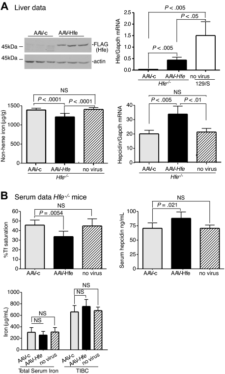
Figure 1.
Liver-specific expression of Hfe lowers iron levels, increases hepcidin expression, and lowers Tf saturation in Hfe−/− mice. Hfe−/− mice (n = 3) were injected with AAV-Hfe (5 × 10¹¹ genome equivalents per mouse; ■ in all panels) or with the same amount of AAV-c virus (▩). Two weeks later, the mice were killed. (A) Liver extracts (100 mg) were analyzed by immunoblots with the use of anti–FLAG-M2 antibody or antiactin. Bands were detected by horseradish peroxidase secondary antibodies and enhanced chemiluminescence. Hfe mRNA was determined by quantitative reverse transcription–PCR (qRT-PCR) and normalized to Gapdh. The levels of Hfe mRNA in AAV-c– and in AAV-Hfe–injected Hfe−/− mice were compared with strain-, sex-, and age-matched control wild-type 129/SvTac (129/S) mice (□) with the use of qRT-PCR. Nonheme iron in livers of uninjected Hfe−/− mice ( ) and of AAV-c– and AAV-Hfe–injected Hfe−/− mice is described in “Nonheme iron assay.” Each sample was measured in triplicate. Hepcidin mRNA in Hfe−/− murine liver was determined by qRT-PCR. (B) Serum data. Percentage of Tf saturation was calculated from total serum iron and total iron-binding capacity (TIBC). The latter 2 parameters were measured as described in “Blood iron analysis.” Three to 4 mice were used in each group. All mice were on a 129/S background. The experiments were repeated with 2 more sets of mice with similar results. Error bars represent SD.

