Abstract
Sepsis and septic acute lung injury are among the leading causes for morbidity and mortality of critical illness. Extracellular adenosine is a signaling molecule implicated in the cellular adaptation to hypoxia, ischemia or inflammation. Therefore, we pursued the role of the A2B adenosine receptor (A2BAR) as potential therapeutic target in endotoxin-induced acute lung injury. We gained initial insight from in vitro studies of cultured endothelia or epithelia exposed to inflammatory mediators showing time-dependent induction of the A2BAR (up to 12.9±3.4-fold, p<0.05). Similarly, murine studies of endotoxin-induced lung injury identified an almost 4.6-fold induction of A2BAR transcript and corresponding protein induction with LPS-exposure. Studies utilizing A2BAR promoter constructs and RNA-protection assays indicated that A2BAR induction involved mRNA stability. Functional studies of LPS-induced lung injury revealed that pharmacological inhibition or genetic deletion of the A2BAR was associated with dramatic increases in lung inflammation and histologic tissue injury. Studies of A2BAR-bone marrow chimeric mice suggested pulmonary A2BAR signaling in lung protection. Finally, studies with a specific A2BAR agonist (BAY 60-6583) demonstrated attenuation of lung inflammation and pulmonary edema in wild-type but not in gene-targeted mice for the A2BAR. These studies suggest the A2BAR as potential therapeutic target in the treatment of endotoxin-induced forms of acute lung injury.
Keywords: Adenosine, A2B, lung injury, sepsis
Introduction
Sepsis is a serious medical condition characterized by a whole-body inflammatory state (called systemic inflammatory response syndrome) and the presence of a known or suspected infection. The body develops this generalized inflammatory reaction as a response to microbes or microbial toxins (such as LPS) circulating in the blood. In the USA, sepsis is the leading cause of death in critically ill patients. As such, sepsis develops in 750,000 people annually, resulting in more than 210,000 mortalities (1). Mortality rates associated with severe sepsis and septic shock are 25 to 30%, and 40 to 70%, respectively (2). One of the main problems for patients with sepsis is the development of respiratory failure. Indeed, up to 40% of patients with sepsis go on to develop respiratory failure in the form of acute lung injury (ALI) or its more severe form acute respiratory distress syndrome (ARDS) (3). ALI is characterized by bilateral pulmonary edema, and severe hypoxia and is considered the leading cause of death in patients suffering from sepsis (4). At present, therapeutic approaches for patients suffering from septic lung injury are mainly symptomatic (1, 3). Therefore, the search for novel and specific therapies to prevent or treat sepsis-associated respiratory failure and endotoxin-induced acute lung injury is an area of intense investigation.
Previous studies indicated that the generation of the extracellular signaling molecule adenosine plays a role in inflammatory cell trafficking and lung inflammation during endotoxin-induced lung injury (5). Adenosine is generated in the extracellular space via enzymatic phosphohydrolysis from its precursor molecules (6). This two step reaction is under the enzymatic control of two enzymes located on the extracellular surface – the ecto-apyrase (conversion of ATP/ADP to AMP, CD39)(7) and the ecto-5′-nucleotidase (CD73, conversion of AMP to adenosine) (8). Studies in LPS-induced acute lung injury and lung inflammation demonstrated that gene-targeted mice for CD39 or CD73 show a more severe degree of lung inflammation than their corresponding controls (5) These studies indicate a potential role for extracellular adenosine signaling in LPS-induced lung injury. Extracellular adenosine can signal through four different adenosine receptors (ARs), the A1AR, A2AAR, A2BAR and A3AR. Previous studies also showed that the dominant adenosine receptor in the lungs is the A2BAR (9) with its expression predominantly on pulmonary epithelia (10), vascular endothelia (11–14) or inflammatory cells (11, 14). Functional studies implicated the A2BAR in hypoxia-driven pulmonary edema (11), or lung inflammation during mechanical ventilation (15). Moreover, a recent study demonstrated a key role of A2BAR-dependent attenuation of hypoxia-driven inflammation of mucosal organs, including the lungs (16). Based on these studies we hypothesize that the A2BAR could represent a potential therapeutic target for endotoxin-induced acute lung injury. Therefore, we utilized a combination of pharmacological and genetic approaches to determine the role of the A2BAR in lung inflammation in LPS induced lung injury.
Material and Methods
Cell culture and inflammatory stimulation
Human Microvascular Endothelial Cells (HMEC-1) and cultured pulmonary epithelial cells (A549 cells, LGC Standards GmbH, Wesel, Germany) were cultured as described previously.(17–20) Primary pulmonary endothelial cells (HMVEC-L, Lonza Walkersville, Inc., Walkersville, MD, USA) were cultured under supplier’s instructions. Cells were grown to full confluency and stimulated with 1μM PGE2 (Sigma, Taufkirchen, Germany), 10 ng/ml Interleukin-1β (PromoKine, Heidelberg, Germany),70 ng/ml Interleukin-4 (PromoKine) and 20 ng/ml Interleukin-6 (PromoKine) for 6, 12 and 24h.
Murine LPS inhalation model
Experiment protocols were approved by the University of Tübingen, Germany, or the University of Colorado Denver USA. They were also in accordance with the German Law on the Protection of Animals and the NIH guidelines for use of live animals. C57BL/6J mice (Charles River Laboratories International Inc., Wilmington, MA, USA), A2BAR−/− mice on a C57BL/6J or age-, gender- and weight-matched littermate controls were bred and genotyped as described previously (12, 21). Mice at age 8 to 12 weeks were exposed to aerosolized LPS in a cylindrical chamber connected to an air nebulizer (MicroAir; Omron Healthcare Inc., Mannheim, Germany). The outlet of the chamber was connected to a vacuum pump producing a constant flow rate. LPS from E. coliB026 (Sigma) was dissolved in 0.9% saline (500 μg/ml), A2BAR antagonist MRS1754 (Tocris, Bristol, UK) was dissolved in 0.9% saline (2.4 μg/ml) and mice were allowed to inhale for 30 min prior to LPS exposure. Control mice were exposed to saline aerosol. In other studies, mice were pre-treated with the A2BAR agonist BAY 60-6583 (2 mg/kg i.p., Bayer Healthcare, Germany) or vehicle (21). Mice were sacrificed 4h after LPS exposure, the remaining blood was removed from the pulmonary circulation by injecting 1 ml of PBS into the right ventricle. Lungs were excised and immediately frozen at −80°C for further analysis.
Human and mouse transcriptional analysis
Total RNA was extracted as described previously (12, 21–28) using the RNA isolation kit NucleoSpin RNA II (Machery & Nagel, Düren, Germany). RNA was washed and the concentration was quantified. cDNA synthesis was performed by using iScript cDNA Synthesis Kit (Bio-Rad Laboratories, Inc, Munich, Germany) according to the manufactor’s instructions. Quantitative real-time reverse transcriptase PCR (RT-PCR) (iCycler; Bio-Rad Laboratories, Inc.) was performed to examine A2BAR expression levels. qPCR master mix contained 1 μM sense and 1μM antisense primers with iQ™ SYBR® Green (Bio-Rad Laboratories, Inc.). To quantify change in A2BAR transcript the following primer sets were used: Sense 5′-ATC TCC AGG TAT CTT CTC -3′, antisense 5′-GTT GGC ATA ATC CAC ACA G -3′. Samples were controlled for β-actin using following primers: Sense 5′-GGA GAA AAT CTG GCA CCA CA -3′, antisense 5′-AGA GGC GTA CAG GGA TAG CA -3′.
To analyze murine A2BAR transcript, lung tissue was excised and total RNA was isolated. A2BAR mRNA levels were quantified using sense primer 5′-GGG CAG CAA CTC AGA AAA CT -3′ and 5′-GGA AGG ACT TCG TCT CTC CA -3′ antisense primer. Murine β-actin expression was evaluated with sense 5′-GGC TCC TAG CAC CAT GAA GA -3′, antisense 5′-TCT GCT GGA AGG TGG ACA G -3′, murine IL-1β expression with sense 5′-GGC AGG CAG TAT CAC TCA TT -3′, antisense 5′-CAC ACC AGC AGG TTA TCA TC -3′, murine IL-6 expression with sense 5′-ACC GCT ATG AAG TTC CTC TC -3′ and antisense 5′-CTC CGA CTT GTG AAG TGG TA -3′ and murine TNF-α expression with sense 5′-CAG GCG GTG CCT ATG TCT CA -3′ and antisense 5′-TCC AGC TGC TCC TCC ACT TG -3′ primers.
A2BAR reporter assay
To measure the transcriptional activity of A2BAR, epithelial cells were plated in 24-well plates at a density of 2.5×10⁴ cell/well and allowed to adhere overnight. The monolayers were then transfected with 0.5μg of either A2BAR-luc promoter reporter (29) or control pGL3 vector, and co-transfected with 0.05μg Renilla reporter vector for 6h using Fugene 6 transfection reagent (Roche) in accordance with the manufacturer’s instructions. Cells were then treated with either PGE2 (1μM), IL-1β (10ng/mL), IL-4 (70ng/mL), or IL-6 (20ng/mL) for 12h. Cells were then washed twice in ice-cold PBS and Dual Luciferase Reporter Assay (Promega) carried out according to manufacturer’s instructions.
A2BAR mRNA Stability Assay
For the mRNA degradation assay, HMECS were plated on 60mm dishes and incubated 37C overnight. Cells were then treated with either 0.5uM PGE2, or vehicle (0.015% EtOH) for 4 h at 37C. Subsequently, cells were then treated with the transcriptional inhibitor 5,6-dichlorobenzimidazole (DRB, 50μM) to prevent de novo transcription of mRNA. RNA was harvested at 0, 1, 2, 3 and 4h post-treatment with DRB using trizol. The degradation rate of A2BAR mRNA was evaluated by real time RT-PCR (n=3) and calculated relative to the amount of mRNA levels at 0h.
ELISA for IL-6 from lung tissue
The snap-frozen lungs were thawed, weighed, transferred to different tubes on ice containing 1 ml of Tissue Protein Extraction Reagent (T-PER; Pierce Biotechnology). The lung tissues were homogenized at 4°C. Lung homogenates were centrifuged at 9,000 g for 10 min at 4°C. Supernatants were transferred to clean microcentrifuge tubes, frozen on dry ice, and thawed on ice. Total protein concentrations in the lung tissue homogenates were determined using a bicinchoninic acid kit (Pierce Biotechnology). Lung tissue homogenates were diluted with 50% assay diluent and 50% T-PER reagent to a final protein concentration of 400 μg/ml. IL-6 levels were evaluated in lung tissue homogenates using a mouse ELISA kit (R&D Systems), in accordance with the manufacturer’s instructions.
Human and mouse protein analysis
Cell culture and mouse tissue samples were normalized for protein levels before applying them in non-reducing conditions to SDS containing polyacrylamide gels. Antibodies used for Western blotting included rabbit polyclonal anti–A2BAR (Santa Cruz Biotechnology, Inc., Santa Cruz, CA, USA) for human and murine A2BAR analysis. Goat polyclonal anti- IL-6 (Santa Cruz Biotechnology, Inc.) and goat polyclonal anti- TNF-α (Santa Cruz Biotechnology, Inc.) was used to analyze murine protein levels. β-actin was stained using rabbit anti- β-actin antibody (Cell Signaling Technology, Inc., Danvers, MA, USA). Blots were washed and species-matched AP-conjugated secondary antibodies were added: Goat anti-rabbit IgG (Santa-Cruz Biotechnology, Inc.) and donkey anti-goat IgG (Santa Cruz Biotechnology, Inc.). Labeled bands from washed blots were developed by using a detection buffer containing BCIP (AppliChem, Darmstadt, Germany) and NBT (AppliChem).
Histopathological evaluation of endotoxin-induced acute lung injury
Following LPS inhalation, mice were euthanized and lungs were fixed by instillation of 10% formaldehyde solution through a tracheal cannula. Lungs were then embedded in paraffin and stained with hematoxylin and eosin. Three random tissue sections from four different lungs in each group were examined by a pathologist who was blinded to the genetic background/treatment of the mice. Lung injury was scored according to the following criteria: 1) alveolar congestion, 2) hemorrhage, 3) infiltration or aggregation of neutrophils in airspace or vessel wall and 4) thickness of the alveolar wall/hyaline membrane formation. For each subject, a 5-point scale was applied: 0, minimal (little) damage; 1+, mild damage; 2+, moderate damage; 3+, severe damage; and 4+, maximal damage. Points were added up and are expressed as median ± range of injury score.
Immunofluorescent staining
After animals were killed, lungs were embedded in paraffin and sectioned. Tissue sections were placed on slides, air-dried, fixed in methanol and subsequently in 4% acetone. Air-dried tissue sections were washed three times in PBS after each step of staining, and blocked with 5% non-fat milk for 20 minutes. Samples were incubated for 60 minutes with the following antibodies: Polyclonal rabbit anti-A2BAR antibody (Santa Cruz Biotechnology, Inc.) at a dilution of 1:1000 as primary antibody, rabbit Ig Fraction (Dako Cytomation, Glostrup, Denmark) as negative control. Alexa Fluor® 488 goat anti-rabbit (Invitrogen, Carlsbad, CA, USA) was used as secondary antibody. Then the slides were covered with DAPI (4′,6-Diamidino-2-phenylindol, Molecular Probes, Inc., Eugene, OR, USA) to perform nuclear counter staining.
Quantification of pulmonary neutrophils and pulmonary edema
Pulmonary infiltration by PMNs was quantified by enzymatic assay for the azurophilic neutrophil granule protein myeloperoxidase (MPO). Whole sections of murine lung were snap frozen at harvest, then thawed and homogenized in PBS/1% Triton®-X-100 (Sigma). Samples were acidified in PBS/Citrate buffer, diluted 1:1 with ABTS. Resulting supernatant was measured at 405 nm. To quantify pulmonary edema, wet-to-dry ratios were measured as described previously.(30) In short, following LPS inhalation lungs were excised en bloc. The weight of tissue samples was obtained immediately to prevent evaporative fluid loss of the tissues. Lungs were than lyophilized for 48 h and the dry weight was measured. Wet-to-dry ratios were then calculated as milligrams of water per milligram of dry tissue.
Generation of bone marrow chimeras
To define the contribution of the myeloid and lung tissue specific A2BAR bone marrow, chimeric mice were generated in which bone marrow was ablated by radiation in WT mice (C57BL/6J) followed by reconstitution with bone marrow derived from previously characterized mice gene-targeted for A2BAR−/− and vice versa. Transplantation efficiencies and details of the protocol were described previously (11, 12, 15, 16). To control for non-specific radiation effects, bone marrow was transplanted from WT → WT and A2BAR−/− → A2BAR−/− mice. Male donor mice (8–10 wk old, 20–25 g) were euthanized, marrow was harvested by flushing the marrow cavity, bone marrow cells were then centrifuged at 400×g for 5 min, re-suspended and counted. Recipient mice (8–10 wk old, 20–25 g) were irradiated with a total dose of 12 Gy from a 137Cs source. Immediately after irradiation, 1×10⁷ bone marrow cells were injected in 0.3 ml 0.9% sodium chloride into the jugular vein of each recipient. The resulting chimeric mice were housed in microisolators for at least 8 weeks before experimentation and were fed with water containing tetracycline (100 mg/l) during the first two weeks following BM transplantation. Preliminary experiments using the same conditioning regimen and transplanting CD45.1+ bone marrow into irradiated CD45.1− mice resulted in >95% chimerism in B cells, neutrophils, and monocytic cells, and ~85% chimerism in CD4+ and Ly6G+ T cells of recipient mice (11, 12, 15, 16). After successful transplantation, mice were again exposed to LPS inhalation, sacrificed and lung damage evaluated as described above.
Statistics
Data are presented as mean ± SD from four to six animals per condition. We performed statistical analysis using the Student t test (two sided, < 0.05). Lung injury score was analyzed with the Kruskal-Wallis rank test. A value of p < 0.05 was considered statistically significant.
Results
In vitro exposure to inflammatory stimuli increases A2BAR transcript and protein levels
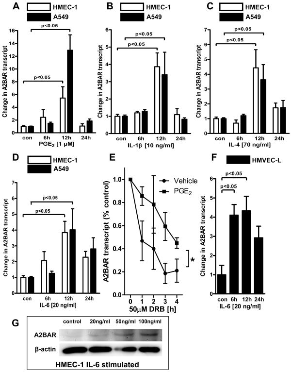
In order to investigate the role of the A2BAR in endotoxin-induced acute lung injury, we first pursued in vitro studies of A2BAR expression following exposure with different inflammatory stimuli. These studies are based on previous work indicating an important contribution of the A2BAR in dampening inflammation caused by hypoxia,(16) and other reports demonstrating transcriptionally regulated pathways for the A2BAR [e.g. involving the transcription factor, hypoxia-inducible factor (HIF)-1α] (11, 18, 20, 22, 29). Based on these studies, we pursued the hypothesis that A2BAR expression is enhanced following exposure to inflammatory stimuli. Previous studies have indicated that pulmonary A2BAR activity includes relevant expression on pulmonary epithelial cells (10) or vascular endothelia (12–14). As such, we modeled this event by exposing cultured pulmonary epithelial cells (A549) or vascular endothelia (HMEC-1) to a panel of inflammatory mediators, including PGE2, IL-1β, IL-4 and IL-6 over a time-course of up to 24h and assessed regulation of A2ABR expression by real-time RT-PCR (Figure 1A–D). In fact, these studies revealed dramatic increases in A2BAR transcript levels with exposure to inflammatory stimuli. To gain insight into the mechanisms of A2BAR induction, we profiled the influence of the above inflammatory mediators on previously characterized A2BAR luciferase reporter constructs (29). These studies revealed no significant changes in promoter activity under any of the conditions tested (data not shown). Based on these findings, we investigated whether A2BAR mRNA is protected against degradation in the absence of de novo transcription (i.e. change in mRNA half-life). Based on the prominent up-regulation of A2BAR transcript in response to PGE2 stimulation (Fig. 1A), we investigated the potential for PGE2 to stabilize A2BAR mRNA. In order to ascertain the effects of PGE2 on the enhancement of A2BAR levels in the absence of de novo synthesis, we evaluated the ability for PGE2 to post-transcriptionally regulate A2BAR levels in HMEC cells. Accordingly, we treated HMEC cells with 0.5μM PGE2, followed by a 4h treatment with 50μM DRB. Using real-time RT-PCR analysis, we found that PGE2 treatment is associated with increased A2BAR mRNA stability (p<0.05, n=3). These findings implicate that PGE2 treatment elicits anti-inflammatory signaling pathways involving enhanced mRNA stability of the A2BAR.
Figure 1. A2B adenosine receptor (A2BAR) expression following inflammatory stimulation in vitro.
(A-D): Pulmonary epithelial cells (A549) or vascular endothelia (HMEC-1) were exposed to indicated concentrations of inflammatory mediators, and A2BAR transcript levels were determined by real-time RT-PCR following 0–24h of exposure time. Data were calculated relative to the internal housekeeping gene (β-actin) and are expressed as mean fold change compared with control (0h of exposure) ± SD at each indicated time (n=4). (E) Degradation curve of A2BAR mRNA in HMEC-1 cells treated with PGE2 and challenged with DRB (n=3; *p<0.005 by ANOVA). (F). A2BAR expression in primary pulmonary endothelial cells treated with IL-6 (HMVEC-L) (n=4). (G) Western-blot analysis of A2BAR protein following 24h of IL-6 stimulation at indicated concentrations. The same blot was stripped and re-probed for murine β-actin to control for loading conditions. One representative of three Western blots is displayed.
Further to our findings in established endothelial cultures of non-pulmonary origin, we sought to determine whether this regulation occurred in a primary pulmonary cell line. Accordingly, we exposed primary pulmonary endothelial cells (HMVEC-L) to IL-6. Consistent with the previous studies of immortalized cell lines, we found time-dependent increases in A2BAR transcript levels in primary pulmonary endothelial cells (Fig. 1F). Similarly, A2BAR protein levels were elevated in a dose-dependent fashion following exposure to IL-6 over 24h (Fig. 1G). Taken together, these data indicate increased A2BAR transcript and protein levels following inflammatory stimulation of cultured pulmonary epithelial cells, vascular endothelia or primary pulmonary endothelial cells –at least in part through post-transcriptional regulation of A2BAR mRNA.
The A2BAR is induced during septic lung injury in vivo
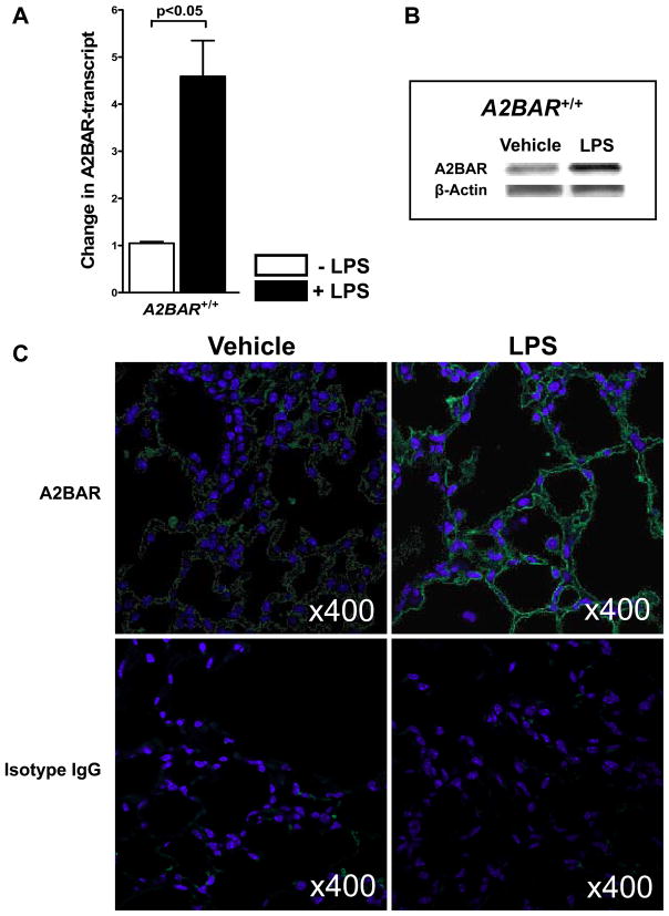
After having shown that inflammatory stimulation of pulmonary cells (endothelia and epithelia) is associated with robust induction of the A2BAR, we next pursued these findings in an in vivo model of endotoxin-induced acute lung injury. For this purpose, we used LPS inhalation. We exposed C57BL/6J mice over 30 min to inhaled LPS in a model system that we had used previously in studies on the role of CD39- and CD73-dependent adenosine generation in endotoxin-induced acute lung injury (5). Control animals underwent similar treatment with inhaled vehicle (vehicle). At 4h following LPS exposure, mice were sacrificed and A2BAR expression was assessed. As shown in Figure 2, A2BAR transcript levels and protein from the lungs of mice exposed to endotoxin were dramatically elevated (Figure 2A and 2B, respectively). Similarly, immunohistochemistry of lungs from mice exposed to endotoxin showed robust increases in A2BAR staining (Figure 2C, green staining) on pulmonary epithelial or endothelial structures. Taken together these studies indicate A2BAR induction following in vivo exposure to inhaled LPS.
Figure 2. A2B adenosine receptor (A2BAR) expression following LPS inhalation in vivo.
Mice were exposed to 30 min of LPS inhalation, and animals were sacrificed after 4h. (A) Pulmonary A2BAR transcript levels were assessed by real-time RT-PCR. Data were calculated relative to internal housekeeping gene (β-actin) and are expressed as mean fold change compared with control (-LPS) ± SD (n=9). Western-blot analysis of A2BAR protein following in vivo exposure to inhaled LPS. The same blot was stripped and re-probed for murine β-actin to control for loading conditions. One representative of four Western blots is displayed. (C) Pulmonary immunohistochemistry for the A2BAR following LPS exposure. Lungs from mice exposed to 30 min of LPS inhalation or vehicle control (Vehicle) were harvested. Sections were stained with antibodies specifical for murine A2BAR (green), or isotype controls. 4′,6-diamidino-2-phenylindole was used for nuclear counterstain (blue) (magnification, 400×). One representative image from 3 pulmonic sections is displayed.
Pharmacological inhibition of A2BAR signaling is associated with enhanced lung inflammation following LPS inhalation
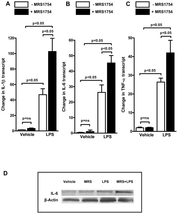
To study a functional role of the A2BAR in septic lung injury, we first pursued pharmacological studies. Here, we used MRS1754, a compound demonstrated to act as a specific antagonist of the A2BAR in vivo (8). Based on other studies showing high expression levels of the A2BAR in pulmonary epithelial cells, we decided to employ an inhaled route of administration. For this purpose, MRS1754 (2.4 μg/ml) was given via nebulizer over 30min, followed by 30 min of LPS inhalation. Lungs were excised after 4h and inflammatory markers were determined. LPS treatment was associated with increases in pulmonary transcript levels of IL-1β (Fig. 3A), IL-6 (Fig. 3B) and TNF-α (Fig. 3C) in vehicle-treated animals. However, inhibition of the A2BAR with MRS1754 dramatically augmented the effects of LPS administration (Fig. 3A-C). Similarly, pulmonary IL-6 protein levels were elevated in conjunction with LPS treatment, and showed additional increases following A2BAR inhibition (Figure 3D). Taken together these data indicate that pretreatment with a pharmacological inhibitor of the A2BAR synergistically enhances endotoxin-induced increases in lung inflammation during acute lung injury.
Figure 3. Influence of inhaled A2B adenosine receptor (A2BAR) antagonist MRS1754 on lung inflammation following LPS inhalation in vivo.
8–12 week old C57BL/6J mice matched in age, gender and weight were pre-exposed to 30 min of inhaled A2BAR antagonist (MRS1754) or vehicle, followed by 30 min of LPS or vehicle inhalation. Animals were sacrificed after 30 min and pulmonary transcript levels of (A) IL-1β, (B) IL-6 or (C) TNF-α were assessed by real-time RT-PCR. Data were calculated relative to internal housekeeping gene (β-actin) and are expressed as mean fold change compared with control (−LPS, −MRS1754) ± SD (n=4–7). (D) Western-blot analysis of IL-6 protein assessed by Western blot. The same blot was stripped and re-probed for murine β-actin to control for loading conditions. One representative of three Western blots is displayed. Note enhanced lung inflammation following A2BAR antagonist treatment following LPS exposure.
Gene-targeted mice for the A2BAR develop a more severe phenotype in endotoxin-induced acute lung injury compared to control animals
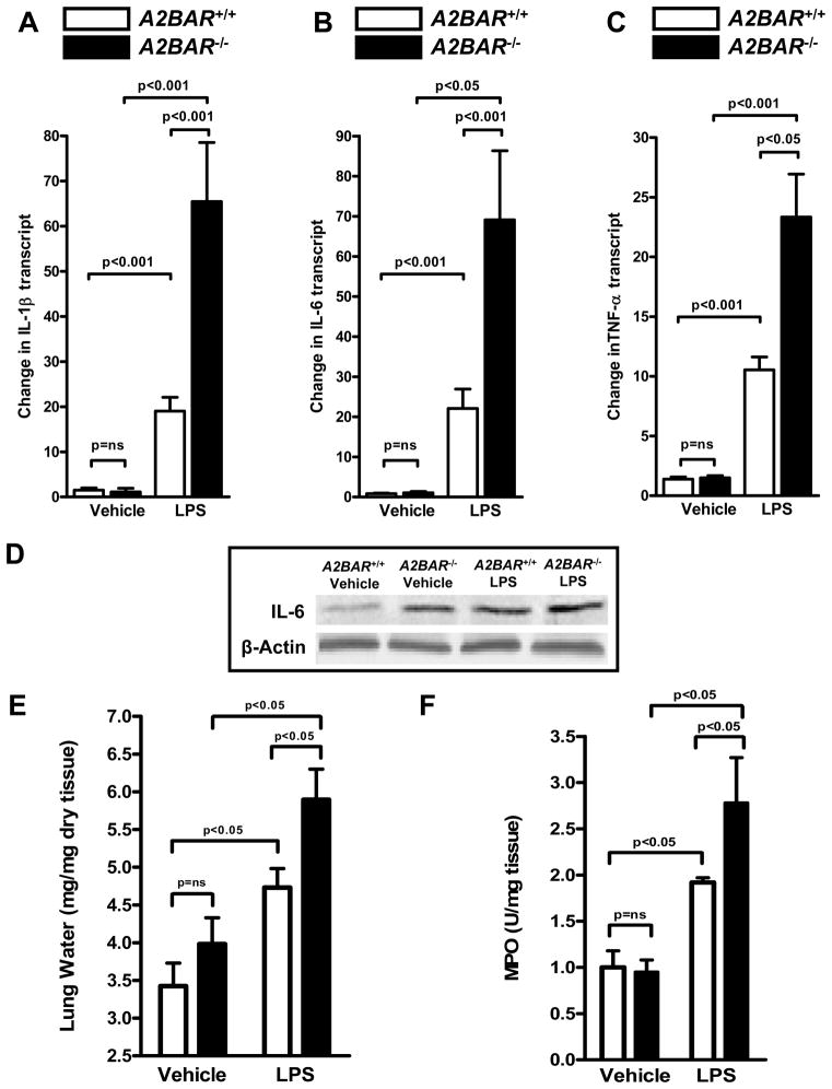
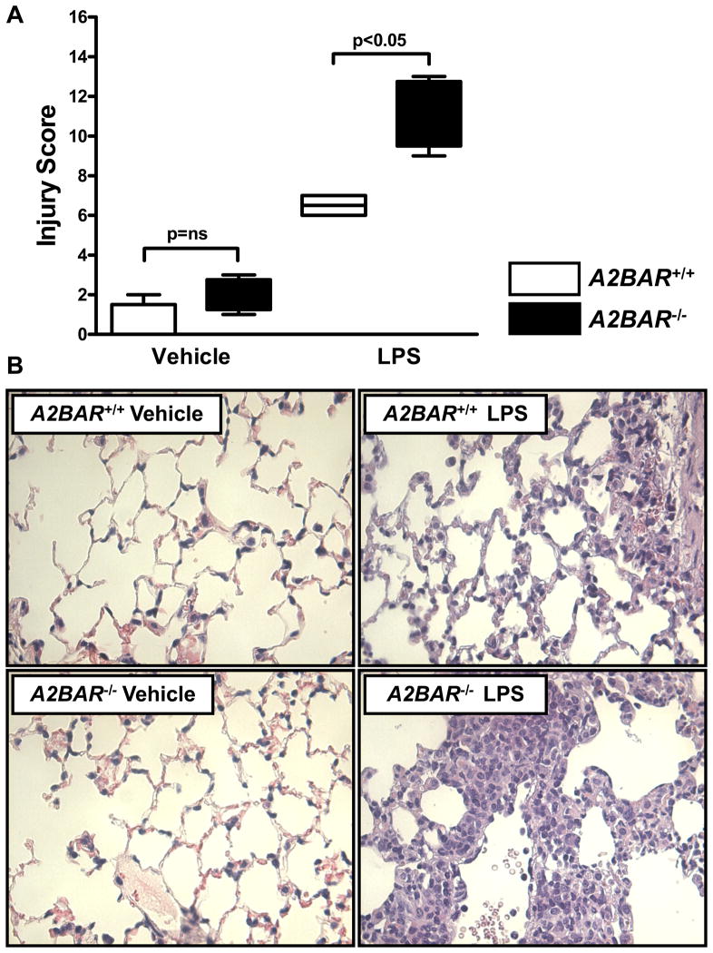
After having shown that pharmacological inhibition of the A2BAR is associated with increased lung inflammation following LPS-inhalation, we next pursued studies in previously characterized mice gene-targeted for the A2BAR (11, 16, 21, 24). For this purpose, wild-type or A2BAR−/− mice matched in age, gender and weight were exposed to aerosolized LPS for 30 min. Consistent with our studies of pharmacological A2BAR inhibition (Figure 3), we found significantly augmented elevations of pulmonary transcript levels for IL-1β (Fig. 4A), IL-6 (Fig 4B), or TNF-α (Fig. 4C) in A2BAR−/− mice following LPS exposure, when compared with wild-type treated animals. Moreover, elevations of IL-6 protein levels following LPS treatment in wild-type mice were dramatically enhanced in A2BAR−/− mice (Fig. 4D). Similarly, LPS treatment induced increases in lung-water in wild-type mice were far more pronounced in A2BAR−/− mice (Figure 4E). Furthermore, LPS-induced increases in MPO activity was enhanced in A2BAR−/− mice (Fig. 4F). To confirm these findings on a structural level, we examined lungs of wild-type or A2BAR−/− mice following LPS exposure histologically. Histological scoring revealed that A2BAR−/− mice exhibited a more severe pulmonary phenotype following LPS exposure than wild-type animals (Figure 5A and B). Taken together, these studies indicate a more severe phenotype of LPS-driven lung injury following genetic-deletion of the A2BAR, and provide indirect evidence for a protective role of A2BAR signaling during acute lung inflammation.
Figure 4. Exposure of gene targeted mice for the A2B adenosine receptor (A2BAR−/− mice) to LPS-induced lung injury.
Endotoxin-induced acute lung injury was induced in 8–12 week old A2BAR−/− mice or littermate controls matched in age, gender and weight by inhalation of LPS over 30 min. Controls were treated with inhaled vehicle. Animals were sacrificed after 4h and pulmonary transcript levels of (A) IL-1β, (B) IL-6 or (C) TNF-α were assessed by real-time RT-PCR. Data were calculated relative to internal housekeeping gene (β-actin) and are expressed as mean fold change compared with control (-LPS) ± SD (n=5–13). (D) Western-blot analysis of IL-6 protein assessed by Western blot. The same blot was stripped and re-probed for murine β-actin to control for loading conditions. One representative of three Western blots is displayed. (E) Assessment of pulmonary edema by measurement of lung water content (n=5–6). (F) Measurement of pulmonary myeloperoxidase (MPO) as indicator for neutrophil accumulation (n=5–9). Note enhanced lung inflammation and pulmonary edema in A2BAR−/− mice following LPS exposure.
Figure 5. Pulmonary histology of gene targeted mice for the A2B adenosine receptor (A2BAR−/− mice) following LPS-exposure.
Endotoxin-induced acute lung injury was induced in 8–12 week old A2BAR−/− mice or littermate controls matched in age, gender and weight by inhalation of LPS over 30 min. Controls were treated with inhaled vehicle. Animals were sacrificed, and pulmonary histology was assessed by a pathologist blinded to the treatment group. (A) Lung injury score (mean ± range, n=4). (B) Representative H&E staining (magnification 400x; one representative slide of 4). Note enhanced lung inflammation and tissue injury in A2BAR−/− mice exposed to LPS.
LPS-induced lung injury in A2BAR bone marrow chimeric mice
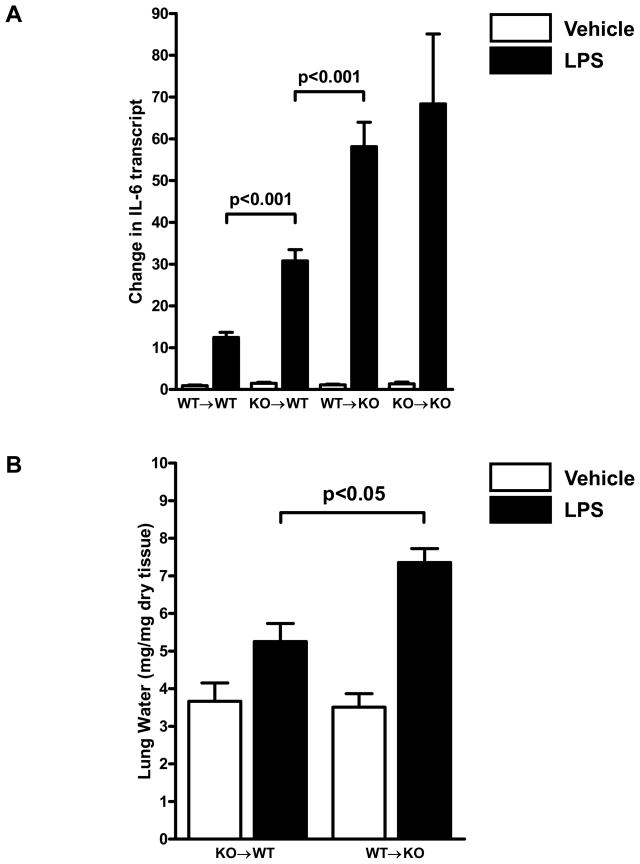
After having demonstrated detrimental effects of genetic deletion or pharmacological inhibition of A2BAR signaling during endotoxin-induced acute lung injury, we were interested to define the contribution of pulmonary versus myeloid A2BAR signaling effects. Previous studies had demonstrated functional A2BAR expression on inflammatory cells (e.g. neutrophils or macrophages) (14, 16) and on pulmonary tissues (e.g. pulmonary epithelia, vascular endothelia) (10, 11, 18, 31). Therefore, we generated A2BAR bone marrow chimeric mice as - we have done previously (11, 12) - to study the contribution of pulmonary versus myeloid A2BARs in endotoxin-induced acute lung injury. As expected, A2BAR+/+→A2BAR+/+ chimeric mice showed a similar degree of LPS-induced increase in lung inflammation as wild-type mice, while A2BAR−/−→A2BAR−/− mice showed a similar phenotype as A2BAR−/− mice (Figure 6A). However, bone marrow chimeric mice expressing the A2BAR exclusively on the pulmonary tissues (A2BAR−/−→A2BAR+/+ chimeric mice) had a significantly lower degree of LPS-induced IL-6 elevation or pulmonary edema than bone marrow chimera with exclusive A2BAR expression on the myeloid lineages (A2BAR+/+→A2BAR−/−) (Fig. 6A, B). Taken together, these studies suggest an important contribution of pulmonary A2BAR signaling in endotoxin-induced acute lung injury and pulmonary inflammation.
Figure 6. Influence of LPS-inhalation on IL-6 levels and pulmonary edema in A2BAR bone marrow chimeric mice.
8 to 12 week old A2BAR bone marrow chimeric mice (A2BAR+/+ → A2BAR+/+, WT → WT; A2BAR−/− → A2BAR+/+, WT → KO; A2BAR+/+ → A2BAR−/−, WT → KO and A2BAR−/− → A2BAR−/−, KO → KO) matched in age, gender and weight were exposed to 30 min of LPS or vehicle inhalation. Animals were sacrificed after 4h and (A) pulmonary transcript levels of IL-6 were determined by real-time RT-PCR. Data were calculated relative to internal housekeeping gene (β-actin) and are expressed as mean fold change compared with control (WT → WT treated with vehicle) ± SD (n=4–7).
A2BAR agonist treatment in endotoxin-induced acute lung injury
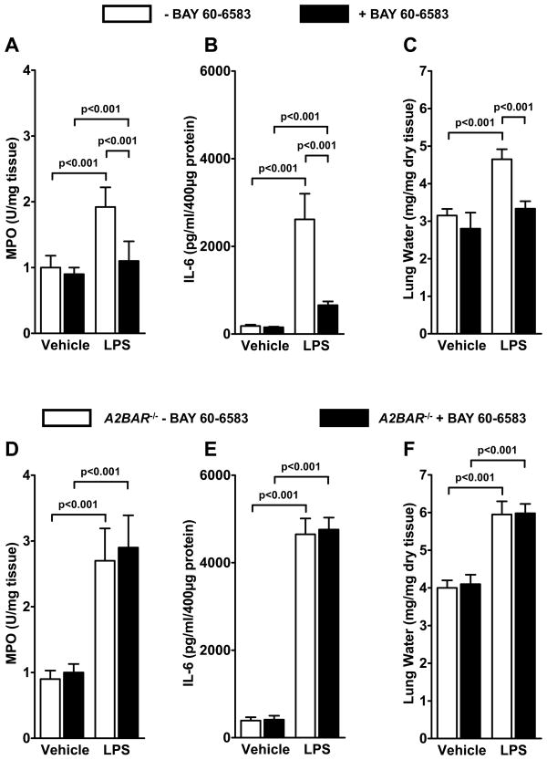
After having shown that pharmacological inhibition or genetic deletion of the A2BAR is associated with a more severe degree of lung inflammation and pulmonary edema, we next pursued the hypothesis that A2BAR agonist treatment will attenuate LPS-induced lung injury. For this purpose, we utilized a recently described non-adenosine like agonist of the A2BAR (BAY 60-6583). As such, previous studies have provided genetic in vivo evidence for A2BAR activity and specificity by showing significant reduction of myocardial infarct size in wild-type, but not in A2BAR−/− mice (21). Therefore, we pretreated mice with BAY 60-6583 30 min prior to LPS exposure (2mg/kg i.p.). Pretreatment with BAY 60-6583 was associated with attenuated pulmonary myeloperoxidase elevations following LPS treatment (Figure 8A) indicating diminished LPS-elicited inflammatory cell accumulation. Similarly, LPS-elicited increases in pulmonary IL-6 levels (Figure 7B) or LPS-dependent increases in lung water (Figure 7C) were significantly attenuated. In contrast, A2BAR treatment was ineffective in abrogating these inflammatory parameters in A2BAR−/ mice (Figure 8D-E), confirming the specificity and efficacy of BAY 60-6583 for the A2BAR. Taken together these studies identify the A2BAR as a pharmacological target for endotoxin-induced acute lung injury.
Figure 7. A2B adenosine receptor (A2BAR) agonist BAY 60-6583 in the treatment of LPS-induced lung injury.
(A-C) 8–12 week old C57BL/6J mice matched in age, gender and weight were pre-treated with A2BAR agonist (BAY 60-6583, 2mg/kg i.p.) or vehicle, followed by 30 min of LPS or vehicle inhalation. Animals were sacrificed after 30 min and pulmonary myeloperoixidase (A), IL-6 protein levels measured by ELISA (B) or lung water content (C) were assessed (n=4). (D-E) Similar studies in gene-targeted mice for the A2BAR (A2BAR−/− mice; n=4). Note attenuated lung inflammation and pulmonary edema with A2BAR agonist treatment only in wild-type mice.
Discussion
Over 40% of patients with sepsis go on to develop acute lung injury, which is the most common cause of death among death in these patients (3). At present, research studies to define novel therapeutic approaches for endotoxin-induced acute lung injury is an area of intense investigation. Based on previous studies showing a potential therapeutic role for signaling events through the A2BAR in attenuating mucosal inflammation (16, 24, 32) we pursued the hypothesis that the A2BAR represents a therapeutic target during LPS-induced lung injury. Indeed, bacterial toxins, such as LPS are a common cause of lung injury in patients suffering from sepsis (33). In the studies presented here, we demonstrated induction of the A2BAR following exposure to inflammatory stimuli in cultured pulmonary epithelia or vascular endothelia in vitro, or in an in vivo model investigating the lungs of mice exposed to LPS inhalation. Surprisingly, A2BAR induction was not associated with enhanced A2BAR promoter activity, but involved alterations in mRNA stability. Functional studies of LPS-driven lung injury utilizing A2BAR agonist treatment or mice following genetic deletion of the A2BAR revealed a higher degree of lung inflammation and pulmonary edema with A2BAR inhibition or deletion, respectively. Moreover, bone marrow chimeric mice for the A2BAR demonstrated a contribution of pulmonary A2BAR signaling in regulating lung inflammation and pulmonary edema. Finally, pretreatment with A2BAR agonist (BAY 60-6583) significantly attenuated lung inflammation and pulmonary edema in wild-type animals, but was ineffective in A2BAR−/− mice. Taken together, such studies indicate a potential role for A2BAR signaling in dampening lung inflammation and pulmonary edema during LPS-induced lung injury.
It has been previously shown that A2BAR expression is upregulated in response to pro-inflammatory cytokines, such as TNFα. In contrast to these findings, the there are few studies to date that show the mechanism of how A2BR protein expression is regulated in response to inflammatory stimuli. As such, pprevious studies had identified transcriptionally regulated alterations of A2BAR expression during hypoxia-elicited inflammation. These studies demonstrated a selective induction of the A2BAR following exposure to ambient hypoxia. In contrast, transcript levels of other ARs were either repressed or unaltered (18). Subsequent studies identified a previously unrecognized binding site for hypoxia-inducible factor (HIF)-1 within the promoter region of the A2BAR (29). Additional studies investigating the promoter activity, functional chromatin binding and HIF loss-of-function studies demonstrated a critical role of HIF-1α in mediating hypoxia-associated induction of the A2BAR (29). Other studies demonstrated HIF-dependent induction of the A2BAR during myocardial ischemia (21, 22). Similarly, a recent study indentified a transcriptionally regulated pathway elicited by hypoxia involving HIF-2α-dependent induction of the A2AAR (34). While these studies demonstrate transcriptionally regulated alterations of AR gene expression, the present studies could not find alterations of A2BAR promoter activity elicited by inflammatory mediators. In contrast, the present studies indicate that increases in A2BAR following exposure to inflammatory stimuli involve alterations in mRNA stability. Further studies are however required to elucidate the signaling mechanisms underpinning the stabilization of A2BAR stabilization.
Similar to the present results, other studies confirmed a role of adenosine generation and signaling in different forms of inflammatory diseases. For example, genetic deletion of CD39 or CD73 – the key enzymes in extracellular adenosine generation from precursor molecules (8, 18) – results in increased lung inflammation and pulmonary edema when exposed to ventilator induced lung injury (30). Similarly, cd39−/− or cd73−/− mice demonstrate signs of increased neutrophil trafficking into the lungs upon LPS exposure. As such, pulmonary CD39 and CD73 transcript levels were elevated following LPS exposure in vivo. Moreover, LPS-induced accumulation of PMN into the lungs was enhanced in cd39−/− or cd73−/− mice, particularly into the interstitial and intra-alveolar compartment. Such increases in PMN trafficking were accompanied by corresponding changes in alveolar-capillary leakage. Similarly, inhibition of extracellular nucleotide phosphohydrolysis with the nonspecific ecto-nucleoside-triphosphate-diphosphohydrolases inhibitor POM-1 confirmed increased pulmonary PMN accumulation in wild-type, but not in gene-targeted mice for cd39 or cd73. Finally, treatment with apyrase or nucleotidase was associated with attenuated pulmonary neutrophil accumulation and pulmonary edema during LPS-induced lung injury (5). Together, such data indicate the likelihood that CD39- and CD73-dependent adenosine production protects from LPS- or ventilator- induced lung injury (5, 30).
Previous research work had identified different ARs in lung protection. Specifically, several studies have pointed towards an important role of A2AAR signaling. Indeed, it has been demonstrated that that A2AAR−/− mice exhibit a more severe phenotype when exposed to different models of inflammation or sepsis (35–37). Similarly, studies of LPS-induced lung injury revealed a contribution of myeloid A2AAR signaling in lung protection (38). Utilizing studies with bone marrow chimeric mice in conjunction with studies of myeloid specific A2AAR deletion, the authors found a critical role of myeloid A2AAR signaling in attenuating PMN trafficking into the lungs. Furthermore, an important role of pulmonary A2BAR signaling in lung protection during mechanical ventilation-induced injury has recently been demonstrated (15). In conjunction with the findings from the present studies, it appears that that LPS induced lung injury could be attenuated by extracellular adenosine signaling events involving A2AARs expressed predominantly on inflammatory cells, and A2BARs expressed predominantly on pulmonary tissues.
In conjunction with the present studies, several other studies indicated the A2BAR in disease models that frequently occur in patients suffering from sepsis. As such, the A2BAR agonist BAY 60-6583 has been implicated in the treatment of intestinal ischemia induced by intermittent ligation of intestinal blood flow, followed by reperfusion (24). Similarly, an anti-inflammatory and tissue protective effect of A2BAR signaling had been observed in models of acute intestinal inflammation (32). Moreover, activation of the A2BAR has been shown to decrease vascular leakage in the setting of hypoxia-induced vascular leakage (11), or acute kidney injury (12). It is important to point out that the relatively selective role of A2BAR signaling in these models may be related to the robust induction of these A2BAR under these conditions. While A2BAR−/− mice appear phenotypically normal and do not exhibit signs of immunologic defects when housed in a pathogen free environment (21), the A2BAR appears to play an important role under disease conditions associated with its induction (11, 15, 18, 21, 22). Moreover, a coordinated response of increased adenosine production (18), attenuated adenosine uptake (39, 40) and decreased intracellular adenosine metabolism (41) may further contribute to the elevation of extracellular adenosine levels, resulting in sufficient adenosine concentrations capable of activating the relatively “adenosine-insensitive” A2BAR. Moreover, recent studies indicate that the neuronal guidance molecule netrin-1 is induced during conditions of inflammatory hypoxia, and may contribute to enhanced extracellular signaling events through the A2BAR (16). Taken together, such studies highlight a potential role for the A2BAR as therapeutic target during sepsis.
In contrast to the beneficial effects of increased adenosine production and signaling during ALI, there is some evidence suggesting a potentially detrimental role of chronically elevated adenosine levels (42–45). For example, levels of adenosine are increased in the lungs of asthmatics (46), and correlate with the degree of inflammatory insult (47). At present, it is not entirely clear weather such elevations of adenosine are part of a protective pathway to dampen lung inflammation, or play a provocative role of adenosine in asthma or chronic obstructive pulmonary disease (48). For example, mice incapable of extracellular adenosine generation (cd73−/− mice) exhibit a more severe phenotype in bleomycin-induced lung injury, indicating a protective role of extracellular adenosine signaling in this chronic model of lung disease.(49) In contrast, adenosine-deaminase (ADA)-deficient mice develop signs of chronic lung inflammation in association with dramatically elevated pulmonary adenosine levels. In fact, ADA-deficient mice die within weeks after birth from severe respiratory distress (50), and pharmacological studies suggest that attenuation of adenosine signaling through the A2BAR may reverse the severe pulmonary phenotypes in ADA-deficient mice (44, 50). To address these findings on a genetic level, a very elegant study examined the contribution of A2BAR signaling in this model via a genetic approach by generating ADA/A2BAR double-knockout mice (51). The authors’ initial hypothesis was that genetic removal of the A2BAR from ADA-deficient mice would lead to diminished pulmonary inflammation and damage. Unexpectedly, ADA/A2BAR double-knockout mice exhibited enhanced pulmonary inflammation and airway destruction. Marked loss of pulmonary barrier function and excessive airway neutrophilia are thought to contribute to the enhanced tissue damage observed. These findings support an important protective role for A2BAR signaling during acute stages of lung disease (51).
Taken together, the present studies indicate a protective role of A2BAR signaling in endotoxin-driven lung injury and suggest a potential role for A2BAR agonists in the treatment of endotoxin-induced acute lung injury. While all the in vivo evidence was established in murine models, it will be an important challenge to translate these findings into a clinical setting. In addition, it will be critical to determine convenient pharmacological approaches to utilize A2BAR agonists, and study potential side effects of these compounds, for example with regard to blood pressure, heart-rate (52) or platelet function (53).
Acknowledgments
Financial Support Information
The present studies were supported by United States National Institutes of Health grant R01-HL092188, Foundation for Anesthesia Education and Research (FAER) Grants and American Heart Association Grants to HKE and TE.
References
- 1.Hotchkiss RS, I, Karl E. The Pathophysiology and Treatment of Sepsis. N Engl J Med. 2003;348:138–150. doi: 10.1056/NEJMra021333. [DOI] [PubMed] [Google Scholar]
- 2.Russell JA. Management of sepsis. N Engl J Med. 2006;355:1699–1713. doi: 10.1056/NEJMra043632. [DOI] [PubMed] [Google Scholar]
- 3.Hudson LD, Milberg JA, Anardi D, Maunder RJ. Clinical risks for development of the acute respiratory distress syndrome. Am J Respir Crit Care Med. 1995;151:293–301. doi: 10.1164/ajrccm.151.2.7842182. [DOI] [PubMed] [Google Scholar]
- 4.Rubenfeld GD, Caldwell E, Peabody E, Weaver J, Martin DP, Neff M, Stern EJ, Hudson LD. Incidence and Outcomes of Acute Lung Injury. N Engl J Med. 2005;353:1685–1693. doi: 10.1056/NEJMoa050333. [DOI] [PubMed] [Google Scholar]
- 5.Reutershan J, Vollmer I, Stark S, Wagner R, Ngamsri KC, Eltzschig HK. Adenosine and inflammation: CD39 and CD73 are critical mediators in LPS-induced PMN trafficking into the lungs. FASEB J. 2009;23:473–482. doi: 10.1096/fj.08-119701. [DOI] [PubMed] [Google Scholar]
- 6.Weissmuller T, Campbell EL, Rosenberger P, Scully M, Beck PL, Furuta GT, Colgan SP. PMNs facilitate translocation of platelets across human and mouse epithelium and together alter fluid homeostasis via epithelial cell-expressed ecto-NTPDases. J Clin Invest. 2008;118:3682–3692. doi: 10.1172/JCI35874. [DOI] [PMC free article] [PubMed] [Google Scholar]
- 7.Enjyoji K, Sevigny J, Lin Y, Frenette PS, Christie PD, Esch JS, 2nd, Imai M, Edelberg JM, Rayburn H, Lech M, Beeler DL, Csizmadia E, Wagner DD, Robson SC, Rosenberg RD. Targeted disruption of cd39/ATP diphosphohydrolase results in disordered hemostasis and thromboregulation. Nat Med. 1999;5:1010–1017. doi: 10.1038/12447. [DOI] [PubMed] [Google Scholar]
- 8.Thompson LF, Eltzschig HK, Ibla JC, Van De Wiele CJ, Resta R, Morote-Garcia JC, Colgan SP. Crucial role for ecto-5′-nucleotidase (CD73) in vascular leakage during hypoxia. J Exp Med. 2004;200:1395–1405. doi: 10.1084/jem.20040915. [DOI] [PMC free article] [PubMed] [Google Scholar]
- 9.Lukashev DE, Smith PT, Caldwell CC, Ohta A, Apasov SG, Sitkovsky MV. Analysis of A2a receptor-deficient mice reveals no significant compensatory increases in the expression of A2b, A1, and A3 adenosine receptors in lymphoid organs. Biochem Pharmacol. 2003;65:2081–2090. doi: 10.1016/s0006-2952(03)00158-8. [DOI] [PubMed] [Google Scholar]
- 10.Cagnina RE, Ramos SI, Marshall MA, Wang G, Frazier CR, Linden J. Adenosine A2B receptors are highly expressed on murine type II alveolar epithelial cells. Am J Physiol Lung Cell Mol Physiol. 2009 doi: 10.1152/ajplung.90553.2008. [DOI] [PMC free article] [PubMed] [Google Scholar]
- 11.Eckle T, Faigle M, Grenz A, Laucher S, Thompson LF, Eltzschig HK. A2B adenosine receptor dampens hypoxia-induced vascular leak. Blood. 2008;111:2024–2035. doi: 10.1182/blood-2007-10-117044. [DOI] [PMC free article] [PubMed] [Google Scholar]
- 12.Grenz A, Osswald H, Eckle T, Yang D, Zhang H, Tran ZV, Klingel K, Ravid K, Eltzschig HK. The Reno-Vascular A2B Adenosine Receptor Protects the Kidney from Ischemia. PLoS Medicine. 2008;5:e137. doi: 10.1371/journal.pmed.0050137. [DOI] [PMC free article] [PubMed] [Google Scholar]
- 13.Yang D, Koupenova M, McCrann DJ, Kopeikina KJ, Kagan HM, Schreiber BM, Ravid K. The A2b adenosine receptor protects against vascular injury. Proc Natl Acad Sci U S A. 2008;105:792–796. doi: 10.1073/pnas.0705563105. [DOI] [PMC free article] [PubMed] [Google Scholar]
- 14.Yang D, Zhang Y, Nguyen HG, Koupenova M, Chauhan AK, Makitalo M, Jones MR, St Hilaire C, Seldin DC, Toselli P, Lamperti E, Schreiber BM, Gavras H, Wagner DD, Ravid K. The A2B adenosine receptor protects against inflammation and excessive vascular adhesion. J Clin Invest. 2006;116:1913–1923. doi: 10.1172/JCI27933. [DOI] [PMC free article] [PubMed] [Google Scholar]
- 15.Eckle T, Grenz A, Laucher S, Eltzschig HK. A2B adenosine receptor signaling attenuates acute lung injury by enhancing alveolar fluid clearance in mice. J Clin Invest. 2008;118:3301–3315. doi: 10.1172/JCI34203. [DOI] [PMC free article] [PubMed] [Google Scholar]
- 16.Rosenberger P, Schwab JM, Mirakaj V, Masekowsky E, Mager A, Morote-Garcia JC, Unertl K, Eltzschig HK. Hypoxia-inducible factor-dependent induction of netrin-1 dampens inflammation caused by hypoxia. Nat Immunol. 2009;10:195–202. doi: 10.1038/ni.1683. [DOI] [PubMed] [Google Scholar]
- 17.Haeberle HA, Durrstein C, Rosenberger P, Hosakote YM, Kuhlicke J, Kempf VA, Garofalo RP, Eltzschig HK. Oxygen-independent stabilization of hypoxia inducible factor (HIF)-1 during RSV infection. PLoS ONE. 2008;3:e3352. doi: 10.1371/journal.pone.0003352. [DOI] [PMC free article] [PubMed] [Google Scholar]
- 18.Eltzschig HK, Ibla JC, Furuta GT, Leonard MO, Jacobson KA, Enjyoji K, Robson SC, Colgan SP. Coordinated adenine nucleotide phosphohydrolysis and nucleoside signaling in posthypoxic endothelium: role of ectonucleotidases and adenosine A2B receptors. J Exp Med. 2003;198:783–796. doi: 10.1084/jem.20030891. [DOI] [PMC free article] [PubMed] [Google Scholar]
- 19.Eltzschig HK, Faigle M, Knapp S, Karhausen J, Ibla J, Rosenberger P, Odegard KC, Laussen PC, Thompson LF, Colgan SP. Endothelial catabolism of extracellular adenosine during hypoxia: the role of surface adenosine deaminase and CD26. Blood. 2006;108:1602–1610. doi: 10.1182/blood-2006-02-001016. [DOI] [PMC free article] [PubMed] [Google Scholar]
- 20.Eltzschig HK, Thompson LF, Karhausen J, Cotta RJ, Ibla JC, Robson SC, Colgan SP. Endogenous adenosine produced during hypoxia attenuates neutrophil accumulation: coordination by extracellular nucleotide metabolism. Blood. 2004;104:3986–3992. doi: 10.1182/blood-2004-06-2066. [DOI] [PubMed] [Google Scholar]
- 21.Eckle T, Krahn T, Grenz A, Kohler D, Mittelbronn M, Ledent C, Jacobson MA, Osswald H, Thompson LF, Unertl K, Eltzschig HK. Cardioprotection by ecto-5′-nucleotidase (CD73) and A2B adenosine receptors. Circulation. 2007;115:1581–1590. doi: 10.1161/CIRCULATIONAHA.106.669697. [DOI] [PubMed] [Google Scholar]
- 22.Eckle T, Kohler D, Lehmann R, El Kasmi KC, Eltzschig HK. Hypoxia-Inducible Factor-1 Is Central to Cardioprotection: A New Paradigm for Ischemic Preconditioning. Circulation. 2008;118:166–175. doi: 10.1161/CIRCULATIONAHA.107.758516. [DOI] [PubMed] [Google Scholar]
- 23.Hart ML, Henn M, Kohler D, Kloor D, Mittelbronn M, Gorzolla IC, Stahl GL, Eltzschig HK. Role of extracellular nucleotide phosphohydrolysis in intestinal ischemia-reperfusion injury. FASEB J. 2008;22:2784–2797. doi: 10.1096/fj.07-103911. [DOI] [PMC free article] [PubMed] [Google Scholar]
- 24.Hart ML, Jacobi B, Schittenhelm J, Henn M, Eltzschig HK. Cutting Edge: A2B Adenosine receptor signaling provides potent protection during intestinal ischemia/reperfusion injury. J Immunol. 2009;182:3965–3968. doi: 10.4049/jimmunol.0802193. [DOI] [PubMed] [Google Scholar]
- 25.Hart ML, Much C, Gorzolla IC, Schittenhelm J, Kloor D, Stahl GL, Eltzschig HK. Extracellular adenosine production by ecto-5′-nucleotidase protects during murine hepatic ischemic preconditioning. Gastroenterology. 2008;135:1739–1750. e1733. doi: 10.1053/j.gastro.2008.07.064. [DOI] [PubMed] [Google Scholar]
- 26.Eltzschig HK, Eckle T, Mager A, Kuper N, Karcher C, Weissmuller T, Boengler K, Schulz R, Robson SC, Colgan SP. ATP release from activated neutrophils occurs via connexin 43 and modulates adenosine-dependent endothelial cell function. Circ Res. 2006;99:1100–1108. doi: 10.1161/01.RES.0000250174.31269.70. [DOI] [PubMed] [Google Scholar]
- 27.Faigle M, Seessle J, Zug S, El Kasmi KC, Eltzschig HK. ATP release from vascular endothelia occurs across Cx43 hemichannels and is attenuated during hypoxia. PLoS ONE. 2008;3:e2801. doi: 10.1371/journal.pone.0002801. [DOI] [PMC free article] [PubMed] [Google Scholar]
- 28.Kohler D, Eckle T, Faigle M, Grenz A, Mittelbronn M, Laucher S, Hart ML, Robson SC, Muller CE, Eltzschig HK. CD39/ectonucleoside triphosphate diphosphohydrolase 1 provides myocardial protection during cardiac ischemia/reperfusion injury. Circulation. 2007;116:1784–1794. doi: 10.1161/CIRCULATIONAHA.107.690180. [DOI] [PubMed] [Google Scholar]
- 29.Kong T, Westerman KA, Faigle M, Eltzschig HK, Colgan SP. HIF-dependent induction of adenosine A2B receptor in hypoxia. Faseb J. 2006;20:2242–2250. doi: 10.1096/fj.06-6419com. [DOI] [PubMed] [Google Scholar]
- 30.Eckle T, Fullbier L, Wehrmann M, Khoury J, Mittelbronn M, Ibla J, Rosenberger P, Eltzschig HK. Identification of ectonucleotidases CD39 and CD73 in innate protection during acute lung injury. J Immunol. 2007;178:8127–8137. doi: 10.4049/jimmunol.178.12.8127. [DOI] [PubMed] [Google Scholar]
- 31.Hasko G, Csoka B, Nemeth ZH, Vizi ES, Pacher P. A(2B) adenosine receptors in immunity and inflammation. Trends Immunol. 2009 doi: 10.1016/j.it.2009.04.001. [DOI] [PMC free article] [PubMed] [Google Scholar]
- 32.Frick JS, MacManus CF, Scully M, Glover LE, Eltzschig HK, Colgan SP. Contribution of adenosine A2B receptors to inflammatory parameters of experimental colitis. J Immunol. 2009;182:4957–4964. doi: 10.4049/jimmunol.0801324. [DOI] [PMC free article] [PubMed] [Google Scholar]
- 33.Levy MM, Fink MP, Marshall JC, Abraham E, Angus D, Cook D, Cohen J, Opal SM, Vincent JL, Ramsay G. 2001 SCCM/ESICM/ACCP/ATS/SIS International Sepsis Definitions Conference. Crit Care Med. 2003;31:1250–1256. doi: 10.1097/01.CCM.0000050454.01978.3B. [DOI] [PubMed] [Google Scholar]
- 34.Ahmad A, Ahmad S, Glover L, Miller SM, Shannon JM, Guo X, Franklin WA, Bridges JP, Schaack JB, Colgan SP, White CW. Adenosine A2A receptor is a unique angiogenic target of HIF-2alpha in pulmonary endothelial cells. Proc Natl Acad Sci U S A. 2009;106:10684–10689. doi: 10.1073/pnas.0901326106. [DOI] [PMC free article] [PubMed] [Google Scholar]
- 35.Ohta A, Sitkovsky M. Role of G-protein-coupled adenosine receptors in downregulation of inflammation and protection from tissue damage. Nature. 2001;414:916–920. doi: 10.1038/414916a. [DOI] [PubMed] [Google Scholar]
- 36.Sitkovsky MV, Lukashev D, Apasov S, Kojima H, Koshiba M, Caldwell C, Ohta A, Thiel M. Physiological control of immune response and inflammatory tissue damage by hypoxia-inducible factors and adenosine A2A receptors. Annual Review of Immunology. 2004;22:657–682. doi: 10.1146/annurev.immunol.22.012703.104731. [DOI] [PubMed] [Google Scholar]
- 37.Thiel M, Chouker A, Ohta A, Jackson E, Caldwell C, Smith P, Lukashev D, Bittmann I, Sitkovsky MV. Oxygenation inhibits the physiological tissue-protecting mechanism and thereby exacerbates acute inflammatory lung injury. PLoS Biol. 2005;3:e174. doi: 10.1371/journal.pbio.0030174. [DOI] [PMC free article] [PubMed] [Google Scholar]
- 38.Reutershan J, Cagnina RE, Chang D, Linden J, Ley K. Therapeutic anti-inflammatory effects of myeloid cell adenosine receptor A2a stimulation in lipopolysaccharide-induced lung injury. J Immunol. 2007;179:1254–1263. doi: 10.4049/jimmunol.179.2.1254. [DOI] [PubMed] [Google Scholar]
- 39.Eltzschig HK, Abdulla P, Hoffman E, Hamilton KE, Daniels D, Schonfeld C, Loffler M, Reyes G, Duszenko M, Karhausen J, Robinson A, Westerman KA, Coe IR, Colgan SP. HIF-1-dependent repression of equilibrative nucleoside transporter (ENT) in hypoxia. J Exp Med. 2005;202:1493–1505. doi: 10.1084/jem.20050177. [DOI] [PMC free article] [PubMed] [Google Scholar]
- 40.Morote-Garcia JC, Rosenberger P, Nivillac NM, Coe IR, Eltzschig HK. Hypoxia-inducible factor-dependent repression of equilibrative nucleoside transporter 2 attenuates mucosal inflammation during intestinal hypoxia. Gastroenterology. 2009;136:607–618. doi: 10.1053/j.gastro.2008.10.037. [DOI] [PubMed] [Google Scholar]
- 41.Morote-Garcia JC, Rosenberger P, Kuhlicke J, Eltzschig HK. HIF-1-dependent repression of adenosine kinase attenuates hypoxia-induced vascular leak. Blood. 2008;111:5571–5580. doi: 10.1182/blood-2007-11-126763. [DOI] [PubMed] [Google Scholar]
- 42.Blackburn MR. Too much of a good thing: adenosine overload in adenosine-deaminase-deficient mice. Trends Pharmacol Sci. 2003;24:66–70. doi: 10.1016/S0165-6147(02)00045-7. [DOI] [PubMed] [Google Scholar]
- 43.Chunn JL, Molina JG, Mi T, Xia Y, Kellems RE, Blackburn MR. Adenosine-Dependent Pulmonary Fibrosis in Adenosine Deaminase-Deficient Mice. J Immunol. 2005;175:1937–1946. doi: 10.4049/jimmunol.175.3.1937. [DOI] [PubMed] [Google Scholar]
- 44.Sun CX, Zhong H, Mohsenin A, Morschl E, Chunn JL, Molina JG, Belardinelli L, Zeng D, Blackburn MR. Role of A2B adenosine receptor signaling in adenosine-dependent pulmonary inflammation and injury. J Clin Invest. 2006;116:2173–2182. doi: 10.1172/JCI27303. [DOI] [PMC free article] [PubMed] [Google Scholar]
- 45.Sun CX, Young HW, Molina JG, Volmer JB, Schnermann J, Blackburn MR. A protective role for the A1 adenosine receptor in adenosine-dependent pulmonary injury. J Clin Invest. 2005;115:35–43. doi: 10.1172/JCI22656. [DOI] [PMC free article] [PubMed] [Google Scholar]
- 46.Driver AG, Kukoly CA, Ali S, Mustafa SJ. Adenosine in bronchoalveolar lavage fluid in asthma. Am Rev Respir Dis. 1993;148:91–97. doi: 10.1164/ajrccm/148.1.91. [DOI] [PubMed] [Google Scholar]
- 47.Huszar E, Vass G, Vizi E, Csoma Z, Barat E, Molnar Vilagos G, Herjavecz I, Horvath I. Adenosine in exhaled breath condensate in healthy volunteers and in patients with asthma. Eur Respir J. 2002;20:1393–1398. doi: 10.1183/09031936.02.00005002. [DOI] [PubMed] [Google Scholar]
- 48.Fozard JR, Hannon JP. Adenosine receptor ligands: potential as therapeutic agents in asthma and COPD. Pulm Pharmacol Ther. 1999;12:111–114. doi: 10.1006/pupt.1999.0191. [DOI] [PubMed] [Google Scholar]
- 49.Volmer JB, Thompson LF, Blackburn MR. Ecto-5′-nucleotidase (CD73)-mediated adenosine production is tissue protective in a model of bleomycin-induced lung injury. J Immunol. 2006;176:4449–4458. doi: 10.4049/jimmunol.176.7.4449. [DOI] [PubMed] [Google Scholar]
- 50.Blackburn MR, Volmer JB, Thrasher JL, Zhong H, Crosby JR, Lee JJ, Kellems RE. Metabolic Consequences of Adenosine Deaminase Deficiency in Mice Are Associated with Defects in Alveogenesis, Pulmonary Inflammation, and Airway Obstruction. J Exp Med. 2000;192:159–170. doi: 10.1084/jem.192.2.159. [DOI] [PMC free article] [PubMed] [Google Scholar]
- 51.Zhou Y, Mohsenin A, Morschl E, Young HW, Molina JG, Ma W, Sun CX, Martinez-Valdez H, Blackburn MR. Enhanced airway inflammation and remodeling in adenosine deaminase-deficient mice lacking the A2B adenosine receptor. J Immunol. 2009;182:8037–8046. doi: 10.4049/jimmunol.0900515. [DOI] [PMC free article] [PubMed] [Google Scholar]
- 52.Koeppen M, Eckle T, Eltzschig HK. Selective deletion of the A1 adenosine receptor abolishes heart-rate slowing effects of intravascular adenosine in vivo. PLoS One. 2009;4:e6784. doi: 10.1371/journal.pone.0006784. [DOI] [PMC free article] [PubMed] [Google Scholar]
- 53.Hart ML, Kohler D, Eckle T, Kloor D, Stahl GL, Eltzschig HK. Direct treatment of mouse or human blood with soluble 5′-nucleotidase inhibits platelet aggregation. Arterioscler Thromb Vasc Biol. 2008;28:1477–1483. doi: 10.1161/ATVBAHA.108.169219. [DOI] [PubMed] [Google Scholar]


