Abstract
Fractionation of the crude extract of the marine cyanobacterium Lyngbya majuscule collected from Panama led to the isolation of malyngolide dimer (1). The planar structure of 1 was determined using 1D and 2D NMR spectroscopy, and HRESI-TOF MS. The absolute configuration was established by chemical degradation followed by chiral GC-MS analyses and comparisons with an authentic sample of malyngolide seco acid (4). Compound 1 showed moderate in vitro antimalarial activity against chloroquine resistant Plasmodium falciparum (W2) (IC50 = 19 µM) but roughly equivalent toxicity against H-460 human lung cell lines. Furthermore, because the closely related cyanobacterial natural product ‘tanikolide dimer’ was a potent SIRT2 inhibitor, compound 1 was evaluated in this assay, but found to be essentially inactive.
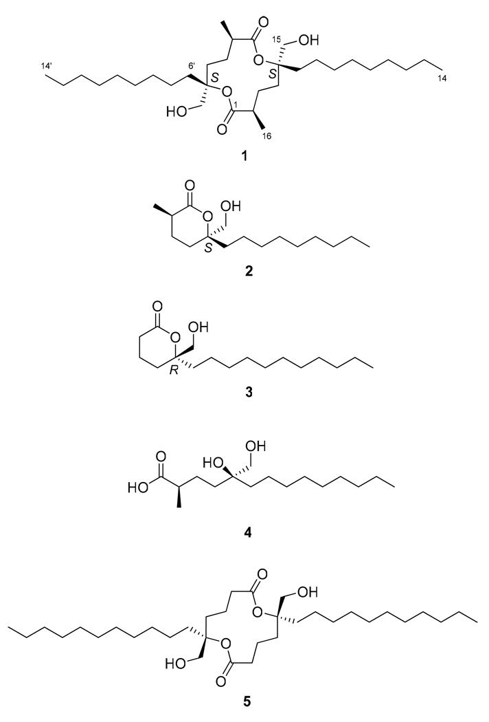
Malyngolide (2) and tanikolide (3) are natural δ-lactones obtained from different strains of the marine cyanobacterium Lyngbya and characterized by a hydroxymethyl group and a long aliphatic chain attached to the δ-position of a six-membered lactone ring. The main differences between compounds 2 and 3 are the presence of a secondary methyl group at C-2 of malyngolide, the number of carbons in the aliphatic chain, and perhaps most interestingly, the absolute configuration of the δ-carbon (2 = 5S; 3 = 5R). Compounds 2 and 3 were isolated from L. majuscula collected in Hawaii and Madagascar, respectively.1,2 Malyngolide (2) displayed antibacterial activity against Mycobacterium smegmatis and Streptococcus pyogenes, while tanikolide (3) showed cytotoxicity against brine shrimp as well as antifungal and molluscicidal activity. Since malyngolide (2) was synthesized for the first time in 1980,3 more than 66 synthetic studies have been published for this compound, and 23 for the more recently isolated tanikolide (3), providing evidence of the remarkable interest by the synthetic community in these compounds.4
Here we report the isolation, structural determination and bioactivity of malyngolide dimer (1) from L. majuscula (Oscillatoriaceae, Gomont ex Gomont 1892), collected at Coiba National Park off the Pacific coast of Panama as a part of our International Cooperative Biodiversity Group (ICBG) drug discovery program.5 Interestingly, we recently discovered and reported the structure of the naturally occurring dimer of tanikolide, tanikolide dimer (5), from a Malagasy collection of this same cyanobacterium,6 and thus the work herein reported indicates an emergent trend in the lipophilic natural products chemistry of these organisms. Furthermore, as tanikolide dimer showed remarkable activity as a SIRT2 inhibitor,7 an HDAC-associated protein with potential as a target for anticancer therapy, isolation of malyngolide dimer (1) provided an opportunity to expand on structure-activity information in this compound class.
HRESI–TOF MS spectra of compound 1 showed a pseudomolecular ion [M + H]+ at m/z 541.4453 and its sodium adduct [M+Na]+ at m/z 563.4286 which corresponded with the molecular formulas C32H61O6 and C32H60O6Na, respectively. However, 13C-NMR resonances were observed for only 16 carbon atoms, indicating that 1 must be a symmetric dimer. The three degrees of unsaturation implied by the molecular formula were accounted for by two carbonyl groups, and one ring. DEPT experiments in combination with HSQC data revealed that each monomer of compound 1 possessed two methyl groups at δC 14.03 (C-14) and 16.9 (C-16), 11 methylenes at δC 67.5 (C-15), 36.6, 31.76, 29.9, 29.42, 29.36, 29.2, 23.5, 22.6 (C-6 to C-13), 25.1, 26.1 (C-3 and C-4), one methine at δC 35.4 (C-2) and two quaternary carbons at δC 175.4 (C-1) and 86.9 (C-5).
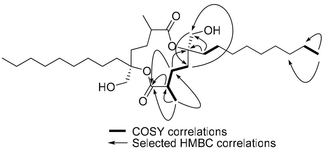
A key structural feature revealed by the 1H NMR analysis was a pair of doublets at δH 3.64 and 3.44 that were assigned as the methylene protons H-15a and H-15b, respectively. A multiplet located at δH 2.41 (m, H-2) a methine proton, was coupled to the secondary methyl group at C-16 and a methylene at C-3. A group of six diastereotopic methylene protons observed between 1.5 and 2.00 ppm and were assigned as δH 1.91 (m, H-3a), δH 1.59 (m, H-3b), 1.99 (ddd, J = 4.0, 14.0, 17.5 Hz, H-4a), 1.74 (dt, J = 4.0, 14.0 Hz, H-4b), 1.68 (m, 1H, H-6a), 1.53 (m, H-6b), by 1H-1H COSY and 1H-13C HMBC (Figure 1). A group of overlapped prominent signals between 1.23 and 1.30 ppm were assigned to the aliphatic methylene chain (H-7 to H-13) and the secondary methyl group H-16 using gHSQC and COSY experiments. Finally a triplet located at δH 0.85 (t, J = 6.5 Hz, H-14) corresponded with the terminal methyl group of the aliphatic chain. The position of the methyl group H3-16 as well as the connectivity of the oxygen-bonded methylene at C-15 and the spin systems formed by protons H-2 to H-4 and H-6 to H-14, to carbon C-5 was determined based on 2J and 3J HMBC correlations (Figure 1). Thus, the overall analysis of the 1D-NMR (1H, 13C, DEPT) and 2D-NMR (gCOSY, gHSQC, gHMBC) data indicated that 1 was structurally related to malyngolide (2).
Figure 1.
Selected 2D NMR correlations for malyngolide dimer (1).
The absolute configurations at carbons C-2 and C-5 were determined by chiral GC-MS analysis. Compound 1 was hydrolyzed under mild basic conditions using barium hydroxide in aqueous methanol to obtain malyngolide seco-acid (4).1 Compound 4 was analyzed directly by chiral GC-MS and gave a single peak at 28.64 min indicating that both monomers of dimer 1 had the same absolute configuration at C-2 and C-5. Compound 4 was then analyzed by co-injection using chiral GC-MS and an authentic sample of natural malyngolide seco-acid obtained from a Papua New Guinea collection of Lyngbya sp.; the chiral standard was shown to have the configuration of 2R and 5S by NMR and optical rotation ([α]D -10.7, c 0.44, CH2Cl2; lit. [α]D - 14.6, c 1, CH2Cl2).1 GC-MS analysis gave only a single peak at 28.67 min indicating that both the authentic and malyngolide dimer-derived synthetic malyngolide seco-acid (4) possessed the same 2R,5S absolute configuration. Therefore, we deduce that 1 possesses a 2R,2’R,5S,5’S stereoconfiguration.
Malyngolide dimer (1) was evaluated for in vitro activity against the chloroquine resistant Plasmodium falciparum strain W2 and showed an IC50 of 19 µM.8 However, compound 1 showed toxicity when evaluated against the H-460 human lung cell line at approximately the same level (9 µM, 110% survival; 55 µM, 10% survival). Because tanikolide dimer (5) had shown considerable potency as a SIRT2 inhibitor (176 nM to 2.4 µM in two different assay formats),6 malyngolide dimer (1) was similarly evaluated, but gave only 30% inhibition at 50 µM. Thus, it appears that one or a combination of the three structural differences between these two dimeric molecules [a secondary methyl group at C-2 and C-2’ in 1, a 5S,5’S configuration in 1 versus a 5R,5’R configuration in 5, and an alkyl chain length of 14 carbons in 1 versus 16 carbons in 5] prevents the effective binding of 1 to an inhibitory site on the SIRT2 protein. Biosynthetically, isolation of malyngolide dimer is intriguing, especially in light of our recent isolation and characterization of the related compound, tanikolide dimer (5),6 suggesting that cyanobacteria possess a generalized capacity to dimerize these polyketide-derived natural products. Indeed, precedence exists for such polyketide dimerization in cyanobacterial natural products as shown by the isolation of swinholide and ankaraholide A from two field collections, both of which are macrocyclic dimeric products of polyketide synthases (PKS).9 Whether the two halves of such dimers are produced by a PKS, released and then dimerized in a separate enzymatic reaction, or dimerization occurs coincidentally with release from the PKS, is an intriguing and interesting area for future investigation. Finally, it is also interesting to note the conservation of configurational fidelity in the tanikolide monomer and dimer series (5R in both cases) versus the malyngolide monomer and dimer series (5S in both cases).
Experimental Section
General Experimental Procedures
Optical rotations were measured with a Jasco P-2000 polarimeter. UV spectra were measured on a Beckman Coulter DU-800 spectrophotometer and IR spectra were recorded on a Nicolet IR 100 FT-IR spectrophotometer. NMR spectra were acquired on Varian Inova 500 MHz and 300 MHz spectrometers and referenced to residual solvent 1H and 13C signals (δH 7.26, δC 77.0 for CDCl3). Low resolution ESIMS spectra were acquired on a Finnigan LCQ Advantage Max mass spectrometer while high accuracy mass measurements were obtained on an Agilent ESI-TOF mass spectrometer. Purification of the compound was carried out on a Phenomenex reversed-phase C-18 solid phase extraction cartridge.
Biological Material Collection and Identification
Reddish filaments of the marine cyanobacterium Lyngbya majuscula were collected from a sandy bottom in 2 m of water by snorkeling near Coiba Island at Coiba National Park, Panama (N 07° 23’ 38.5”, W 81° 40’ 16.5”). The samples were stored in 1:1 EtOH/H2O and frozen at −20 °C. Voucher specimens are available from WHG as collection number PAC-03/12/06D37. The cyanobacterium was microscopically identified as L. majuscula Harvey ex Gomont based on the current taxonomic systems of Hoffmann 199410 and Komárek et al. 2005.11 The filaments of PAC-03-12-06-D37 were cylindrical, approximately 25 µm in width with cells organized in trichomes enclosed by distinct sheaths. The cells were disk-shaped, approximately 20 µm in width and 3 µm in length, and displayed no constrictions at the cell cross-walls. The terminal cells were rounded without the presence of calyptras.
Extraction and Isolation Procedures
A 4 L collection of L. majuscula was extracted with 2:1 CH2Cl2/MeOH and concentrated to dryness in vacuo to give 5.5 g of crude extract. VLC fractionation of the extract using a gradient with 0–100% EtOAc in hexanes followed by 0–100% of MeOH in EtOAc, yielded nine fractions (A– I). Fraction E, eluted with 60% MeOH in EtOAc (200 mg) was further purified using a reversed-phase C-18 solid phase extraction cartridge eluted with 50–100% MeOH in H2O to yield 7 fractions. Fraction E3 eluted with 70% MeOH and gave 66.0 mg of malyngolide dimer (1) as a glassy yellow solid.
Malyngolide dimer (1)
Yellow glassy solid; [α]25D – 9.2 (c = 3, CHCl3); UV (MeOH) λmax (log ε) 208 (3.73); IR (film) νmax 3415 (br), 2930, 2855, 1714, 1461, 1377, 1332, 1251, 1210, 1068 cm−1; 1H NMR (CDCl3, 500 MHz) δ 3.64 (2H, d, J = 12.0 Hz, H-15a and H-15a’), 3.44 (2H, d, J = 12.0 Hz, H-15b and H-15b’), 2.41 (2H, m, H-2 and H-2’), 1.99 (2H, ddd, J = 4.0, 14.0, 17.5, H-4a and H-4a’), 1.91 (2H, m, H-3a and H-3a’), 1.74 (2H, dt, J = 4.0, 14.0, H-4b and H-4b’), 1.68 (2H, m, H-6a and H-6a’), 1.59 (2H, m, H-3b and H-3b’), 1.53 (2H, m, H-6b and H-6b’), 1.24 (34H, m, H2-7 to H2-13, H2-7’ to H2-13’, H3-16 and H3-16’), 0.85 (6H, t, J = 6.5 Hz, H3-14 and H3-14’); 13C NMR (CDCl3, 125 MHz, * indicates these signals may be interchanged) δ 175.4 (C, C-1 and C-1’), 86.9 (C, C-5 and C-5’), 67.5 (CH2, C-15 and C-15’), 36.6 (CH2, C-6 and C-6’), 35.4 (CH, C-2 and C-2’), 31.7 (CH2, C-12 and C-12’), 29.9* (CH2, C-8 and C-8’), 29.42* (CH2, C-9 and C-9’), 29.36* (CH2, C-10 and C-10’), 29.2* (CH2, C-11 and C-11’), 26.14 (CH2, C-4 and C-4’), 25.13 (CH2, C-3 and C-3’), 23.5 (CH2, C-7 and C-7’), 22.6 (CH2, C-13 and C-13’), 16.99 (CH3, C-16 and C-16’), 14.0 (CH3, C-14 and C-14’); ESIMS m/z (%) 239 (77), 211 (56), 155 (41), 143 (75), 115 (31); HRESI-TOFMS m/z [M + H]+ 541.4453 (calcd for C32H60O6, 541.4468), [M + Na]+ 563.4286 (calcd for C32H60O6Na, 563.4288).
Basic hydrolysis of compound 1
A solution of 5.5 mg of 1 and 30 mg of Ba(OH)2 in 1 mL of 20% aq MeOH was reacted for 72 h at 4 °C. The MeOH was evaporated and the aqueous suspension acidified to pH 4 with dilute HCl. Extraction with CHCl3 gave 5.5 mg of malyngolide seco acid (4), isolated as a white glassy solid; [α]25 D – 7.9 (c = 0.3, CH2Cl2); 1H NMR (CDCl3, 500 MHz) δ 3.48 (2H, m, H2-15), 2.48 (1H, m, H-2), 1.48 (6H, m, H2-3, H2-4, H2-6), 1.27 (14H, m, H2-7 to H2-13), 1.21 (3H, d, J = 7.0 Hz, H3-16), 0.88 (3H, t, J = 6.9 Hz, H3-14); HRESI-TOFMS m/z [M + Na]+ 311.2192 (calcd for C16H32O4Na, 311.2193).
Biological Activity
Antiplasmodial activity was determined in a chloroquine-resistant P. falciparum strain (W2) utilizing a microfluorimetric assay to measure the inhibition of the parasite growth based on the detection of the parasitic DNA by intercalation with PicoGreen.7 P. falciparum was cultured according to the methods described by Trager and Jensen.12 The parasites were maintained at 2% haematocrit in flat-bottom flasks (75 mL) with RPMI 1640 medium (GibcoBRL) supplemented with 10% human serum.
Cytotoxicity was measured in NCI H-460 human lung tumor cells with cell viability being determined by MTT reduction.13 Cells were seeded in 96-well plates at 6000 cells/well in 180 µL of medium. After 24 h, the test chemicals were dissolved in DMSO and diluted into medium without fetal bovine serum and then added at 20 µg/well. DMSO was less than 0.5% of the final concentration. After 48 h, the medium was removed and cell viability determined.
The measurement of SIRT2 inhibitory activity followed the procedures outlined in Heltweg et al.14 except that the SIRT2 enzyme was overexpressed and its activity assayed as described in Uciechowska et al.15
Supplementary Material
Acknowledgment
We gratefully acknowledge the Government of Panama (ANAM) for permission to make these collections, N. Engene for taxonomic identification of the specimen, and J. Wingerd for cytotoxicity assays. M.G. thanks SENACYT, Panama for a postdoctoral fellowship, and K.T. thanks the Growth Regulation & Oncogenesis Training Grant NIH/NCI (T32CA009523-24) at UCSD for a fellowship. This work was supported by the Fogarty International Center’s International Cooperative Biodiversity Groups program (grant number TW006634) and NIH CA52955.
Footnotes
Supporting Information Available. 1H NMR, 13C NMR, and 2D NMR spectra in CDCl3 for malyngolide dimer (1) and malyngolide seco acid (4). This material is available free of charge via the Internet at http://pubs.acs.org.
References and Notes
- 1.Cardellina RH, Moore RE. J. Org. Chem. 1979;44:4039–4042. [Google Scholar]
- 2.Singh IP, Milligan KE, Gerwick WH. J. Nat. Prod. 1999;62:1333–1335. doi: 10.1021/np990162c. [DOI] [PubMed] [Google Scholar]
- 3.Sakito Y, Tanaka S, Asami M, Mukaiyama T. Chem. Lett. 1980:1223–1226. [Google Scholar]
- 4.Trost BM, Tang W, Schulte JL. Org. Lett. 2000;2:4013–4015. doi: 10.1021/ol006599p. [DOI] [PubMed] [Google Scholar]
- 5.Linington RG, Clark BR, Trimble EE, Almanza A, Urena L-D, Kyle DE, Gerwick WH. J. Nat. Prod. 2009;72:14–17. doi: 10.1021/np8003529. [DOI] [PMC free article] [PubMed] [Google Scholar]
- 6.Gutiérrez M, Andrianasolo EH, Shin WK, Goeger DE, Yokochi A, Schemies J, Jung M, France D, Cornell-Kennon S, Lee E, Gerwick WH. J. Org. Chem. 2009;74:5267–5275. doi: 10.1021/jo900578j. [DOI] [PMC free article] [PubMed] [Google Scholar]
- 7.Smith JS. Trends Cell Biol. 2002;12:404–406. doi: 10.1016/s0962-8924(02)02342-5. [DOI] [PubMed] [Google Scholar]
- 8.Corbett Y, Herrera L, Gonzalez J, Cubilla L, Capson TL, Colley PD, Kursar TA, Romero LI, Ortega-Barrıa E. Am. J. Trop. Med. Hyg. 2004;70:119–124. [PubMed] [Google Scholar]
- 9.Andrianasolo EH, Gross H, Goeger D, Musafija-Girt M, McPhail K, Leal RM, Mooberry SL, Gerwick WH. Org. Lett. 2005;7:1375–1378. doi: 10.1021/ol050188x. [DOI] [PubMed] [Google Scholar]
- 10.Hoffmann L. Belg. J. Bot. 1994;127:79–86. [Google Scholar]
- 11.Komárek J, Anagnostidis K. Süsswasserflora von Mitteleuropa 19/2. Germany: Elsevier/Spektrum, Heidelberg; 2005. pp. 1–759. [Google Scholar]
- 12.Trager W, Jensen JB. Science. 1976;193:673–675. doi: 10.1126/science.781840. [DOI] [PubMed] [Google Scholar]
- 13.Manger RL, Leja LS, Lee SY, Hungerford JM, Hokama Y, Dickey RW, Granade HR, Lewis R, Yasumoto T, Wekell MM. J. AOAC Int. 1995;78:521–527. [PubMed] [Google Scholar]
- 14.Heltweg B, Trapp J, Jung M. Methods. 2005;36:332–337. doi: 10.1016/j.ymeth.2005.03.003. [DOI] [PubMed] [Google Scholar]
- 15.Uciechowska U, Schemies J, Neugebauer RC, Huda E-M, Schmitt ML, Meier R, Verdin E, Jung M, Sippl W. ChemMedChem. 2008;3:1965–1976. doi: 10.1002/cmdc.200800104. [DOI] [PubMed] [Google Scholar]
Associated Data
This section collects any data citations, data availability statements, or supplementary materials included in this article.


