Abstract
We have previously shown that forced expression of CDK4 in mouse skin (K5CDK4 mice) results in increased susceptibility to squamous cell carcinomas (SCC) development in a chemical carcinogenesis protocol. This protocol induces skin papilloma development causing a selection of cells bearing activating Ha-ras mutations. We have also demonstrated that myc-induced epidermal proliferation and oral tumorigenesis (K5Myc mice) depends on CDK4 expression. Biochemical analysis of K5CDK4 and K5Myc epidermis as well as skin tumors showed that keratinocyte proliferation is mediated by CDK4 sequestration of p27Kip1 and p21Cip1, and activation of CDK2. Here, we studied the role of CDK2 in epithelial tumorigenesis. In normal skin loss of CDK2 rescues CDK4-induced, but not myc-induce epidermal hyperproliferation. Ablation of CDK2 in K5CDK4 mice results in decrease incidences and multiplicity of skin tumors as well as malignant progression to SCC. Histopathological analysis showed that K5CDK4 tumors are drastically more aggressive than K5CDK4/CDK2−/− tumors. On the other hand, we show that CDK2 is dispensable for myc-induced tumorigenesis. In contrast to our previous report K5Myc/CDK4−/− mice, K5Myc/CDK2−/− mice developed oral tumors with the same frequency as K5Myc mice. Overall we have established that ras-induced tumors are more susceptible to CDK2 ablation than myc-induced tumors, suggesting that the efficacy of targeting CDK2 in tumor development and malignant progression is dependent on the oncogenic pathway involved.
Keywords: CDK2, CDK4, Myc, skin, tumorigenesis
Introduction
Normal cell growth and differentiation requires precise control of the mechanisms that govern the entry into, passage through, and exit from the cell cycle. Progress through the G1 phase of the mammalian cell cycle is mediated by D-type cyclins (D1, D2 and D3), which associate and activate CDK4 and CDK6 kinases, resulting in their catalytic activation and substrate recognition (1, 2). CDK2 is considered a unique kinase which binds to cyclin E regulating S phase entry. The pRb family of proteins, pRb, p107 and p130, are key substrates for G1 cyclin/CDK complexes, and negatively regulate the passage of cells from G1 to S phase (2). The Cip/Kip family of CDK-inhibitors, p21Cip1, p27Kip1 and p57Kip2, form inactive complexes with CDK2-Cyclin E and CDK2-Cyclin A. The Cip/Kip family members p21Cip1 and p27Kip1 also bind CDK4,6/Cyclin D complexes but do not interfere with their kinase activities (3, 4). Thus, cyclin D-CDK4 may sequester p27Kip1, controlling the amount of p27Kip1 available for inhibition of CDK2 activity. We and others have demonstrated that indirect activation of CDK2 occurs by sequestration of p27Kip1 upon forced expression of CDK4 (5-7). The role of CDK2 in cell proliferation has been supported by several founder reports in this field. A dominant-negative form of CDK2 prevents growth of cells in culture (8), and microinjection of antibodies against CDK2, cyclin E, or cyclin A block initiation of DNA synthesis in mammalian cells (9-11). However, in the last few years the concept that CDK2 is crucial for control entry into S phase was challenged when two independent groups reported the generation of CDK2−/− mice (12, 13). These mice are viable, develop normally and show defects in meiosis, but not in mitosis. Also, CDK2 appears to be dispensable for cell cycle inhibition and tumor suppression mediated by p27Kip1 and p21Cip1 (14). Likewise, inhibition of CDK2 through expression of dominant-negative (DN) CDK2 or antisense oligonucleotides did not prevent cell proliferation of cancer cells in vitro (15). On the other hand, genetic ablation of CDK4 has deep effect on normal cell proliferation and inhibition of tumorigenesis. CDK4−/− mice are viable, but showed reduced size suggesting growth retardation in several tissues. In fact, severe defects leading to degeneration of pancreatic islet and proliferative defects in the pituitary gland were observed (16-19). In addition, we and others have demonstrated inhibition of tumor development in CDK4−/− mice (20-22).
Altogether, these data have lead to the general concept that CDK4, but not CDK2 is an adequate target for therapeutic intervention. However, our previous studies demonstrated that induction of keratinocyte proliferation by forced expression of CDK4 or myc is follow by CDK2 activation (7, 20, 23). Moreover, we also demonstrated that ablation of CDK4 results in the reduction of CDK2 activity in K5Myc/CDK4−/− epidermis due to redistribution of p21Cip1 and p27Kip1 (23). These results suggest that CDK2 activity is in part implicated in CDK4-mediated keratinocyte proliferation and skin tumorigenesis.
To investigate this hypothesis, we generated K5CDK4/CDK2−/− and K5Myc/CDK2−/− compound mice. These models were used to study the role of CDK2 in ras- and myc-induced tumorigenesis. In this manuscript we demonstrate that CDK2 plays an important role in CDK4-induced keratinocyte proliferation. This study also shows that CDK2 contributes to the progression of mouse skin tumors carrying Ha-ras activating mutations. In addition, a decreased number of skin tumors and a strong reduction of malignant progression to squamous cell carcinomas (SCCs) were clearly observed in K5CDK4/CDK2−/− mice. However, we found that myc-induced tumorigenesis and keratinocyte proliferation was not responsive to CDK2 ablation. Overall we have established that ras-induced tumors are more susceptible to CDK2 ablation than myc-induced tumors, suggesting that the efficacy of targeting CDK2 in tumor development and malignant progression is dependent on the oncogenic pathway involved.
Materials and Methods
Mouse Experiments and Pathological Analysis
K5CDK4, K5Myc and CDK2-null mice were generated as previously described (7, 13, 24). K5CDK4/CDK2−/− and K5Myc/CDK2−/− compound mice were obtained by interbreeding the respective K5-transgenic mice with CDK2+/− and further K5-transgenic/CDK2+/− were backcrossed with CDK2+/−. For two-stage carcinogenesis experiments newborn mice were initiated at day 1 after birth by application of 50 μg of DMBA in 50 μl of acetone on dorsal surface (dorsal mouse back). At day 21, mice received 2.5 μg of TPA in 200 μl of acetone twice a week for 25 weeks. Skin tumors were counted once a week until the end of the experiment at 40 weeks. Malignant progression to SCC was determined by macroscopic observation and further confirmed by histopathological analysis of paraffin-embedded H&E stained cross sections.
Western Blots and Kinase Assays
For Immunoblots protein lysates were collected from epidermal skin scrapes with RIPA lysis buffer, 150 mM NaCl, 1.0% IGEPAL, 0.5% DOC, 0.1% SDS, 50mM Tris (pH 8.0). For immunoblot analysis of skin tumors, papillomas were snap frozen in liquid nitrogen and crushed with a pestle and mortar. Homogenates were sonicated and centrifuged at 14,000 r.p.m at 4°C. Supernatants were boiled in 2X lameli sample buffer for western blot analysis or stored at −80C. To asses CDK2 and CDK1 kinase activities proteins were extracted and immunoprecipitated in NP-40 lysis buffer; Tris [pH 7.5], 150 mM NaCl, 0.5% NP-40, 50 mM NaF, 1 mM Na3VO4, 1 mM DTT, 1 mM PMSF. For CDK4 kinase activity proteins were extracted and immunoprecipitated with Tween 20 buffer; 50 mM HEPES, 150 mM NaCl, 1 mM EDTA, 2.5 mM EGTA, 10% Glycerol, 0.1% Tween 20, 1mM NaF, 1 mM Na3VO4 and 1 mM DTT. Briefly, 250ug of protein lysates were immunoprecipitated with 2.5 ug of antibodies against CDK2 (M-20), CDK4 (C-22), or CDK1 (C-19) (Santa Cruz Biotechnology, Santa Cruz, CA, USA) for 2 hr at 4°C, then incubated with 35 μl of protein-A-agarose beads. Beads were washed twice each with IP buffer and kinase buffer (50 mM HEPES [pH 7], 10 mM MgCl2, 5 mM MnCl2). Then, 30 μl of kinase buffer, 1 μg of pRb or histone H1 (Upstate Biotechnology Inc., Charlottesville, VA.) substrate, 5 Ci of [γ-32P]ATP (6,000 Ci/mmol), 1 mM DTT, and 5 μM ATP were added to the bead pellet and incubated for 30 min at 30°C. SDS sample buffer was added, and each sample was boiled for 3 min to stop reaction and electrophoresed through polyacrylamide gels. Western blot and kinase assays bands were quantified using UN-SCAN-IT gel ™ version 6.1 software for windows.
Immunostaining
For immunofluorescence tissue cross sections of formalin fixed skin tumors were permeablized using citrate antigen retrieval buffer, blocked with 10% normal serum and stained with antibodies for keratin 5 (Covance Research Products, Berkeley, CA, USA) and keratin 13 (Novus Biological, Littleton, CO, USA) followed by incubation with Alexafluor secondary antibodies (Molecular Probes). Epithelial cell proliferation was measured by intraperitoneal injection of BrdU 30min before the mice were sacrificed by CO2 asphyxiation. BrdU incorporation was detected by immunohistochemical staining of paraffin-embedded skin sections with mouse anti-BrdU (Ab-2) monoclonal antibody (Calbiochem, San Diego, CA, USA), biotin-conjugated anti-mouse antibody (Vector Laboratories, Inc., Burlingame, CA, USA) and an avidin–biotin–peroxidase kit (Vectastain Elite, Vector Laboratories, Inc.) with diaminobenzidine as chromogen.
Statistical analysis
Statistical analysis was performed using GraphPad Prism 4 Software (GraphPad Software, San Diego, CA, USA)
Results
Biochemical and histological evaluation of K5CDK4 / CDK2−/− mice
CDK2 expression has been largely implicated in induction of cell proliferation (8, 9, 11), however, ablation of CDK2 do not affect cell cycle progression in mice, but rather the meiotic process (12, 13). We previously demonstrated that overexpression of CDK4 in mouse skin results in increase epidermal proliferation associated with elevated CDK4 and CDK2 kinase activity, presumable due to sequestration of p27Kip1 and p21Cip1 (7, 20). In order to investigate the role of CDK2 in CDK4-induced epidermal proliferation and malignant transformation, we have generated K5CDK4/CDK2−/− compound mice.
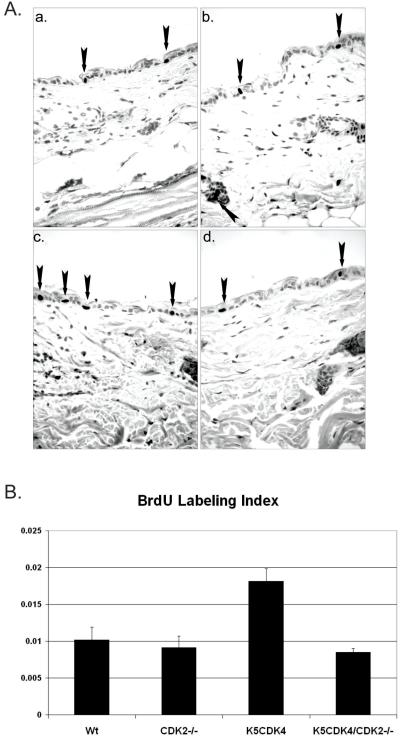
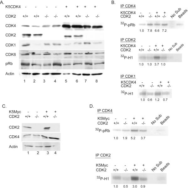
Consistent with our previous results, analysis of epidermal proliferation shows a two-fold increase in the number of S-phase cells (BrdU-positive) in K5CDK4 epidermis compared with wild type mice (Figure 1). On the other hand, CDK2-null mice do not show a difference in the level of keratinocyte proliferation compared with wild type mice, however, ablation of CDK2 in K5CDK4 background (K5CDK4/CDK2−/− mice) diminishes proliferation triggered by CDK4 overexpression (K5CDK4 mice) (Figure 1B). Biochemical analysis of mouse epidermis shows that CDK2 ablation does not affect transgenic expression of CDK4 in K5CDK4/CDK2−/− compound mice which remain higher compared to wild type littermate (Figure 2A). Moreover, loss of CDK2 expression is not compensated for increased levels of endogenous CDK4 or CDK6 which although heterogeneous among siblings of same genotype, the levels of expression were similar between CDK2−/− and wild type mice (Figure 2A). Consistent with previous description of our K5CDK4 model, we did not observe differences in Rb protein levels. In addition, we did not detect changes in protein mobility consistent with pRb-phosphorylation (Figure 2A) (7). In vitro kinase assay shows a 3.7-fold increase of CDK2 activity in epidermis from K5CDK4 mice (Figure 2B) (7). K5CDK4 mice shows increased CDK4 kinase activity (6.6-fold) compared with wild type mice and importantly, CDK2 ablation does not reduce this kinase activity (K5CDK4/CDK2−/− mice, 7.2-fold increase) (Figure 2B). Interestingly, the kinase activity of endogenous CDK4 is also augmented by 7.8-fold in CDK2−/− epidermis compared with wild type siblings (Figure 2B). At present, we do not know the molecular mechanism underlying this increase of CDK4 activity, but whether this is a compensatory mechanism that is functional in keratinocytes and/or other CDK2−/− cells warrant further investigations. Recently, it has been demonstrated that CDK1 can compensate the loss of CDK2 by binding to cyclin E and cyclin A, however, we did not observed increase CDK1 kinase activity in CDK2−/− or K5CDK4/CDK2−/− mice (25-27). In fact, a mild reduction in CDK1 kinase activity was observed in keratinocyte lacking CDK2 expression (Figure 2B). We conclude that ablation of CDK2 does not affect normal keratinocyte proliferation, but reduces CDK4-induced hyperproliferation in mouse skin.
Figure 1.
Epidermal proliferation in K5CDK4/CDK2−/− compound mice. A. BrdU incorporation of representative paraffin skin sections from wild type (a), CDK2−/− (b), K5CDK4 (c), and K5CDK4/CDK2−/− (d) mice at 40X magnification. Arrows indicate BrdU-positive cells. B. Bar graph indicates quantification of BrdU labeling index from interfollicular epidermis (BrdU positive-cells / Total number of interfollicular cells).
Figure 2.
Biochemical analysis of K5CDK4/CDK2−/− and K5Myc/CDK2−/− mice epidermis. (A) Immunoblots analysis of epidermal protein lysates from age matched wild type (1-2), CDK2−/− (3-4), K5CDK4 (5-6) and K5CDK4/CDK2−/− (7-8) mice. Antibodies against CDKs and pRb are indicated on the left column. Immunoblot for actin used as loading control. (B) CDK kinase assays of epidermal protein lysates from age matched wild type, CDK2−/−, K5CDK4 and K5CDK4/CDK2−/− mice were carried out with antibodies against CDK4 (IP CDK4), CDK2 (IP CDK2) and CDK1 (IP CDK1) using pRb and histone H1 peptides as substrates. Fold increase in CDK kinase activities gauged in comparison to wild type activity. (C) Immunoblots analysis of wild type (1), CDK2−/− (2), K5-Myc (3) and K5-Myc/CDK2−/− (4) epidermal lysates for CDK2, CDK4 and actin as loading control. (D) CDK kinase assay of epidermal lysates from age matched wild type, CDK2−/−, K5Myc and K5Myc/CDK2−/− mice were carried out with antibodies against CDK4 (IP CDK4) and CDK2 (IP CDK2) using pRb and histone H1 peptides as substrates, respectively. Fold increase in CDK kinase activities gauged in comparison to wild type activity. Control “Beads”, immunoprecipitation using normal rabbit IgG and K5CDK4 (B) or K5Myc (D) lysates. Control “No Sub”, kinase reaction without pRb or H1 recombinant protein substrates.
CDK2 deficiency reduces skin tumor development and malignant progression
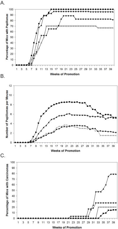
We previously demonstrated that forced expression of CDK4 results in increase malignant progression to skin squamous cell carcinomas (SCC) in a two-stage carcinogenesis protocol (20). This protocol induces skin papilloma development by a single application of a carcinogen follow by bi-weekly treatment with a tumor promoter causing a selection of cells bearing Ha-ras mutations. In order to investigate the role of CDK2 in K5CDK4 skin tumorigenesis, we assessed the response of K5CDK4/CDK2−/− to the two-stage carcinogenesis protocol. Four group of mice (K5CDK4, CDK2−/−, K5CDK4/CDK2−/− and wild type) (20 newborns each) were treated with 50 μg/200μl of DMBA followed by two applications per week of 2.5 μg of TPA after being weaned (21 days old). TPA treatment was continued for 25 weeks and the incidence and multiplicity of papillomas and SCCs were scored in each group for 40 weeks. Note that no spontaneous skin tumors were detected in untreated mice. The incidence of papilloma formation reaches a plateau at ~15 weeks in K5CDK4 and wild type mice, 100% and 97%, respectively (Figure 3A). A reduced tumor incidence was observed in CDK2−/− and K5CDK4/CDK2−/−, with each group reaching a plateau of 82% and 70%, respectively (Figure 3A). Ablation of CDK2 also results in decreased number of tumors per mouse (multiplicity). At 20 weeks of TPA promotion the four groups of mice reached a plateau where reduced papilloma multiplicity was observed in K5CDK4 mice compared with wild type littermates (8.5 and 6 papillomas per mouse respectively) (p<0.05; Mann-Whitney U test) (Figure 3B). CDK2 ablation results in the development of fewer papillomas in both CDK2−/− and K5CDK4/CDK2−/− mice (3.5 tumors/mouse) compared with wild type and K5CDK4 mice (p<0.05; Mann-Whitney U test) (Figure 3B).
Figure 3.
Effect of CDK4 overexpression and CDK2 deletion on induced tumorigenesis in mouse skin. A. Percentage of mice with at least one papilloma (tumor incidence), within 40 weeks of the bi-weekly administration of 2.5 μg TPA. B. Average number of papillomas per mouse (tumor multiplicity) within 40 weeks of bi-weekly promotion with 2.5 μg TPA. C. Percentage of mice with squamous cell carcinomas (SCC incidence) within 40 weeks of bi-weekly promotion with 2.5 μg TPA. Wt (■), CDK2 (x), K5CDK4 (▲) and K5CDK4/CDK2−/− (●) mice.
Consistent with our previous studies, SCCs develop rapidly and in greater number in K5CDK4 transgenic mice (Figure 3C) (20). The first SCC appeared at 22 weeks of promotion and 70% of K5CDK4 mice had developed SCC by 40 weeks of promotion. In sharp contrast, CDK2 ablation leads to a strong reduction of malignant progression since only 28% of K5CDK4/CDK2−/− mice developed SCCs (p<0.05) (Figure 3C). K5CDK4/CDK2−/− mice also exhibit an increased SCC latency period, where the first SCC was observed at 29 weeks of promotion. SCC incidence was similar between CDK2−/− and wild type groups where 15-20% of mice develop SCCs (Figure 3C). Histopathological analysis was performed with skin tumors of 30-40 weeks of promotion, papillomas and SCCs were graded according to a modified Broders classification system (28, 29). This classification of SCCs breaks down this category into four type of tumors: 1) SCC I: well differentiated SCC, most of the tumor mass composed of differentiated cells; 2) SCC II: moderately differentiated with approximately 50% of differentiating cells; 3) SCC III: poor differentiated tumor with little keratinization and 4) SCC IV: poorly differentiated tumors with marginal or no keratinization, including spindle cell carcinomas (28, 30). Most of the wild type tumors analyzed were regular papillomas (60%) and 40% were classified as SCC I. On the other hand, analyzed skin tumors from K5CDK4 mice were classified as SCC I (45%), SCC II (25%) and SCC III (5%) and papillomas (25%) (Table 1). These data are consistent with our previous observation that forced expression of CDK4 increase the rate of malignant conversion (20). In sharp contrast, only 43% of the K5CDK4/CDK2−/− tumors analyzed were classified as SCC and all of them were well differentiated (SCC I) and no SCC grade II or III were observed in this group. Finally, CDK2−/− mice showed a spectrum of tumors similar to wild type littermates (Table 1).
Table 1.
Histopathological analysis of skin tumors
| Mice |
No of tumors/group |
No (%) of tumors classified as:* |
|||
|---|---|---|---|---|---|
| Papillomaa | SCC Ib | SCC IIc | SCC IIId | ||
| Wild type | 23 | 14 (60) | 9 (40) | 0 (0) | 0 (0) |
| CDK2−/− | 12 | 8 (66) | 4 (33) | 0 (0) | 0 (0) |
| K5CDK4 | 20 | 5 (25) | 9 (45) | 6 (25) | 1 (5) |
| K5CDK4/CDK2−/− | 14 | 8 (57) | 6 (43) | 0 (0) | 0 (0) |
χ2, p< 0.05.
No atypia in basal layers.
Well differentiated SCC, most of the tumor mass composed of differentiated cells.
Moderately differentiated with approximately 50% of differentiating cells.
Poorly differentiated tumor with little keratinization, approximately 10-25% of the tumor composed of differentiating cells.
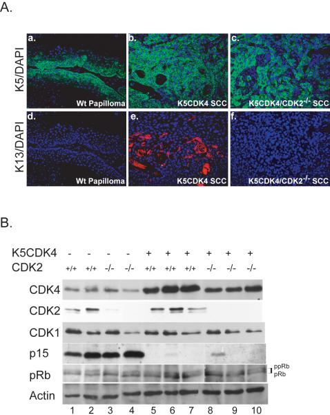
Consistently, K5CDK4 carcinomas showed strong positive immunofluorescence staining for keratin 13 (K13), a malignancy marker in squamous epithelia (31), whereas SCC from K5CDK4/CDK2−/− mice were K13-negative (Figure 4A), further confirming that SCC from K5CDK4/CDK2−/− mice were well differentiated SCC and less aggressive than K5CDK4 SCC.
Figure 4.
Keratin 13 expression and biochemical analysis of mouse skin tumors. (A) Expression of normal differentiation marker keratin 5 (K5) (green) was detected by immunofluorescence in all paraffin cross sections of wild type papillomas (a), K5CDK4 SCC (b), and K5CDK4/CDK2−/− SCC (c). Note that while the normal pattern of K5 expression (basal cell layer), as seen in wt papillomas, is lost in SCCs. SCC from K5CDK4 mice (e) stain positive for keratin 13 (K13) (red), marker associated with malignant progression. Magnifications at 20X; Dapi (blue) used as nuclear counter stain.
(B) Protein lysates from 30 week papillomas obtained from wt (lines 1, 2), CDK2−/− (lines 3, 4), K5CDK4 (lines 5, 6, 7) and K5CDK4/CDK2−/− (lines 8, 9, 10) mice were separated by SDS-PAGE, transferred to nitrocellulose membrane and blotted for CDK4, CDK2, CDK1, p15Ink4b and pRb. Actin was used as loading control. Hyperphosphorylation (ppRb) and hypophosphorylation (pRb) of retinoblastoma protein was denoted on the right.
Biochemical analysis of skin tumors from the four genotypes indicates that ablation of CDK2 results in a mild reduction in the transgenic expression of CDK4 in K5CDK4/CDK2−/− compound mice (Figure 4B, lines 5-7 and 8-10). The mechanism and consequences of this mild reduction in transgenic CDK4 expression warrants further investigation. Moreover, no compensation by CDK1 was observed in CDK2−/− tumors, and all tumors showed similar level of pRb phosphorylation (Figure 4B). Interestingly, p15Ink4b expression was reduced in K5CDK4 tumors independently of the CDK2 status (Figure 4B). We hypothesized that CDK4 expression increases Smad3 phosphorylation resulting in inhibition of p15Ink4b expression. Consistent with this hypothesis Liu et al. have shown that CDK4 and CDK2 phosphorylation of Smad3 inhibits its antiproliferative effects (32, 33). Whether reduction of p15Ink4 levels take part in the increase susceptibility of K5CDK4 mice to malignant transformation warrant further investigation. Finally, similar levels of CDK4 kinase activity were also observed in K5CDK4 and K5CDK4/CDK2−/− tumors (data no shown).
We conclude that lack of CDK2 expression decreases papilloma development and more importantly abolishes malignant progression induced by forced overexpression of CDK4 in ras-dependent tumorigenesis.
Lack of CDK2 expression does not inhibit oral tumor development
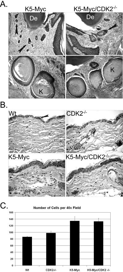
Overexpression of the murine c-myc gene in the basal cell layer of epithelium (K5Myc mice) results in epidermal hyperplasia and hypertrophy (20, 24). In addition, a high incidence of spontaneous tumors was observed in the oral mucosa of K5Myc transgenic mice (20, 24). These results showed that myc acts as an oncogene in the stratified epithelium, but the mechanisms leading to the malignant phenotype are not fully understood. Interestingly, K5Myc mice exhibit elevated CDK4 protein levels and ablation of CDK4 inhibits myc tumorigenic activities in epithelial tissues and reduces CDK2 activity, presumably through redistribution of p27Kip1 that bind and inhibit CDK2 (20). In order to investigate whether CDK2 mediates the oncogenic activities of myc, we developed K5Myc transgenic mice that lack the expression of CDK2 (K5Myc/CDK2−/− mice). These mice, along with K5Myc, CDK2−/− and wild type siblings, were analyzed for the development of spontaneous tumors. Histological analysis of mice bearing the myc transgene revealed tumors of oral mucosa (Figure 5A). These tumors were classified as SCC consisting of islands, cords of neoplastic epithelial cells showing a variable degree of squamous differentiation with occasional keratin pearls in the centers. The invasive margins of the tumor are surrounded by proliferative fibrous connective tissue (Figure 5A). Oral tumors were observed in K5Myc mice as young as 8 weeks old with an incidence of ~95%. In sharp contrast to our previously published K5Myc/CDK4−/− mice (20), K5Myc/CDK2−/− mice develop tumors with similar frequency as K5Myc mice (93%) (Figure 5A). Cell proliferation analysis of the oral cavity tumors did not reveal differences between the two genotypes (data no shown). Wild type and CDK2−/− littermates did not show any signs of spontaneous tumor development up to 18 months of age. Thus, mice with genetic ablation of CDK2 remain fully susceptible to myc-driven tumorigenesis of the oral mucosa. Collectively, these data indicate that CDK2 does not play a relevant role in the development of the spontaneous oral tumors observed in K5Myc mice.
Figure 5.
Analysis of oral tumors and epidermis from K5-Myc/CDK2−/− compound mice. A. H&E stained paraffin sections of 3 month old decalcified mouse heads from K5-Myc and K5-Myc/CDK2−/− mice show similar levels of fibrosis and areas of invasion, denoted by arrows, top panel. Oral SCC formation, consisting of neoplastic epithelial cell cords with a variable degree of squamous differentiation and occasional keratin pearls (K) in the centers. B. Paraffin sections of skin from K5Myc/CDK2−/− and control littermates stained with H&E. Arrow indicates epidermis. C. Quantification of the total number of nucleated cells per 40X field. Original magnification at 20X (A) and 40X (B); K, keratin pearls; De, dentin.
CDK2 is dispensable for c-myc mediated keratinocyte proliferation
We also asked whether CDK2 mediates the epidermal hyperproliferative phenotype triggered by myc overexpression. Thus, we analyzed the epidermis of K5Myc and K5Myc/CDK2−/− mice. The skin of K5Myc mice exhibit epidermal hyperplasia (increase cell number) and increased keratinocyte proliferation (increase number of BrdU-positive cells) (Figure 5B and C). Abrogation of CDK2 expression (K5Myc/CDK2−/−) does not result in reversion on these parameters and the hyperkeratosis (accumulation of keratinized cells in the epidermal surface) characteristic of myc overexpression still persists (Figure 5B).
Several reports have demonstrated that myc induces the transcription of CDK4 (34-36). Consistent with these results, we demonstrated that CDK4 and myc overexpression results in CDK2 activation through sequestration of p27Kip1 and p21Cip1 by CDK4 (7, 20). Furthermore, CDK2 activity was inhibited in K5Myc/CDK4−/− epidermis, most likely due to redistribution of the CDK2 inhibitors p27Kip1 and p21Cip1 (20). In order to evaluate whether expression and kinase activity of CDK4 was affected by the loss of CDK2 expression in K5Myc epidermis, we studied protein expression and in vitro kinase activity in epidermis from K5Myc/CDK2−/− compound mice. Protein extracts from mouse epidermis were subject to western blot analysis for CDK4 and CDK2. Consistent with our previous results, the levels of CDK4 was two-fold higher in K5Myc compared to wild type epidermis and absence of CDK2 does not modify CDK4 expression (Figure 2C). As previously reported the kinase activity of CDK4 increases 5.2-fold in K5Myc epidermis, compared to wild type epidermis. Elevation in CDK4 kinase activity was also observed in K5Myc/CDK2−/− epidermis (3.7-fold) compared to wild type mice (Figure 2D). As expected increased CDK2 kinase activity was observed in K5Myc mice (3-fold) due to sequestration of p27Kip1 and p21Cip1 by elevated levels of CDK4 whereas a baseline activity was observed in the other three genotypes (Figure 2D) (20). Collectively, these results indicate that CDK2 is dispensable for myc-induce epidermal proliferation.
DISCUSSION
The concept that CDK2 is essential for controlled entry into S phase and the onset of DNA replication was challenged a few years ago when two independent groups reported the generation of CDK2−/− mice (12, 13). These mice are viable, develop normally and only show meiotic defects. These results were surprising, as CDK2 was thought to be required for promoting G1/S transition. In addition, it was also demonstrated that cancer cells can proliferate in absence of CDK2 (15). Thus, the data accumulated in the last few years has brought the concept that CDK2 is not an adequate target for therapeutic intervention. In this report we prove that CDK2 inhibition can still be considerate as an adequate target for therapeutic intervention depending on the particular oncogenic pathway involved.
We have previously shown that forced expression of CDK4 in mouse skin induces keratinocyte proliferation and malignant progression in a ras-dependent skin tumorigenesis model (7, 20). Here, we have examined the skin of CDK2−/− mice which is structural and functional similar to that of wild type siblings. It has been reported that CDK1 activation compensates for the absence of CDK2 in thymocytes, although this mechanism depends on the presence of p27Kip1 (25). However, biochemical analysis of CDK2−/− epidermis shows reduction rather than activation of CDK1. We also observed increase activity of CDK4 in the epidermis of CDK2−/− mice (Figure 2B). Since p27Kip1 stabilize CDK4/D-type cyclin complexes (37), it was possible that in absence of CDK2, redistribution of p27Kip1 increase CDK4/D-type cyclin/p27Kip1 complex formation. Nevertheless, biochemical analysis did not show increased formation of those complexes (data no shown). Whether the increase in CDK4 activity compensates for the absence of CDK2 in the epidermis merits further investigation. The importance of the CDK4-CDK2 link in keratinocyte proliferation was demonstrated by the activation of CDK2, through titration of p27/p21, by forced expression of CDK4 (7, 23) (Figure 2B) and the reduction of the total number of proliferative cells in K5CDK4/CDK2−/− epidermis compared to K5CDK4 mice. Therefore, activation of both, CDK4 and CDK2 kinases are necessary for epidermal hyperproliferation observed in K5CDK4 mice (Figure 1).
We also studied whether CDK2 is required for CDK4-induced malignant progression to SCCs (20). In our experimental setting overexpression of CDK4 reduces papilloma multiplicity, although the number of mice with papilloma (incidence) was similar between K5CDK4 and wild type mice. Lack of CDK2 leads to a significant reduction of papilloma incidence and multiplicity in K5CDK4/CDK2−/− and CDK2−/− mice. More importantly, lack of CDK2 expression in K5CDK4 mice (K5CDK4/CDK2−/− mice) results in a severe reduction of CDK4-induced malignant progression to SCCs. Therefore, lack of CDK2 affects papilloma development and more importantly CDK2 expression is required for CDK4-induced malignant progression. Histopathological analysis of tumors from the four genotypes confirms the role of CDK2 in malignant progression. Tumors from K5CDK4 mice were classified as regular papillomas and SCC I, II, and III; whereas most of the K5CDK4/CDK2−/− tumors were regular papillomas and no SCCs II or III were observed. In addition, K5CDK4 carcinomas showed areas of strong positive immunofluorescence staining for keratin 13 (K13), a malignancy marker in squamous epithelia (31), whereas SCC from K5CDK4/CDK2−/− mice were K13-negative.
We have previously demonstrated that cyclin D1 and CDK4 expression plays an important role in papilloma development (38-41). We determined that cyclin D1 expression is not affected in CDK2−/− papillomas (data no shown). The latter confirms that reduced papilloma formation and progression in CDK2−/− and K5CDK4/CDK2−/− compound mice is not due to altered D-type cyclin levels. We also observed a mild reduction of the expression of transgenic CDK4 in tumors from K5CDK4/CDK2−/− mice compared with K5CDK4 siblings (Figure 4B). However, the CDK4 protein levels were still higher than wild type mice. Thus, the reduction of transgenic-CDK4 is not responsible for the reduction in the number of papillomas and SCC in K5CDK4/CDK2−/− mice. Supporting these results we previously reported, that two independent K5CDK4 transgenic lines exhibiting different levels of CDK4 protein expression develop SCCs at a similar rate (20). Similar to CDK1 expression in epidermis, CDK1 protein level was variable among tumors from the same genotype, but there are no relevant differences in CDK1 levels among the four genotypes. Interesting, p15Ink4b expression was reduced in K5CDK4 mice independently of the CDK2 status. p15Ink4b transcription can be down-regulated via phosphorylation and inactivation of Smad3 by CDK4 (32, 33), but decrease p15Ink4b was observed in both K5CDK4 and K5CDK4/CDK2−/−, suggesting that its downregulation is not responsible for the CDK4-induced malignant progression.
The present study allows us to define an important role of CDK2 in malignant progression. Interestingly, Duensing et al. determined that CDK2 is dispensable for normal centrosome duplication, but required for oncogene-induced centrosome overduplication (42). Thus, lack of CDK2 could also abrogate the centrosome overduplication in epithelial tumor cells protecting the cells against genetic instability, a mechanism involved in malignant progression. The present study also shows a reduction in the total number of chemically induced tumors in both CDK2−/− and K5CDK4/CDK2−/− mice suggesting that CDK2 may play an important role in cell survival. To this effect Huang et al have recently shown that CDK2 influences survival of cells under genotoxic stress by inhibiting the forkhead transcription factor, FOXO1. Importantly, CDK2 phosphorylate and inhibits the pro-apoptotic function of FOXO1 (43, 44). Thus, CDK2 ablation could restore or increase FOXO1 activity leading to reduce tumorigenesis.
We have previously demonstrated that CDK4 ablation, but not cyclin D2, inhibits myc-mediated oral tumorigenesis (23). Here we show that deletion of CDK2 in a K5Myc background does not affect tumor development as 93% of K5Myc/CDK2−/− mice developed oral tumors. These tumors were classified as SCCs showing a variable degree of squamous differentiation. No difference in tumor incidences or the degree of differentiation was observed between K5Myc and K5Myc/CDK2−/− mice as opposed to K5Myc/CDK4−/− mice which did not develop tumors (23). Immunostaining analysis of myc-induced oral tumors revealed equal levels of proliferation (BrdU-positive cells) in the epithelial compartment of the oral tumors from K5Myc and K5Myc/CDK2−/− mice (data no shown). Thus, it is clear that lack of CDK2 does not affect the proliferation mediated by myc. To determine the effect of myc in other tissues, we also analyzed mouse epidermis from K5Myc and K5Myc/Cdk2−/− mice. In contrast to the reduced proliferation mediated by lack of CDK4, ablation of CDK2 did not result in suppression of the epidermal phenotype observed in K5Myc mice. As we previously reported, overexpression of CDK4 was observed in K5Myc epidermis and it was not affected by CDK2 ablation (23, 36). Consequently, lack of CDK2 cannot suppress the epidermal phenotype mediated by myc suggesting that CDK4 plays a unique role in myc-induced keratinocyte proliferation (45, 46). Thus, our results suggest that CDK2 is expendable for myc-induce proliferation not only in the gingival epithelium, but as a general characteristic for myc-induced proliferation in epithelial tissues. In addition to inducing proliferation, myc can also drive apoptosis which serves as a barrier to unchecked cellular proliferation (47, 48). In this sense, it was recently described that the removal of CDK2 suppressed the ability of myc to induce apoptosis (49). Thus, it is possible that loss of CDK2 reduces the myc-mediated apoptosis supporting tumor development.
In summary, our data suggest that the efficacy of targeting CDK2 in tumor development and malignant progression is dependent on the specific oncogenic pathway involved. Similarly, other groups have shown that different oncogenic pathways respond differently to the inhibition of cell cycle regulators. For instance, cyclin D1-deficient mice are resistant to breast cancers induced by ras, but remains fully sensitive to other oncogenic pathways such as those driven by myc or Wnt-1 (50). Also, CDK1 inhibition induces apoptosis in tumor cell lines transformed with myc, but not Ras or a variety of other activated oncogenes (51).
Whether loss of CDK2 inhibits tumorigenesis mediated by other pathways such us neu or wnt remains to be determined. Overall we have established that ras/CDK4-induced tumors are more sensitive to CDK2 inhibition than myc-induced tumors. Therefore, the suitability of CDK2 as a target for therapeutic intervention must be considered in the context of the particular pathway affected.
Acknowledgements
We would like to thank Juan C. Santiago for technical support, the Laboratory Animal Resources (LAR), and the CVM histology service personnel. We specially thank to Dr. Mariano Barbacid for provide the CDK2-null mice. We thank to Christopher Sistrunk for help editing this paper. This study is supported by NIH Grant CA90864 and CA116328.
REFERENCES
- 1.Sherr CJ. D-type cyclins. Trends Biochem Sci. 1995;20:187–90. doi: 10.1016/s0968-0004(00)89005-2. [DOI] [PubMed] [Google Scholar]
- 2.Weinberg RA. The retinoblastoma protein and cell cycle control. Cell. 1995;81:323–30. doi: 10.1016/0092-8674(95)90385-2. [DOI] [PubMed] [Google Scholar]
- 3.Blain S, Montalvo E, Massague J. Differential interaction of the cyclin-dependent kinase (cdk) inhibitor p27Kip1 with cyclin A-cdk2 and cyclin D2-cdk4. J Biol Chem. 1997;272:25863–72. doi: 10.1074/jbc.272.41.25863. [DOI] [PubMed] [Google Scholar]
- 4.Labaer J, Garret MD, Stevenson LF, et al. New functional activities for the p21 family of CDK inhibitors. Genes & Dev. 1997;11:847–62. doi: 10.1101/gad.11.7.847. [DOI] [PubMed] [Google Scholar]
- 5.Bouchard C, Thieke K, Maier A, et al. Direct induction of cyclin D2 by Myc contributes to cell cycle progression and sequestration of p27. Embo J. 1999;18(19):5321–33. doi: 10.1093/emboj/18.19.5321. [DOI] [PMC free article] [PubMed] [Google Scholar]
- 6.Perez-Roger I, Kim SH, Griffiths B, Sewing A, Land H. Cyclins D1 and D2 mediate myc-induced proliferation via sequestration of p27(Kip1) and p21(Cip1) Embo J. 1999;18(19):5310–20. doi: 10.1093/emboj/18.19.5310. [DOI] [PMC free article] [PubMed] [Google Scholar]
- 7.Miliani de Marval P, Gimenez-Conti I, LaCava M, Martinez L, Conti C, Rodriguez-Puebla M. Transgenic Expression of CDK4 results in epidermal hyperplasia and severe dermal fibrosis. Am J Pathology. 2001;159(1):369–79. doi: 10.1016/S0002-9440(10)61703-8. [DOI] [PMC free article] [PubMed] [Google Scholar]
- 8.van den Heuvel S, Harlow E. Distinct roles for cyclin-dependent kinases in cell cycle control. Science. 1993;262(5142):2050–4. doi: 10.1126/science.8266103. [DOI] [PubMed] [Google Scholar]
- 9.Ohtsubo M, Theodoras A, Schumacher J, Roberts J, Pagano M. Human cyclin E, a nuclear protein essential for the G1-toS phase transition. Mol Cell Biol. 1995;15:1559–71. doi: 10.1128/mcb.15.5.2612. [DOI] [PMC free article] [PubMed] [Google Scholar]
- 10.Pagano M, Pepperkok R, Verde F, Ansorge W, Draetta G. Cyclin A is required at two points in the human cell cycle. EMBO J. 1992;11(3):961–71. doi: 10.1002/j.1460-2075.1992.tb05135.x. [DOI] [PMC free article] [PubMed] [Google Scholar]
- 11.Tsai LH, Lees E, Faha B, Harlow E, Riabowol K. The cdk2 kinase is required for the G1-to-S transition in mammalian cells. Oncogene. 1993;8(6):1593–602. [PubMed] [Google Scholar]
- 12.Berthet C, Aleem E, Coppola V, Tassarollo L, Kaldis P. Cdk2 Knockout Mice Are Viable. Current Biology. 2003;13:1775–85. doi: 10.1016/j.cub.2003.09.024. [DOI] [PubMed] [Google Scholar]
- 13.Ortega S, Prieto I, Odajima J, et al. Cyclin-dependent kinase 2 is essential for meiosis but not for mitotic cell division in mice. Nat Genet. 2003;35(1):25–31. doi: 10.1038/ng1232. [DOI] [PubMed] [Google Scholar]
- 14.Martin A, Odajima J, Hunt SL, et al. Cdk2 is dispensable for cell cycle inhibition and tumor suppression mediated by p27(Kip1) and p21(Cip1) Cancer Cell. 2005;7(6):591–8. doi: 10.1016/j.ccr.2005.05.006. [DOI] [PubMed] [Google Scholar]
- 15.Tetsu O, McCormick F. Proliferation of cancer cells despite CDK2 inhibition. Cancer Cell. 2003;3(3):233–45. doi: 10.1016/s1535-6108(03)00053-9. [DOI] [PubMed] [Google Scholar]
- 16.Rane SG, Dubus P, Mettus RV, et al. Loss of Cdk4 expression causes insulin-deficient diabetes and Cdk4 activation results in B-islet cell hyperplasia. Nature Genetic. 1999;22:44–52. doi: 10.1038/8751. [DOI] [PubMed] [Google Scholar]
- 17.Tsutsui T, Hesabi B, Moons DS, et al. Targeted disruption of CDK4 delays cell cycle entry with enhanced p27(Kip1) activity. Mol Cell Biol. 1999;19(10):7011–9. doi: 10.1128/mcb.19.10.7011. [DOI] [PMC free article] [PubMed] [Google Scholar]
- 18.Moons DS, Jirawatnotai S, Parlow AF, Gibori G, Kineman RD, Kiyokawa H. Pituitary hypoplasia and lactotroph dysfunction in mice deficient for cyclin-dependent kinase-4. Endocrinology. 2002;143(8):3001–8. doi: 10.1210/endo.143.8.8956. [DOI] [PubMed] [Google Scholar]
- 19.Moons DS, Jirawatnotai S, Tsutsui T, et al. Intact follicular maturation and defective luteal function in mice deficient for cyclin-dependent kinase-4. Endocrinology. 2002;143(2):647–54. doi: 10.1210/endo.143.2.8611. [DOI] [PubMed] [Google Scholar]
- 20.Miliani de Marval PL, Macias E, Conti CJ, Rodriguez-Puebla ML. Enhanced malignant tumorigenesis in Cdk4 transgenic mice. Oncogene. 2004;23:1863–73. doi: 10.1038/sj.onc.1207309. [DOI] [PMC free article] [PubMed] [Google Scholar]
- 21.Rodriguez-Puebla ML, Miliani de Marval PL, LaCava M, Moons DS, Kiyokawa H, Conti CJ. cdk4 Deficiency Inhibits Skin Tumor Development but Does Not Affect Keratinocyte Proliferation. Am J of Pathology. 2002;161(2):405–11. doi: 10.1016/S0002-9440(10)64196-X. [DOI] [PMC free article] [PubMed] [Google Scholar]
- 22.Zou X, Ray D, Aziyu A, et al. Cdk4 disruption renders primary mouse cells resistant to oncogenic transformation, leading to Arf/p53-independent senescence. Genes Dev. 2002;16(22):2923–34. doi: 10.1101/gad.1033002. [DOI] [PMC free article] [PubMed] [Google Scholar]
- 23.Miliani de Marval PL, Macias E, Rounbehler R, et al. Lack of cyclin-dependent kinase 4 inhibits c-myc tumorigenic activities in epithelial tissues. Mol Cell Biol. 2004;24(17):7538–47. doi: 10.1128/MCB.24.17.7538-7547.2004. [DOI] [PMC free article] [PubMed] [Google Scholar]
- 24.Rounbehler RJ, Schneider-Broussard R, Conti CJ, Johnson DG. Myc lacks E2F1’s ability to suppress skin carcinogenesis. Oncogene. 2001;20(38):5341–9. doi: 10.1038/sj.onc.1204691. [DOI] [PubMed] [Google Scholar]
- 25.Aleem E, Kiyokawa H, Kaldis P. Cdc2-cyclin E complexes regulate the G1/S phase transition. Nat Cell Biol. 2005;7(8):831–6. doi: 10.1038/ncb1284. [DOI] [PubMed] [Google Scholar]
- 26.Bashir T, Pagano M. Cdk1: the dominant sibling of Cdk2. Nat Cell Biol. 2005;7(8):779–81. doi: 10.1038/ncb0805-779. [DOI] [PubMed] [Google Scholar]
- 27.Kaldis P, Aleem E. Cell Cycle Sibling Rivalry: Cdc2 vs. Cdk2. Cell Cycle. 2005;4(11) doi: 10.4161/cc.4.11.2124. [DOI] [PubMed] [Google Scholar]
- 28.Klein-Szanto A. Pathology of Neoplasia and Preneoplasia in Rodents. EULEP; Schattauer: 1997. Melanotic and non-melanotic tumours of the rodent skin; pp. 1–18. [Google Scholar]
- 29.Klein-Szanto A, Conti C. Toxicology pathology of the skin. In: Hascheck, Roussaeaunx, Walling, editors. Handbook of Toxicology Pathology. Academic Press; San Diego: 2002. pp. 85–120. [Google Scholar]
- 30.Klein-Szanto A, Larcher F, Bonfil R, Conti C. Multistage chemical carcinogenesis protocols produce spindle cell carcinomas of the mouse skin. Carcinogenesis. 1989;10:2169–72. doi: 10.1093/carcin/10.11.2169. [DOI] [PubMed] [Google Scholar]
- 31.Nischt R, Roop DR, Mehrel T, et al. Aberrant expression during two-stage mouse skin carcinogenesis of a type I 47-kDa keratin, K13, normally associated with terminal differentiation of internal stratified epithelia. Mol Carcinog. 1988;1(2):96–108. doi: 10.1002/mc.2940010205. [DOI] [PubMed] [Google Scholar]
- 32.Liu F. Smad3 phosphorylation by cyclin-dependent kinases. Cytokine Growth Factor Rev. 2006;17(1-2):9–17. doi: 10.1016/j.cytogfr.2005.09.010. [DOI] [PubMed] [Google Scholar]
- 33.Liu F, Matsuura I. Inhibition of Smad antiproliferative function by CDK phosphorylation. Cell Cycle. 2005;4(1):63–6. doi: 10.4161/cc.4.1.1366. [DOI] [PubMed] [Google Scholar]
- 34.Augenlicht LH, Wadler S, Corner G, et al. Low-level c-myc amplification in human colonic carcinoma cell lines and tumors: a frequent, p53-independent mutation associated with improved outcome in a randomized multi-institutional trial. Cancer Res. 1997;57(9):1769–75. [PubMed] [Google Scholar]
- 35.Erisman MD, Rothberg PG, Diehl RE, Morse CC, Spandorfer JM, Astrin SM. Deregulation of c-myc gene expression in human colon carcinoma is not accompanied by amplification or rearrangement of the gene. Mol Cell Biol. 1985;5(8):1969–76. doi: 10.1128/mcb.5.8.1969. [DOI] [PMC free article] [PubMed] [Google Scholar]
- 36.Hermeking H, Rago C, Schuhmacher M, et al. Identification of CDK4 as a target of c-Myc. Proc Natl Acad Sci. 2000;97(5):2229–34. doi: 10.1073/pnas.050586197. [DOI] [PMC free article] [PubMed] [Google Scholar]
- 37.Cheng M, Olivier P, Diehl J, et al. The p21(Cip1) and p27(Kip1) CDK ‘inhibitors’ are essential activators of cyclin D-dependent kinases in murine fibroblasts. EMBO J. 1999;18(6):1571–83. doi: 10.1093/emboj/18.6.1571. [DOI] [PMC free article] [PubMed] [Google Scholar]
- 38.Robles A, Rodriguez-Puebla M, Glick A, et al. Reduced skin tumor development in Cyclin D1 deficient mice highlights the oncogenic ras pathway in vivo. Genes & Dev. 1998;12:2469–74. doi: 10.1101/gad.12.16.2469. [DOI] [PMC free article] [PubMed] [Google Scholar]
- 39.Rodriguez-Puebla ML, LaCava M, Gimenez-Conti IB, Jonhson DG, Conti CJ. Deregulated Expression of Cell-Cycle proteins during Premalignant Progression in SENCAR Mouse Skin. Oncogene. 1998;17:2251–8. doi: 10.1038/sj.onc.1202131. [DOI] [PubMed] [Google Scholar]
- 40.Robles AI, Conti CJ. Early overexpression of cyclin D1 protein in mouse skin carcinogenesis. Carcinogenesis. 1995;16:781–6. doi: 10.1093/carcin/16.4.781. [DOI] [PubMed] [Google Scholar]
- 41.Bianchi AB, Fischer SM, Robles AI, Rinchik EM, Conti CJ. Overexpression of cyclin D1 in mouse skin carcinogenesis. Oncogene. 1993;8:1127–33. [PubMed] [Google Scholar]
- 42.Duensing A, Liu Y, Tseng M, Malumbres M, Barbacid M, Duensing S. Cyclin-dependent kinase 2 is dispensable for normal centrosome duplication but required for oncogene-induced centrosome over duplication. Oncogene. 2006;25(20):2943–9. doi: 10.1038/sj.onc.1209310. [DOI] [PMC free article] [PubMed] [Google Scholar]
- 43.Huang H, Tindall DJ. CDK2 and FOXO1: a fork in the road for cell fate decisions. Cell Cycle. 2007;6(8):902–6. doi: 10.4161/cc.6.8.4122. [DOI] [PubMed] [Google Scholar]
- 44.Huang H, Regan KM, Lou Z, Chen J, Tindall DJ. CDK2-dependent phosphorylation of FOXO1 as an apoptotic response to DNA damage. Science. 2006;314(5797):294–7. doi: 10.1126/science.1130512. [DOI] [PubMed] [Google Scholar]
- 45.Dang CV. c-Myc target genes involved in cell growth, apoptosis, and metabolism. Mol Cell Biol. 1999;19(1):1–11. doi: 10.1128/mcb.19.1.1. [DOI] [PMC free article] [PubMed] [Google Scholar]
- 46.Dang CV, Resar LM, Emison E, et al. Function of the c-Myc oncogenic transcription factor. Exp Cell Res. 1999;253(1):63–77. doi: 10.1006/excr.1999.4686. [DOI] [PubMed] [Google Scholar]
- 47.Pelengaris S, Khan M, Evan G. c-MYC: more than just a matter of life and death. Nat Rev Cancer. 2002;2(10):764–76. doi: 10.1038/nrc904. [DOI] [PubMed] [Google Scholar]
- 48.Yin XY, Grove L, Datta NS, Katula K, Long MW, Prochownik EV. Inverse regulation of cyclin B1 by c-Myc and p53 and induction of tetraploidy by cyclin B1 overexpression. Cancer Res. 2001;61(17):6487–93. [PubMed] [Google Scholar]
- 49.Deb-Basu D, Aleem E, Kaldis P, Felsher DW. CDK2 is required by MYC to induce apoptosis. Cell Cycle. 2006;5(12):1342–7. doi: 10.4161/cc.5.12.2859. [DOI] [PubMed] [Google Scholar]
- 50.Yu Q, Geng Y, Sicinski P. Specific protection against breast cancers by cyclin D1 ablation. Nature. 2001;411(6841):1017–21. doi: 10.1038/35082500. [DOI] [PubMed] [Google Scholar]
- 51.Goga A, Yang D, Tward AD, Morgan DO, Bishop JM. Inhibition of CDK1 as a potential therapy for tumors over-expressing MYC. Nat Med. 2007 doi: 10.1038/nm1606. [DOI] [PubMed] [Google Scholar]


