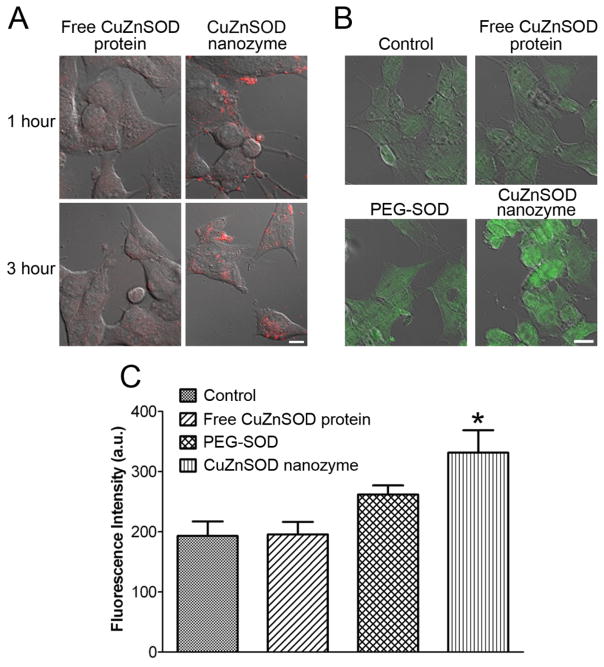
Figure 2. CuZnSOD nanozyme penetrates CATH.a neuronal cell membrane to increase intracellular levels of CuZnSOD protein.
(A) Representative confocal microscopy images showing rhodamine fluorescence in CATH.a neurons following 1 or 3 hours of incubation with rhodamine-labeled free CuZnSOD protein or CuZnSOD nanozyme (400 U/mL). (B) Representative confocal microscopy images showing CuZnSOD immunofluorescence staining in control CATH.a neurons and neurons treated (3 hours) with free CuZnSOD protein, PEG-SOD, or CuZnSOD nanozyme (400 U/mL). (C) Summary data (n=4 separate cultures per group) of CuZnSOD immunofluorescence in control CATH.a neurons (n=96 neurons) and neurons treated (3 hours) with free CuZnSOD protein (n=81 neurons), PEG-SOD (n=75 neurons) or CuZnSOD nanozyme (n=66 neurons). *p<0.05 vs. control and free CuZnSOD. a.u.= arbitrary units. Magnification bar in (A) and (B) equals 10 μm.

