Abstract
Therapeutic vaccination against idiotype is a promising strategy for immunotherapy of B-cell malignancies. We have previously shown that CDR3-based DNA immunization can induce immune response against lymphoma and explored this strategy to provide protection in a murine B-cell lymphoma model. Here we performed vaccination employing as immunogen a naked DNA fusion product. The DNA vaccine was generated following fusion of a sequence derived from tetanus toxin fragment C to the VHCDR3109−116 epitope. Induction of tumor-specific immunity as well as ability to inhibit growth of the aggressive 38C13 lymphoma and to prolong survival of vaccinated mice has been tested. We determined that DNA fusion vaccine induced immune response, elicited a strong protective antitumor immunity, and ensured almost complete long-term tumor-free survival of vaccinated mice. Our results show that CDR3-based DNA fusion vaccines hold promise for vaccination against lymphoma.
1. Introduction
Lymphomas represent the fifth most common malignancies. Each year, approximately 55 000 new cases are diagnosed with non-Hodgkin's lymphomas (NHLs) in the United States [1]. Despite current therapeutic strategies including chemotherapy, transplantation, and passive immunotherapy with monoclonal antibodies, many lymphoma patients remain incurable. The recent years have witnessed the development of a variety of promising immunotherapies for treating patients with B-cell NHLs. Vaccine strategies targeting NHLs have largely focused on using the idiotype (Id) of the tumor immunoglobulin (Ig) individually expressed on the surface of malignant B cells as tumor-specific antigen (Ag). After decades of work, some clear evidence of clinical efficacy in phase I/II trials using Id protein vaccines has accumulated, despite results from phase III trials seem disappointing [2, 3]. Furthermore, streamlined production of these patient-specific vaccines is required for eventual clinical application.
Several strategies are being developed to improve these results and include optimization of antigen delivery and presentation as well as enhancement of antitumor T cell function.
DNA vaccines have emerged as a novel lymphoma vaccine formulation for antigen-specific immunotherapy [4]. Such a method is an attractive and effective approach for active therapeutic vaccination since it does not require the production and isolation of a purified protein for each patient, a process that is expensive, laborious, and time-consuming. The protein is endogenously produced and secreted, which may result in more efficient antigen presentation in both classes I and II major histocompatibility complex (MHC) pathways resulting in enhanced anti-Id immune responses. In addition to their safety, stability, ease of production, DNA vaccines are highly flexible, allowing coexpression of several types of antigens and immunological proteins [5]. Furthermore, the performance of DNA vaccines may be improved by in vivo electroporation (EP) as a safe and efficient method of in vivo delivery resulting in greatly enhanced DNA uptake, protein expression levels, and degree of local inflammation [6]. DNA vaccination has been applied to therapy of experimental murine lymphomas (for reviews, see Hurvitz and Timmerman [7] and Neelapu et al. [8]). DNA vaccines that express either the tumor-derived Id or the tumor variable (V) regions in a single-chain Fv conformation (scFv) have been constructed. However, due to the weak immunogenicity in most cases, their effectiveness depend on carrier proteins or adjuvant proteins linked to the Id structures [9–14].
Idiotypic antigenic determinants lying mainly within the complementary-determining regions (CDRs) 3 have been considered a “hot spot” of particular interest for construction of subunit vaccines [15–18]. Vaccines including only this minimal antigenic domain were proved to induce antibody response [19, 20].
We demonstrated that DNA immunization of outbred mice with different patient-derived VHCDR3 peptides elicited antibodies able to recognize native antigens on individual patient's tumor cells [20]. Recently, our group has shown the tumor protective effects recruited by CDR3-based DNA vaccines in a poorly immunogenic, highly aggressive murine B-cell lymphoma model. A DNA vaccine containing a VHCDR3 epitope of the 38C13 B-cell lymphoma [21], administered in combination with the VLCDR3-encoding plasmid, provided tumor protection and long-term tumor-free survival in 60% of syngeneic mice [22]. In the current study to enhance the potency of this vaccination platform, we used the DNA fusion vaccine design encoding tumor Ags linked to pathogen-derived sequences, aimed to provide CD4+ T cell help. Engagement of CD4+ T helper (TH) cells has been shown to play a major role in linking and coordinating innate and adaptive immune responses [4, 23]. Many attempts to incorporate exogenous helper antigens into DNA vaccine design to break tolerance to self (tumor) antigens and to improve efficacy by helper T cells have been described [24–28]. Fusion protein of tetanus toxin fragment C (TTFrC) first domain to human CEA-derived peptide provided activating signals required for DNA vaccines against weak Ags [25].
Based on such finding, we generated a DNA vaccine consisting of a fusion between a sequence belonging to TTFrC and the VHCDR3109−116 epitope already described [22]. Here we present data on the antitumor efficacy of the CDR3-based DNA fusion vaccine delivered by intramuscular electroporation in a B-cell lymphoma model.
2. Materials and Methods
2.1. Cell Lines
38C13, a carcinogen-induced B-cell lymphoma in the C3H/HeN murine strain, expresses an IgM/κ surface antigen, is MHC II− [21], and was cultured in RPMI 1640, 10% heat-inactivated FBS, 2 mM L-glutamine, 100 U/mL penicillin, 100 U/mL streptomycin, and 50 μM β-Mercaptoethanol. This culture medium is referred to as the complete medium throughout this study. 38C13 cell line was used for tumor challenge experiments.
2.2. Construction of DNA Vaccines
Tetanus toxin (TT) fragment-encoding DNA was amplified by PCR from chromosomal DNA of recombinant Streptococcus gordonii strain GP1253 (kindly provided by Dr. Pozzi, University of Siena, Italy) [29]. The primers (forward 5′-CCG CTC GAG TCA ACA CCA ATT CCA TTT TC-3′ and reverse 5'-CCC AAG CTT TGT CCA TCC TTC ATC TGT-3′), containing the restriction sites XhoI and HindIII (in bold), respectively, were designed to amplify the sequence coding for amino acids 856–1313 of tetanus toxin gene (GenBank Accession No. X04436). The TT fragment spans from aa 856 to aa 1313 (H-chain) and included 9 amino acids of fragment B (aa 856–864).
The amplified fragment was inserted in the cloning vector pUC19, and the resulting recombinant plasmid was named pUC-TT856-1313. Sequencing of the cloned TT fragment revealed three-point mutations (already present in the TT-expressing recombinant S. gordonii strain), which lead to three amino acids substitutions in the protein sequence: N919D, N998D, and M1240V.
The plasmid pUC-TT856-1313 was used as template for the amplification of the partial TT fragment sequence (TT933-1126). The fusion vaccines pTT933-1126-VHCDR3 and pTT933-1126-VLCDR3 were assembled by PCR amplification using the TT933 forward primer (5′-CTA GCT AGC GCC ACC ATG GTT ATA GTG CAT AAA-3′, NheI site underlined) in combination with either the TT1126VHCDR3 reverse primer (5′-ATAGTTTAGCGGCCGCTTAAATGTAGTCAAAGTACCCTTCGTATGTATCATATCGTAAAG-3′, NotI site underlined) or the TT1126 VLCDR3 reverse primer (5′-ATAGTTTAGCGGCCGCTTATCCAAACGTGTACAGATTATCATACTGTAGACATGTATCATATCGTAAAG-3′, NotI site underlined). The VH CDR3 sequence specifies the 8-mer H-2KK “anchor modified” YEGYFDYI109-116 epitope of the murine B-cell lymphoma 38C13 Id, while the VL sequence expressed the 11-mer peptide starting with the Cysteine88 (i.e., Cys104 in the IMGT unique numbering [30]) and encompassing the CDR3 and the conserved Phenylalanine and Glycine residues of framework (FR)4 [22]. The reverse primer overlapped the TT933-1126 carboxyl region and contained an overhang encoding the 38C13 Id peptides, fusing it to TT933-1126 C terminus. A DNA fragment encoding the TT933-1126 sequence alone was also obtained by means of the TT933 forward primer together with TT1126 reverse primer (5′-ATAGTTTAGCGGCCGCTTATGTATCATATCGTAAAG-3′, NotI site underlined). The TT933 forward primer also encoded the Kozak consensus sequence and an ATG start codon.
The expression plasmid pRC110-NTS-IL-2 [22] was used as recipient for cloning of the recombinant fragments under the RSV promoter. The resulting PCR products were ligated into pRC110-NTS-IL-2 as NheI-NotI fragments.
All constructs were sequenced, and plasmid DNA was purified for vaccination using a QIAfilter Giga Kit Endotoxin-free (Qiagen S.p.A., Italy).
2.3. Mice, DNA Vaccination, and Tumor Challenge Protocols
Male C3H/HeN (H-2KK) mice, 8- to 9-week old, were obtained from Charles River Italia S.p.A. (Calco, Italy) and maintained in the Animal Facility at the “Sacro Cuore” Catholic University of Rome, Italy. All animal experiments, including anaesthetic procedures, were conducted in accordance with protocols approved by the Italian Ministry of Health. For protective experiments, on day 0 anesthetized mice (ketamine-Domitor mixture; pTTVHCDR3 group, n = 7; pTT group, n = 6) were vaccinated with a total of 50 μg DNA plasmid in 150 mM phosphate saline buffer into two sites of posterior muscle legs and received booster injection 3 weeks later. Both vaccinations were followed by electroporation with BTX ECM 830 Pulse Generator (Harvard Apparatus, MA, USA) at 175 V/cm, 10 ms square-wave pulses, 1 Hz. Muscles were pretreated with bovine hyaluronidase as reported elsewhere [31]. Unimmunized (naïve) mice (n = 6) received a mock vaccination by injection with phosphate saline buffer. Serum samples were collected by tail bleeding 3 weeks after priming and 2 weeks after boosting injections. All groups were challenged 2 weeks after the booster vaccination by intraperitoneal injection of 2 × 10² 38C13 tumor cells.
In the therapeutic setting, on day 0 C3H/HeN mice were challenged i.p. with a lethal dose of 38C13 (2 × 10²) tumor cells. DNA electrotransfer was performed 4 days after challenge and repeated 11 days later, with a total of 80 μg DNA plasmids pTTVHCDR3/pTTVLCDR3 or with 50 μg of pTT (6 mice/experimental group). Unimmunized mice (n = 5) received a mock vaccination. EP settings were the same used in the prophylactic experiments.
Clinical evidence of tumor and mouse survival were monitored and compared between groups. Animals were checked for visible abdominal tumors and tumor development was monitored daily by abdominal palpation. Animals were checked daily thereafter for mortality.
2.4. Peptide Synthesis
The native peptides NH2-DPNYYDGSYEGYFDYWAQGTTL-COOH (IgM 38C13VH 101–122) and NH2-MHTAVYYCAKGAQGASLGKAYFFDCWGQGTQVTVSS-COOH (VH CDR3-PA; [20]) were synthesized by Primm (Primm S.r.l., Italy) and dissolved in the suggested buffer prior to use.
2.5. Anti-Idiotype Antibody Detection by ELISA
ELISA plates were coated with 50 μg/mL of VH 101–122 peptide or VH CDR3-PA as irrelevant peptide and incubated o.n. at 4°C. Plates were quenched at r.t. for 2 hours with 3% BSA. Mice sera, diluted 1 : 100 in PBS 1X/0,1% BSA/0,05% Tween 20, were added and incubated for 2 hours at r.t. Reactive antibodies were detected with sheep antimouse IgG HRP-conjugated (1 : 5000 diluted, Amersham Biosciences, Italy). Plates were then developed by adding ABTS substrate (Sigma-Aldrich S.r.l., Italy). Absorbance was read at 405 nm using ELISA microplate reader. All measurements of antibody levels in individual animals were determined in triplicate.
2.6. Ex Vivo Intracellular IFN-γ Assay
Mice (3 animals/experimental group) were culled 1 week after booster DNA injection and spleens were removed. Spleens were perfused with 10 mL RPMI 1640 culture medium, cell suspension were passed through 100 μm nylon cell strainer (BD Falcon, BD Biosciences Europe, Belgium) to remove large cells aggregates, and then centrifuged at 1,000 rpm for 10 minutes. Cells were resuspended in 1 mL medium, counted, centrifuged a second time and then resuspended in 90% FBS/10%DMSO and cryopreserved until assessment.
To assess priming of CD8+ T cells, splenocytes harvested from groups of immunized mice were processed for detection on intracellular IFN-γ. Cells (2 × 10⁶/well) were incubated for 5 hours at 37°C in 24-well plates in 2 mL complete medium supplemented with 2 mM sodium pyruvate, 1% nonessential amino acids (1% of 100x stock). Splenocytes were stimulated with 100 μg/mL VH 101–122 in the presence of 1 μL/mL cell culture of Golgi Plug (BD Biosciences Europe, Belgium). Following incubation, stimulated cells were washed twice and Fc receptors were blocked by incubation with rat antimouse CD16/CD32 (Fc Block; BD Biosciences Pharmingen, CA, USA) for 30 minutes. Samples were processed to label surface CD8 (PerCP antimouse CD8a—clone 53–6.7) and subsequently fixed and permeabilized. Cells were stained with Alexa Fluor 488 antimouse IFN-γ (clone XMG1.2) for intracellular labelling and analyzed by FACSCalibur using Cell Quest Pro software (BD, CA, USA). Data collection was gated on live spleen lymphocytes by forward and side angle scatter, utilised to exclude dead cells, debris, nonlymphoid cell, and cell aggregates. Values indicated in the FACS plots refer to double positive cells (CD8+ IFN-γ+) as percentage of total lymphocytes population. Statistical markers were set using unlabelled cells as reference. Typically, less then 0.08% positive cells were detected beyond the statistical marker in the above negative controls. Fluorochromes-conjugated Abs were purchased from Biolegend, CA, USA.
2.7. Statistical Analysis
Data from ELISA assay were analysed by unpaired, two-tailed t-test. Survival analyses were performed using the Kaplan-Meier method and the log-rank test. Tumor-bearing animal proportions and intracellular cytokine staining proportions were compared by X2 analysis (MedCalc Software, Mariakerke, Belgium).
3. Results
3.1. DNA Vaccines and Experimental Design
In this study a DNA fusion vaccine containing pathogen-derived sequence as an immunoenhancer element was generated. The H-2KK MHC class I binding motif-guided “Epitope prediction” (SYFPEITHI database, http://www.syfpeithi.de [32]) was applied to a TT fragment that spans from aa 856 to aa 1313 (GenBank Acc. no. X04436). An amino acids region was selected (TTFrC933-1126) which overlaps some of CD4+ T-cell epitopes (the TT947-967 epitope, the TT1084-1099 epitope, TT1058-1077 epitope) present on the microbial toxin sequence [33–35]. Furthermore, this TTFrC portion should lack of potentially competing epitopes as regards VHCDR3109-116 epitope, avoiding phenomenon of immunodominance [36].
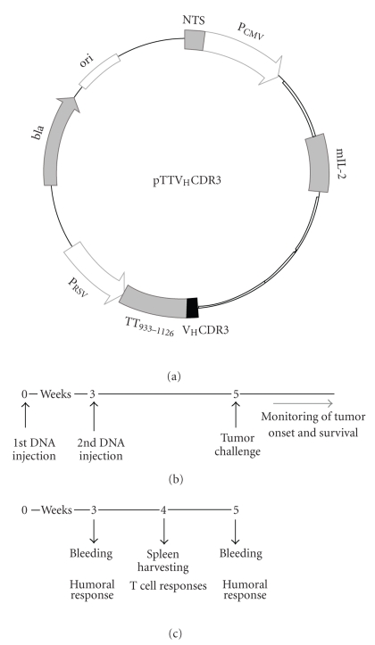
To construct the DNA vaccine, the amplified fragment TT933-1126VHCDR3109-116 was generated after PCR reactions, as described in Section 2. This fragment was cloned into pRC110-NTS backbone vector [22], and the recombinant plasmid designed as pTTVHCDR3, as reported in Figure 1(a). Additionally our plasmid encodes murine IL-2 as cytokine adjuvant. Likewise, the recombinant plasmid pTTVLCDR3 was obtained by cloning the amplified fragment TT933-1126VLCDR388-98 in the same backbone vector. A plasmid encoding the TT933-1126 sequence alone in the same backbone vector was also obtained and named pTT.
Figure 1.
DNA fusion vaccine schematic structure (a). Experimental design of protective (b) and immune responses (c) studies is showed. C3H/HeN mice were immunized twice at 3-week intervals by intramuscular injection followed by electroporation. Unimmunized (naïve) mice received a mock vaccination by injection with phosphate saline buffer. Two weeks after boosting mice were injected i.p. with 2 × 10² 38C13 cells.
Plasmid DNA vaccination was performed using the RSV promoter driving TT933-1126VHCDR3109-116 expression plasmid (pTTVHCDR3), while pTT was used as control vaccine. Naïve mice received a mock vaccination by injection with phosphate saline buffer. Our DNA vaccination protocol consists of two DNA injections both associated with electroporation [22]. Experimental design of protective and immunological studies is showed in Figures 1(b) and 1(c), respectively.
3.2. Antibody Response Analysis
The levels of antibody response specific to DNA fusion vaccine were evaluated in mice following intramuscular immunization. Humoral immune response elicited after pTTVHCDR3 or pTT immunizations was assayed by ELISA for VH peptide (D101–L122) reactive antibodies.
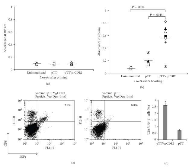
We wondered whether the immunization regimen might influence the immune outcome. Individual blood samples were collected from mice (pTTVHCDR3 group, n = 7; control groups, n = 6) 3 weeks after DNA priming and 2 weeks after boosting injections. The response profile for each vaccine group has been depicted in Figure 2. ELISA test failed to detect antibody titers when performed with mice sera collected after priming as well as analyses of individual sera within the pTTVHCDR3 group revealed no noticeable differences compared to unimmunized and pTT control groups (Figure 2(a)).
Figure 2.
Immune responses elicited after pTTVHCDR3 and pTT immunizations. Humoral immunity was assayed by ELISA for mice sera VH peptide (D101-L-122) reactive antibodies 3 weeks after priming (a) and 2 weeks after boosting. (b) Unimmunized mice represent the control group. Each marker indicates a value from a single mouse; group means are represented by a horizontal bar. Experimental groups (pTTVHCDR3 group, n = 7; control groups, n = 6) were compared by unpaired, two-tailed t-test. (c) The frequency of IFN-γ-positive CD8+ T cells was assessed ex vivo by intracellular cytokine staining. Splenic lymphocytes were harvested 1 week after booster injection, stimulated with VH peptide (D101-L-122), and assayed for IFN-γ production on gated T lymphocytes. Representative flow cytometric plots from pooled mice (3 animals/experimental groups) splenocytes are shown. Numbers in FACS plots refer to CD8+ IFN-γ+ cells as a percentage of the total T cells population. (d) Data were pooled from two identical independent experiments to indicate the mean percentage of CD8+ T cells producing IFN-γ in response to VH peptide. An X2 test for the comparison of the two proportions, expressed as a percentage, was performed. Error bars: SEM. (∗)denotes a statistically significant value (P<.0001).
Two weeks after boosting, mice immunized with pTTVHCDR3 DNA vaccine showed sera positive for antibodies directed against the VH peptide (D101–L122) (Figure 2(b)). Compared with pTT control vaccine and unimmunized groups, the pTTVHCDR3 vaccine group antibody level was statistically significant (P = .0045 and P = .0014, resp.).
The lack of antibody response after priming suggests that boosting is critical for antibody induction. Our data essentially confirmed that immunization schedule was critical for this Ag system [37].
Reactivity of mouse sera against a CDR3 irrelevant peptide (VH CDR3-PA [22]) generated no significant response (Table 1).
Table 1.
Reactivity of mouse sera against a CDR3 control (irrelevant) peptide as assessed by ELISA.
| Mice groups | Vaccine formulation | Absorbance (nm)1 | P2A versus B |
|---|---|---|---|
| A | pTTVHCDR3 | 0.159 ± 0.161 | NS |
| B | pTT | 0.049 ± 0.208 |
1 Control values belonging to unimmunized mice were subtracted.
2 Unpaired, two-tailed t-test.
NS: not significant.
3.3. Induction of IFN-γ Producing CD8+ T Cells by Fusion DNA Vaccine
To investigate whether our vaccination strategy could induce positive CD8+ T cell responses to VHCDR3 epitope, C3H/HeN mice (n = 3) were vaccinated with the same DNA dose and regimen. Splenic lymphocytes were harvested 1 week after booster injection and processed for their ability to induce VH peptide (D101–L122) positive IFN-γ-producing T cells responses. Flow cytometry analyses in Figure 2 showed the percentage of CD8+ T cells producing IFN-γ. Splenocytes isolated from pTTVHCDR3 vaccinated mice generated a significantly higher frequency of IFN-γ-positive CD8+ T cell precursors compared to mice vaccinated with the pTT control vaccine (P < .0001). A graphical representation of the number of VH peptide(D101–L122)-positive CD8+ T cells is depicted in Figures 2(c) and 2(d). Thus, our data suggest that vaccination with pTTVHCDR3 induces priming of CD8+ T lymphocytes.
3.4. Prophylactic and Therapeutic Experiments
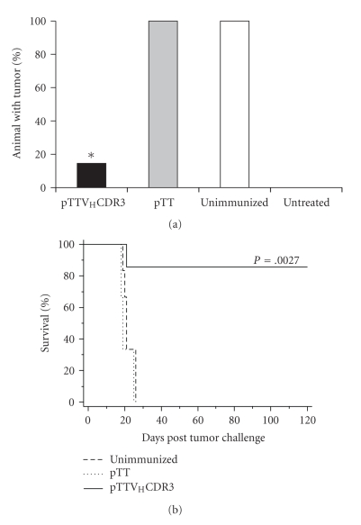
To address the protective tumor immunity of pTTVHCDR3 DNA vaccine, we performed prophylactic vaccination experiments. The details of the immunization protocol and the tumor challenge are described in Figure 1(b). The immunization regimen was previously developed for another CDR3-based vaccine formulation and proved to be efficacious [22]. Two weeks after the last DNA electrotransfer, animals (pTTVHCDR3 group, n = 7; control groups, n = 6) were challenged intraperitoneally (i.p.) with a lethal dose of 38C13 tumor B-cells. The development of i.p. lymphoma was monitored for each mouse and the protective efficacy of fusion vaccine was evaluated in terms of survival of mice over the next 120 days. Immunization with the pTTVHCDR3 DNA significantly impacted tumor growth and ensured long-term tumor-free survival of about 85% of treated mice (P = .0027) (Figures 3(a) and 3(b)). Cohorts vaccinated with the pTT control vaccine or phosphate buffer showed poor lymphoma resistance, with all mice showing median survival time of 19 days.
Figure 3.
In vivo antitumor effects generated by immunization with pTTVHCDR3 vaccine. (a) Tumor resistance. (*) denotes a statistically significant value (P < .001) by X2 analysis when comparing the pTTVHCDR3 group (n = 7) to all other groups (n = 6). (b) Representative long-term tumor-free survival. Survival analyses were performed using the Kaplan-Meier method and the log-rank test (P = .0027).
The potent prophylactic antitumor effect prompted us to assess the therapeutic vaccination against established 38C13 tumor. Therefore, based on our previous data [22] and recent findings (manuscript in preparation), we evaluated the combined effect of VHCDR3 and VLCDR3 peptides fused to TT933-1126 FrC portion in a therapeutic setting. Four days after challenging C3H/HeN mice (6 mice/experimental group) with a tumorigenic dose of 38C13 cells, DNA electrotransfer with pTTVHCDR3/pTTVLCDR3 or with pTT was performed and repeated 11 days later. Even though the timing of tumor onset was similar for the plasmids injected mice and the control mice, at days 18–22 postchallenge all untreated and pTT control mice succumbed. DNA vaccination with pTTVHCDR3/pTTVLCDR3 resulted in a trend toward a prolongation of life span through day 35 posttumor challenge, although the delay in death rate was not statically significant (Table 2).
Table 2.
Therapeutic vaccination induces life span prolongation.
| Mice group | Vaccine formulation | Survival % | Log-rank P-values versus | |||
|---|---|---|---|---|---|---|
| (n./tot) | Group C | Group B | ||||
| Day 15 | Day 22 | Day 28 | ||||
| A | pTTVHCDR3/pTTVLCDR3 | 100 | 50 | 50 | 0.083 | 0.115 |
| (6/6) | (3/6) | (3/6) | ||||
| B | pTT | 100 | 0 | 0.408 | — | |
| (6/6) | (0/6) | |||||
| C | Unimmunized | 80 | 0 | — | — | |
| (4/5) | (0/5) | |||||
4. Discussion
We have previously developed a DNA-based vaccine containing the 8-mer H-2KK “anchor modified” YEGYFDYI109-116 epitope of VHCDR3 sequence of the murine 38C13 B-cell lymphoma. The VHCDR3 epitope has been shown to be protective in combination with the VLCDR3 peptide in a murine tumor protection experiment [22].
In the current study, we aim to gain insights into the enhancement of the effectiveness of the VHCDR3-based DNA vaccine in terms of specific immune responses and tumor protection in mice.
Induction of potent immune responses against self-tumor antigens is not an easy task. Fusion of the antigen with foreign universal TH epitopes (such as tetanus toxoid epitopes) has been shown to brake the tolerance to self-antigen and render a weak tumor antigen more immunogenic.
Engagement of CD4+TH cells has been shown to play a major role in linking and coordinating innate and adaptive immune responses [4, 23].
Thus, a DNA fusion vaccine was generated following fusion of a sequence derived from TTFrC to the VHCDR3109-116 epitope to help immune responses against the tumor antigen. Vaccine efficacy was assayed in a highly aggressive and weakly immunogenic murine model of B-cell lymphoma.
We demonstrated that the fusion DNA vaccine pTTVHCDR3 was able to induce detectable levels of antibodies against the peptide encompassing the VHCDR3 sequence. Humoral immune response could not be achieved by first plasmid electrotransfer suggesting that boosting is critical for antibody induction for this antigen system.
Furthermore, plasmid-driven TTVHCDR3 immunization resulted in the induction of significantly higher frequency of IFN-γ-producing CD8+ T cell precursors as compared to control group.
Prophylactic vaccination with pTTVHCDR3 DNA vaccine through the intramuscular route in combination with electroporation strongly affected the onset of highly aggressive 38C13 B-cell lymphoma. Inhibition of lymphoma growth led to significant and long-lasting protection from tumor in syngeneic mice with about 85% surviving, compared to naïve animals or those given the pTT control vaccine. This study demonstrates that fusion of exogenous protein to tumor-specific epitope converted an ineffective vaccine, namely, pVH [22], into one with considerable activity.
The potent prophylactic antitumor effect prompted us to assess the tumor immunity in a therapeutic setting, which is more clinically relevant. Preliminary data obtained by using this DNA platform strategy provide proof of principle for the treatment of already established tumor in our model. Further enhancement of the potency of CDR3-based DNA vaccines is necessary in a therapeutic scenario; experiments testing new combinations of other crucial cytokines are under evaluation.
Attempts to identify the mechanism of Id-induced antitumor immunity to malignant B-cells have yielded variable results. Despite results from early clinical trials with Id vaccines suggest that both humoral and cellular immune responses may be independently associated with tumor regression and improved progression-free survival [38–42], the relative importance of the antibody and cell mediated immune response is still uncertain. Experiments are currently ongoing to explore the relative role of cellular versus humoral immunity for vaccine efficacy in our system.
The functional insertion of microbial sequence within the DNA vaccine was aimed to stimulate CD4+ T cell help that is critical for inducing and maintaining an effective CTL response [23, 43]. Deeper analyses are needed to explore the role, if any, of VHCDR3 peptide-specific CD8+ T cells precursors in the generation of immune responses via CD4+ T cell-mediated mechanisms. The involvement of CD4+ T helper lymphocytes at the effector phase of anti-tumor responses is coherent with TH cell-dependent “DCs licensing” [44] required for optimal vaccine efficacy, in the absence of MHC class II molecules expression by tumor cells [28, 45]. Licensed DCs presenting peptides from both TTFrC portion and tumor antigen can be able to activate the large repertoire of anti-TTFrC CD4+ T cells. Hence, by ligand-receptor pairs interactions, “DCs licensing” mechanism supports priming and boosting of the weak tumor-derived peptides response [4].
Lastly, employing electrotransfer for the delivery of a DNA vaccine should improve the availability of the antigen, since EP increases and prolongs protein expression level and also results in recruitments of infiltrating inflammatory cells to the administration site [6].
5. Conclusions
This study demonstrates that fusion of exogenous protein to tumor-specific epitope converted an ineffective vaccine into one with considerable activity. Immune responses recruited by CDR3-based DNA fusion vaccine involve anti-Id antibody production and suggest the possible contribution of CD8+ T lymphocytes.
A vaccination protocol consisting of a naked DNA priming and boosting is attractive by virtue of ease and less time-consuming production. Furthermore, safety is also achieved since adverse immune response can be avoided (i.e., immunity generated against some viral vectors).
The CDR3-based DNA fusion vaccines strategy may prove to be a highly useful approach against B-cell lymphoma. Optimal integration of active immunization approaches into standard therapies suggests DNA vaccination as an effective treatment to eradicate minimal residual diseases during clinical remission following standard chemotherapy in lymphoma patients.
Acknowlegments
The authors are grateful to Dr. Ivano Condò (University of Tor Vergata, Rome, Italy) for helpful discussion and critical reading of the paper. They are grateful to Dr. Gianni Pozzi (University of Siena, Italy) providing the TTFrC-expressing Streptococcus gordonii strain GP1253 and to Dr. Arya Biragyn (National Institute on Aging, Baltimore, Maryland, USA) for kind gift of 38C13 cell line. They are grateful to the technical staff of the Animal Facility at the “Sacro Cuore” Catholic University of Rome, Italy, for their assistance. This work was supported by MUR Grant FIRB 2006 (RBIP0695BB), by Bio-Ker S.r.l. research contract (M. Rinaldi) and by the Italian Banca delle Marche. Sandra Iurescia and Daniela Fioretti have been supported by MUR Grant FIRB 2006 (RBIP0695BB) and by Bio-Ker S.r.l. S. Iurescia and D. Fioretti contributed equally to the authorship of this work. V. M. Fazio and M. Rinaldi contributed equally as senior authors.
References
- 1.Jemal A, Tiwari RC, Murray T, et al. Cancer statistics, 2004. CA: A Cancer Journal for Clinicians. 2004;54(1):8–29. doi: 10.3322/canjclin.54.1.8. [DOI] [PubMed] [Google Scholar]
- 2.Houot R, Levy R. Vaccines for lymphomas: idiotype vaccines and beyond. Blood Reviews. 2009;23(3):137–142. doi: 10.1016/j.blre.2008.09.001. [DOI] [PubMed] [Google Scholar]
- 3.Koumarianou A, Kountourakis P, Economopoulos T. Vaccine strategies in the treatment of low-grade non-Hodgkin lymphoma. Journal of Steroid Biochemistry and Molecular Biology. 2008;109(3–5):230–232. doi: 10.1016/j.jsbmb.2008.03.008. [DOI] [PubMed] [Google Scholar]
- 4.Rice J, Ottensmeier CH, Stevenson FK. DNA vaccines: precision tools for activating effective immunity against cancer. Nature Reviews Cancer. 2008;8(2):108–120. doi: 10.1038/nrc2326. [DOI] [PubMed] [Google Scholar]
- 5.Kutzler MA, Weiner DB. DNA vaccines: ready for prime time? Nature Reviews Genetics. 2008;9(10):776–788. doi: 10.1038/nrg2432. [DOI] [PMC free article] [PubMed] [Google Scholar]
- 6.Ahlen G, Soderholm J, Tjelle T, et al. In vivo electroporation enhances the immunogenicity of hepatitis C virus nonstructural 3/4A DNA by increased local DNA uptake, protein expression, inflammation, and infiltration of CD3+ T cells. Journal of Immunology. 2007;179(7):4741–4753. doi: 10.4049/jimmunol.179.7.4741. [DOI] [PubMed] [Google Scholar]
- 7.Hurvitz SA, Timmerman JM. Current status of therapeutic vaccines for non-Hodgkin’s lymphoma. Current Opinion in Oncology. 2005;17(5):432–440. doi: 10.1097/01.cco.0000174040.52427.83. [DOI] [PubMed] [Google Scholar]
- 8.Neelapu SS, Lee S-T, Qin H, Cha S-C, Woo AF, Kwak LW. Therapeutic lymphoma vaccines: importance of T-cell immunity. Expert Review of Vaccines. 2006;5(3):381–394. doi: 10.1586/14760584.5.3.381. [DOI] [PubMed] [Google Scholar]
- 9.Benvenuti F, Burrone OR, Efremov DG. Anti-idiotypic DNA vaccines for lymphoma immunotherapy require the presence of both variable region genes for tumor protection. Gene Therapy. 2000;7(7):605–611. doi: 10.1038/sj.gt.3301133. [DOI] [PubMed] [Google Scholar]
- 10.Biragyn A, Tani K, Grimm MC, Weeks S, Kwak LW. Genetic fusion of chemokines to a self tumor antigen induces protective, T-cell dependent antitumor immunity. Nature Biotechnology. 1999;17(3):253–258. doi: 10.1038/6995. [DOI] [PubMed] [Google Scholar]
- 11.King CA, Spellerberg MB, Zhu D, et al. DNA vaccines with single-chain Fv fused to fragment C of tetanus toxin induce protective immunity against lymphoma and myeloma. Nature Medicine. 1998;4(11):1281–1286. doi: 10.1038/3266. [DOI] [PubMed] [Google Scholar]
- 12.Spellerberg MB, Zhu D, Thompsett A, King CA, Hamblin TJ, Stevenson FK. DNA vaccines against lymphoma: promotion of anti-idiotypic antibody responses induced by single chain Fv genes by fusion to tetanus toxin fragment C. Journal of Immunology. 1997;159(4):1885–1892. [PubMed] [Google Scholar]
- 13.Syrengelas AD, Chen TT, Levy R. DNA immunization induces protective immunity against B-cell lymphoma. Nature Medicine. 1996;2(9):1038–1041. doi: 10.1038/nm0996-1038. [DOI] [PubMed] [Google Scholar]
- 14.Zhu D, Rice J, Savelyeva N, Stevenson FK. DNA fusion vaccines against B-cell tumors. Trends in Molecular Medicine. 2001;7(12):566–572. doi: 10.1016/s1471-4914(01)02126-8. [DOI] [PubMed] [Google Scholar]
- 15.Baskar S, Kobrin CB, Kwaka LW. Autologous lymphoma vaccines induce human T cell responses against multiple, unique epitopes. Journal of Clinical Investigation. 2004;113(10):1498–1510. doi: 10.1172/JCI20312. [DOI] [PMC free article] [PubMed] [Google Scholar]
- 16.Campbell MJ, Carroll W, Kon S, et al. Idiotype vaccination against murine B cell lymphoma. Humoral and cellular responses elicited by tumor-derived immunoglobulin M and its molecular subunits. Journal of Immunology. 1987;139(8):2825–2833. [PubMed] [Google Scholar]
- 17.Hansson L, Rabbani H, Fagerberg J, Osterborg A, Mellstedt H. T-cell epitopes within the complementarity-determining and framework regions of the tumor-derived immunoglobulin heavy chain in multiple myeloma. Blood. 2003;101(12):4930–4936. doi: 10.1182/blood-2002-04-1250. [DOI] [PubMed] [Google Scholar]
- 18.Watanabe A, Raz E, Kohsaka H, et al. Induction of antibodies to a κ V region by gene immunization. Journal of Immunology. 1993;151(5):2871–2876. [PubMed] [Google Scholar]
- 19.Lim SY, Laxmanan S, Stuart G, Ghosh SK. Anti-lymphoma immunity: relative efficacy of peptide and recombinant DNA vaccine. Cancer Detection and Prevention. 2001;25(5):470–478. [PubMed] [Google Scholar]
- 20.Rinaldi M, Ria F, Parrella P, et al. Antibodies elicited by naked DNA vaccination against the complementary-determining region 3 hypervariable region of immunoglobulin heavy chain idiotypic determinants of B-lymphoproliferative disorders specifically react with patients’ tumor cells. Cancer Research. 2001;61(4):1555–1562. [PubMed] [Google Scholar]
- 21.Bergman Y, Haimovich J, Melchers F. An IgM-producing tumor with biochemical characteristics of a small B lymphocyte. European Journal of Immunology. 1977;7(8):574–579. doi: 10.1002/eji.1830070815. [DOI] [PubMed] [Google Scholar]
- 22.Rinaldi M, Fioretti D, Iurescia S, et al. Anti-tumor immunity induced by CDR3-based DNA vaccination in a murine B-cell lymphoma model. Biochemical and Biophysical Research Communications. 2008;370(2):279–284. doi: 10.1016/j.bbrc.2008.03.076. [DOI] [PubMed] [Google Scholar]
- 23.Janssen EM, Lemmens EE, Wolfe T, Christen U, Von Herrath MG, Schoenberger SP. CD4+ T cells are required for secondary expansion and memory in CD8+ T lymphocytes. Nature. 2003;421(6925):852–856. doi: 10.1038/nature01441. [DOI] [PubMed] [Google Scholar]
- 24.Lund LH, Andersson K, Zuber B, et al. Signal sequence deletion and fusion to tetanus toxoid epitope augment antitumor immune responses to a human carcinoembryonic antigen (CEA) plasmid DNA vaccine in a murine test system. Cancer Gene Therapy. 2003;10(5):365–376. doi: 10.1038/sj.cgt.7700574. [DOI] [PubMed] [Google Scholar]
- 25.Rice J, Elliott T, Buchan S, Stevenson FK. DNA fusion vaccine designed to induce cytotoxic T cell responses against defined peptide motifs: implications for cancer vaccines. Journal of Immunology. 2001;167(3):1558–1565. doi: 10.4049/jimmunol.167.3.1558. [DOI] [PubMed] [Google Scholar]
- 26.Stevenson FK, Rice J, Ottensmeier CH, Thirdborough SM, Zhu D. DNA fusion gene vaccines against cancer: from the laboratory to the clinic. Immunological Reviews. 2004;199:156–180. doi: 10.1111/j.0105-2896.2004.00145.x. [DOI] [PubMed] [Google Scholar]
- 27.Tymciu S, Durieux-Alexandrenne C, Wijkhuisen A, et al. Enhancement of antibody responses in DNA vaccination using a vector encoding a universal T-helper cell epitope. DNA and Cell Biology. 2004;23(6):395–402. doi: 10.1089/104454904323145281. [DOI] [PubMed] [Google Scholar]
- 28.Williams BB, Wall M, Miao RY, et al. Induction of T cell-mediated immunity using a c-Myb DNA vaccine in a mouse model of colon cancer. Cancer Immunology, Immunotherapy. 2008;57(11):1635–1645. doi: 10.1007/s00262-008-0497-2. [DOI] [PMC free article] [PubMed] [Google Scholar]
- 29.Medaglini D, Ciabattini A, Spinosa MR, et al. Immunization with recombinant Streptococcus gordonii expressing tetanus toxin fragment C confers protection from lethal challenge in mice. Vaccine. 2001;19(15-16):1931–1939. doi: 10.1016/s0264-410x(00)00434-5. [DOI] [PubMed] [Google Scholar]
- 30.Lefranc M-P, Pommie C, Ruiz M, et al. IMGT unique numbering for immunoglobulin and T cell receptor variable domains and Ig superfamily V-like domains. Developmental and Comparative Immunology. 2003;27(1):55–77. doi: 10.1016/s0145-305x(02)00039-3. [DOI] [PubMed] [Google Scholar]
- 31.McMahon JM, Signori E, Wells KE, Fazio VM, Wells DJ. Optimisation of electrotransfer of plasmid into skeletal muscle by pretreatment with hyaluronidase—increased expression with reduced muscle damage. Gene Therapy. 2001;8(16):1264–1270. doi: 10.1038/sj.gt.3301522. [DOI] [PubMed] [Google Scholar]
- 32.Rammensee H-G, Bachmann J, Emmerich NPN, Bachor OA, Stevanovic S. SYFPEITHI: database for MHC ligands and peptide motifs. Immunogenetics. 1999;50(3-4):213–219. doi: 10.1007/s002510050595. [DOI] [PubMed] [Google Scholar]
- 33.Panina-Bordignon P, Tan A, Termijtelen A, Demotz S, Corradin G, Lanzavecchia A. Universally immunogenic T cell epitopes: promiscuous binding to human MHC class II and promiscuous recognition by T cells. European Journal of Immunology. 1989;19(12):2237–2242. doi: 10.1002/eji.1830191209. [DOI] [PubMed] [Google Scholar]
- 34.Demotz S, Barbey C, Corradin G, Amoroso A, Lanzavecchia A. The set of naturally processed peptides displayed by DR molecules is tuned by polymorphism of residue 86. European Journal of Immunology. 1993;23(2):425–432. doi: 10.1002/eji.1830230219. [DOI] [PubMed] [Google Scholar]
- 35.James EA, Bui J, Berger D, Huston L, Roti M, Kwok WW. Tetramer-guided epitope mapping reveals broad, individualized repertoires of tetanus toxin-specific CD4+ T cells and suggests HLA-based differences in epitope recognition. International Immunology. 2007;19(11):1291–1301. doi: 10.1093/intimm/dxm099. [DOI] [PubMed] [Google Scholar]
- 36.Chen W, McCluskey J. Immunodominance and immunodomination: critical factors in developing effective CD8+ T-cell-based cancer vaccines. Advances in Cancer Research. 2006;95:203–247. doi: 10.1016/S0065-230X(06)95006-4. [DOI] [PubMed] [Google Scholar]
- 37.Hakim I, Levy S, Levy R. A nine-amino acid peptide from IL-1beta augments antitumor immune responses induced by protein and DNA vaccines. Journal of Immunology. 1996;157(12):5503–5511. [PubMed] [Google Scholar]
- 38.Hsu FJ, Caspar CB, Czerwinski D, et al. Tumor-specific idiotype vaccines in the treatment of patients with B- cell lymphoma—long-term results of a clinical trial. Blood. 1997;89(9):3129–3135. [PubMed] [Google Scholar]
- 39.Hsu FJ, Benike C, Fagnoni F, et al. Vaccination of patients with B-cell lymphoma using autologous antigen-pulsed dendritic cells. Nature Medicine. 1996;2(1):52–58. doi: 10.1038/nm0196-52. [DOI] [PubMed] [Google Scholar]
- 40.Kwak LW, Campbell MJ, Czerwinski DK, Hart S, Miller RA, Levy R. Induction of immune responses in patients with B-cell lymphoma against the surface-immunoglobulin idiotype expressed by their tumors. The New England Journal of Medicine. 1992;327(17):1209–1215. doi: 10.1056/NEJM199210223271705. [DOI] [PubMed] [Google Scholar]
- 41.Neelapu SS, Baskar S, Gause BL, et al. Human autologous tumor-specific T-cell responses induced by liposomal delivery of a lymphoma antigen. Clinical Cancer Research. 2004;10(24):8309–8317. doi: 10.1158/1078-0432.CCR-04-1071. [DOI] [PubMed] [Google Scholar]
- 42.Timmerman JM, Czerwinski DK, Davis TA, et al. Idiotype-pulsed dendritic cell vaccination for B-cell lymphoma: clinical and immune responses in 35 patients. Blood. 2002;99(5):1517–1526. doi: 10.1182/blood.v99.5.1517. [DOI] [PubMed] [Google Scholar]
- 43.Zhang S, Zhang H, Zhao J. The role of CD4 T cell help for CD8 CTL activation. Biochemical and Biophysical Research Communications. 2009;384(4):405–408. doi: 10.1016/j.bbrc.2009.04.134. [DOI] [PubMed] [Google Scholar]
- 44.Smith CM, Wilson NS, Waithman J, et al. Cognate CD4+ T cell licensing of dendritic cells in CD8+ T cell immunity. Nature Immunology. 2004;5(11):1143–1148. doi: 10.1038/ni1129. [DOI] [PubMed] [Google Scholar]
- 45.Leitner WW, Baker MC, Berenberg TL, Lu MC, Yannie JP, Udey MC. Enhancement of DNA tumor vaccine efficacy by gene gun mediated codelivery of threshold amounts of plasmid-encoded helper antigen. Blood. 2009;113(1):37–45. doi: 10.1182/blood-2008-01-136267. [DOI] [PMC free article] [PubMed] [Google Scholar]


