
Figure 4.
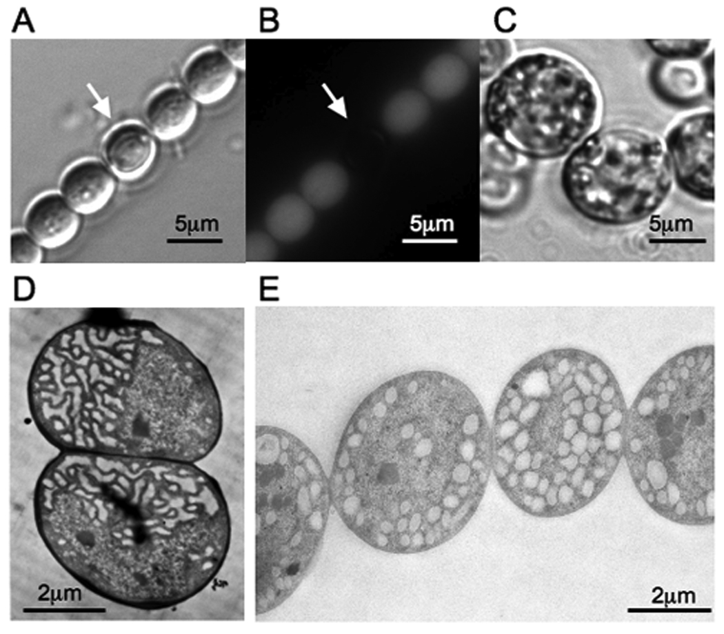
Images of differentiated cell types of N. punctiforme. Vegetative filaments containing heterocysts are shown under light (A) and fluorescence (B) microscopy. The white arrow indicates the position of heterocyst. Akinete cell morphology is shown under light microscopy (C). Electron micrographs of vegetative and akinete cell types are shown in panels D and E, respectively.