Abstract
Vaccinia virus (VV) vaccination is used to immunize against smallpox and historically was considered to have been successful if a skin lesion formed at the vaccination site. While antibody responses have been widely proposed as a correlate of efficacy and protection in humans, the role of cellular and humoral immunity in VV-associated skin lesion formation was unknown. We therefore investigated whether long-term residual humoral and cellular immune memory to VV, persisting 30 years after vaccination, could control VV-induced skin lesion in revaccinated individuals. Here, we have shown that residual VV-specific IFN-γ+TNF-α+ or IFN-γ+IL-2+ CD4+ lymphocytes but not CD8+ effector/memory lymphocytes expressing a skin-homing marker are inversely associated with the size of the skin lesion formed in response to revaccination. Indeed, high numbers of residual effector T cells were associated with lower VV skin lesion size after revaccination. In contrast, long-term residual VV-specific neutralizing antibody (NAbs) titers did not affect skin lesion formation. However, the size of the skin lesion strongly correlated with high levels of NAbs boosted after revaccination. These findings demonstrate a potential role for VV-specific CD4+ responses at the site of VV-associated skin lesion, thereby providing new insight into immune responses at these sites and potentially contributing to the development of new approaches to measure the efficacy of VV vaccination.
Introduction
The persistence of a long-lasting immune memory against vaccinia and smallpox viruses has been a subject of considerable debate, and its efficacy in protection remains unclear in the absence of circulating smallpox virus throughout the world (1–3). A significant proportion of the population has never been vaccinated against smallpox, and the level of immunity remains variable in people who have been vaccinated. The use of attenuated pox vectors for therapeutics and preventive vaccines requires a better understanding of the mechanism of immune control of vaccinia virus (VV) and of vaccine efficacy (1–4). For many years, the “take” was assessed on the presence of the skin lesion, which was considered as a marker of positive and successful vaccination. Scarification of human skin with live VV normally is induced on day 3 at the site of scarification, an itching vesicle that evolves toward a skin lesion containing virus particles surrounded by a strong erythema. This dries at day 10, leaving a scar. Jenner noted that when individuals were vaccinated against smallpox (variolization), no dissemination of skin lesion was observed after subsequent infection at a distant site (5). This step forward by E. Jenner had a crucial impact on the measurement of vaccine efficacy. Recently, it has been shown that fewer adverse reactions were observed in previously vaccinated individuals compared with vaccinia-naive volunteers, which may be due to immunological memory (6). Recent observation has also shown that previously vaccinated individuals have a diminished skin erythema response when compared with nonvaccinated individuals (7). These observations suggest that smallpox vaccination leads to long-term retained immunity that can be detected clinically. However, the mechanism is still unknown.
Immunization by scarification using vaccinia-based vaccines generates a potent induction of immune responses and particularly high levels of neutralizing antibody (NAb) and cell-mediated immune responses (8). Immunity to VV involves both T and B cells, since defects in either compartment increased the risk of complication (9), although patients with T cell defects are far more susceptible to VV infection than those with B cell defects (9). However, this does not overshadow the importance of antibody responses that are involved in the inhibition of virus infection (10, 11). Further identification of protective immune response has therefore become a fundamental issue in the understanding of immune responses as well as in future vaccination strategies.
Currently, quantitative measurement of T and B cell immunity has gained acceptance as the marker of vaccination efficacy (12–14). Hammarlund et al. showed that more than 90% of volunteers vaccinated 25–77 years ago still maintained humoral and cellular responses against vaccinia (15). In addition, we have shown that long-term proliferative memory response persisted in more than 70% of vaccinated individuals 25 years after the end of the vaccination era (16). However, the persistence of memory responses depends on several types of memory cells that can be distinguished as follows: (a) rapid effector immune responses as measured by short-term production of IFN-γ by T cells (effector memory) and (b) memory T cells with proliferative capacity (central memory) (16). However, the specific role of humoral and cellular immune responses remains unknown in the control of VV-induced skin lesion. One could thus question the relationship between skin lesions after subsequent VV vaccination and vaccinia-specific humoral and cellular responses in distant past–vaccinated and in revaccinated individuals. This question remains crucial to further progress in measurement of attenuated VV vaccination efficacy and whether boosting vaccination is necessary for future trials.
Results
Induction of high titers of NAbs after VV vaccination correlates with large skin lesion formation.
We have investigated the consequences of residual and boosted T and B cell responses on skin lesion formation in long-term vaccinated volunteers after revaccination. We thus extended our previous study of residual immunity against VV to study the behavior of skin lesion after VV revaccination in volunteers with a previous history of vaccination against smallpox virus (16). The IMMUVAR study cohort was composed of 184 volunteers, aged 25–68 years, previously vaccinated and revaccinated from March 2003 to November 2006 with live unattenuated VV derived from the Lister strain (Pourquier vaccine). Three immunological parameters were studied: VV-specific NAb titers, cellular immune responses as defined by frequencies of VV-specific IFN-γ–producing T cells, and the proliferative capacity of VV-specific T cells. Success in vaccination was evaluated by measurement of humoral and cellular immune memory as well as the visual inspection of the typical skin lesions at the vaccination site (17). The lesion was measured as the diameter (in mm) of the pustule at 8 days after revaccination.
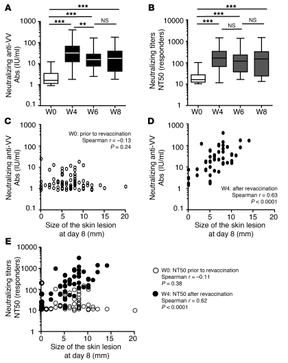
First, the kinetics of NAb responses was followed in 36 previously vaccinated volunteers over 8 weeks. Results are presented as NAbs titers in IU/ml (Figure 1A) and 50% neutralizing titer (NT50) scores (Figure 1B). The residual levels of NAbs (IU/ml) measured at baseline (median, 1.6 IU/ml; interquartile range [IQR], 1.15–3.45) were highly amplified at week 4 (W4) (median, 32.15 IU/ml; IQR, 12.50–67.75; P < 0.0001), W6 (median, 15.50 IU/ml; IQR, 7.60–28; P < 0.0001), and W8 (median, 17.85 IU/ml; IQR, 4.35–39.5; P < 0.0001) after revaccination. NT50 scores of responders are also presented as shown in Figure 1B. Statistical analysis also showed a significant increase in NT50 scores at W4–W8 compared with baseline residual NAb titers (W0) (Figure 1B).
Figure 1. Induction of high titers of neutralizing Ab after vaccination correlates with large cutaneous lesion formation.
Neutralizing VV-specific antibody (NAb) titers (IU/ml) (A) and NT50 scores (B) in previously vaccinated volunteers at different time points after revaccination (kinetics from W0 to W8 were available for n = 36): W0, W4, W6, W8. The box-and-whisker plots show the median values and the 10th, 25th, 75th, and 90th percentiles. Statistical analyses were performed using the Wilcoxon matched pairs test; **P < 0.001, ***P < 0.0001. Dot plots of the size of skin lesions at day 8 (in mm) and the amplitude of NAb responses (IU/ml) in previously vaccinated individuals at W0 (n = 85) (C) and recently revaccinated individuals at W4 (n = 53) (D). (E) Dot plots of the size of skin lesions at day 8 (in mm) and the neutralizing titers of VV-specific Abs (NT50 scores) in previously vaccinated individuals at W0 (n = 55) (white circles) and recently revaccinated individuals at W4 (n = 53, black circles). Each circle represents data from 1 individual. All data are shown in the dot plot. Statistical analyses were performed using the Spearman test.
In addition, we studied the relationship between anti-VV NAb levels (IU/ml and NT50 scores) prior to and at W4 after vaccination, as well as the size of the skin lesion (Figure 1, C–E). Interestingly, the amplitude of NAb titers (IU/ml) prior to vaccination did not correlate with the size of the pustule at day 8 after revaccination (Spearman test r = –0.13, P = 0.24) (Figure 1C), demonstrating that NAbs did not control VV dissemination at the vaccination site. In contrast, a significant correlation (Spearman r = 0.63, P < 0.0001) (Figure 1D) was observed between NAb titers at W4 and the size of the skin lesion at day 8, suggesting that the levels of VV-specific NAbs were determined by the local levels of virus production and inflammation at the site of vaccination. In addition, we also analyzed the relationship between NAb responses and the size of the skin lesions (at day 8) for the same individuals with available data on W0 and W4 (n = 41). We found statistical data similar to that in Figure 1, B and C, with Spearman r = –0.10 at W0, P = 0.52 and Spearman r = 0.57, P < 0.0001 at W4.
As depicted in Figure 1E, statistical analyses were also performed using NT50 scores of responders. This latter analysis confirmed a significant correlation between NT50 scores of VV-specific Ab responses at W4 after revaccination and the size of skin lesion; however, residual VV-specific NAbs (NT50 scores) did not participate in the control of skin lesion formation.
Thus, we found an unexpected absence of relationship between preexisting NAb responses long-term after previous vaccination and the size of skin lesion after revaccination against smallpox.
The size of skin lesion after VV vaccination is controlled by residual effector T cells prior to vaccination.
A similar approach was used to study the relationship between T cell immunity and skin lesion formation. As described previously (16), 23% and 70% of previously vaccinated volunteers of the IMMUVAR cohort displayed detectable levels of IFN-γ–producing memory T cells and proliferative T cell responses, respectively. The frequency of VV-specific IFN-γ–producing cells (W0) and of VV-specific proliferating cells (W0) in previously vaccinated volunteers was significantly higher than that of unvaccinated healthy volunteers (Figure 2A and Figure 3A) (P = 0.0011 and P < 0.0001, respectively), in accordance with our previous study (16). Revaccination boosted effector/memory and proliferative T cell responses as measured by IFN-γ ELISpot assays (Figure 2A) and [3H]thymidine incorporation 4 weeks after revaccination, respectively (Figure 3A). The threshold of IFN-γ–producing memory T cells was set at greater than 50 spot-forming units (SFU)/million PBMCs, and proliferative T cell responses were considered responders when the index of proliferation was greater than 3.
Figure 2. Control of skin lesion formation by residual effector/memory T cell responses.
(A) IFN-γ ELISpot assays (SFU/million PBMCs) were performed prior to and after revaccination at W0 and W4 (n = 65). Assays were also performed in control unvaccinated individuals (Unvac, n = 10). Levels of unstimulated cells were subtracted as background. The threshold of positivity of VV-specific IFN-γ+ T cell frequency was set greater than 50 SFU/million PBMCs after subtraction of background values. Data represent the median values and the 10th, 25th, 75th, and 90th percentiles. The P value between groups was calculated using Wilcoxon matched pairs test; **P < 0.001, ***P < 0.0001. (B and C) Dot plots of the size of the skin lesions at day 8 (in mm) and IFN-γ ELISpot responses at W0 (B) and W4 (C). Each circle represents data from 1 individual. All data are shown in the figures. Statistical analyses were performed using the Spearman test.
Figure 3. Control of skin lesion formation by residual proliferative memory T cell responses.
(A) Index of proliferation to VV was calculated prior to and after revaccination (n = 83) at W0 and W4. Assays were also performed in control unvaccinated individuals (n = 10). The threshold of a positive response was set at an index of proliferation of at least 3. Data represent the median values and the 10th, 25th, 75th, and 90th percentiles. The P value between groups was calculated using the Wilcoxon matched pairs test; ***P < 0.0001. (B and C) Dot plots of VV-specific T cell proliferation index at W0 (B) and W4 (C). Each circle represents data from 1 individual. Statistical analyses were performed using the Spearman test.
The frequency of VV-specific IFN-γ–producing memory T cells was significantly raised from a median of 10 SFU/million PBMCs (IQR, 1–30) prior to vaccination to a median of 117 SFU/million of PBMCs (IQR, 38–308) (P < 0.0001) after revaccination (Figure 2A). We found a significant correlation (Spearman r = –0.35, P = 0.0003) between the numbers of residual IFN-γ–producing memory T cells prior to vaccination and the size of the skin lesion at day 8 after revaccination (Figure 2B). Statistical analysis performed after removal of the largest lesion (>15 mm) gave similar P values and Spearman r for all immunological parameters tested, including NAb responses (W4: P < 0.0001, Spearman r = 0.63), VV-specific IFN-γ–producing T cells (W0: P < 0.0006, Spearman r = –0.34), and proliferative T cell responses (W0: P = 0.01, Spearman r = –0.22). In contrast (Spearman r = 0.02, P = 0.82), the level of IFN-γ–producing memory T cell responses after revaccination did not correlate with the size of the pustule at day 8 after revaccination (Figure 2C). The same effect was observed with the proliferative memory responses to VV rising from a median proliferation index of 7.5 (IQR, 3.1–21.4) to a median of 24.6 (IQR, 9.3–57.5) (P < 0.0001) (Figure 3A). We found that proliferative memory T cell responses prior to revaccination weakly correlated with the size of the skin lesion (Spearman r = –0.23, P = 0.009) (Figure 3B) but not the proliferative memory T cell responses measured after revaccination (Spearman r = 0.06, P = 0.52) (Figure 3C).
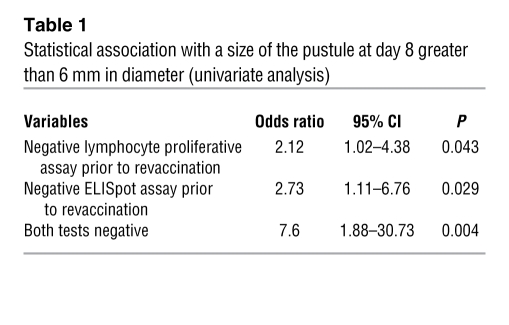
Further analysis of responders and nonresponders for cellular immune responses to VV were performed as followed: for ELISpot assays, individuals were considered positive responders if levels were greater than 50 vaccinia-specific SFU/million PBMCs. For proliferation assays, individuals were considered positive responders if the index of proliferation was greater than 3. We thus performed additional univariate analysis as shown below (Table 1). Size of the pustule (>6 mm) at day 8 was associated with a negative ELISpot assay (<50 SFU/million PBMCs) before revaccination (P = 0.029), with a negative T cell proliferation assay (<3 index of proliferation) before revaccination (P = 0.043), with both tests negative (P = 0.004). The absence of detectable cellular immunity prior to revaccination was associated with a size of the pustule greater than 6 mm in diameter at day 8.
Table 1 .
Statistical association with a size of the pustule at day 8 greater than 6 mm in diameter (univariate analysis)
Thus, both statistical analysis of the relationship taking into account quantitative cellular responses (Figures 2 and 3) and analysis of responder and nonresponder individuals (Table 1) showed a relationship between diminished size of skin lesion at the site of inoculation after revaccination and high systemic effector/memory T cell responses in long-term vaccinated individuals.
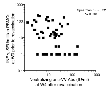
Our data demonstrate for the first time to our knowledge the crucial role of residual VV-specific T cell responses, but not of residual NAb levels, in the control of virus dissemination at the site of reimmunization in individuals who have been vaccinated during childhood. These data suggested that the residual effector/memory VV-specific T cells influencing the size of the skin lesion also influence the NAb response. In this regard, we also found a slight correlation between residual T cell responses prior to vaccination and lower NAbs 1 month after vaccination (n = 52, Spearman r = –0.32, P = 0.018) (Figure 4), further suggesting the impact of residual T cell response on boosting of the humoral responses after live virus vaccination.
Figure 4. Influence of long-term residual VV virus effector/memory T cell response on the boosting of humoral responses after VV vaccination.
IFN-γ ELISpot assays (SFU/million PBMCs) were performed prior to revaccination (W0) and neutralizing VV-specific antibody (NAb) titers (IU/ml) were determined at W4 after revaccination (n = 52). Each symbol represents data from 1 individual. Statistical analyses were performed using the Spearman test.
Residual IFN-γ+TNF-α+ and IFN-γ+IL-2+ long-term effector/memory CD4+ lymphocytes that express the cutaneous homing marker cutaneous lymphocyte–associated antigen and cytotoxic effector molecule CD107a control VV skin lesion formation after smallpox vaccination in previously vaccinated humans.
The functional profile of CD4+ and CD8+ T cells is clearly more diverse than the IFN-γ production, and the importance of polyfunctionality in the protection against viral infections remains uncertain. Multiparametric flow cytometric assays were performed to further determine the importance of subpopulations of both residual and post-revaccination VV-specific CD3+CD4+ or CD8+ T cells with regard to IL-2, IFN-γ, and TNF-α production (Figure 5) in 23 previously vaccinated volunteers at W0. PBMC samples were available for only a few individuals (n = 23) for further analyses. Data from all PBMCs tested were analyzed and shown without previous selection. In addition, there were no statistically significant differences between the IFN-γ ELISpot data of the 23 subjects compared with the rest of the cohort (P = 0.86 for IFN-γ ELISpot assays at day 0 and P = 0.9 for the size of the pustule at day 8).
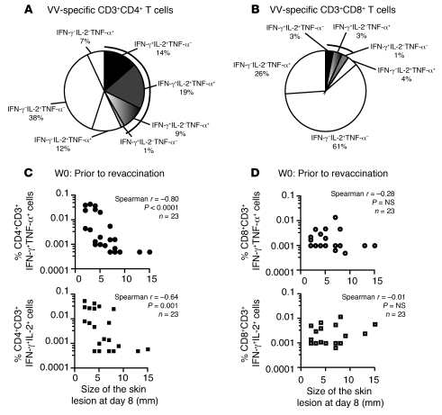
Figure 5. Residual IFN-γ+TNF-α+ long-term effector/memory CD4 lymphocytes control VV skin lesion formation after viral challenge in humans.
(A and B) PBMCs from previously vaccinated volunteers were stimulated for 16 hours with VV and harvested for flow cytometric intracellular cytokine staining. Boolean gating using FlowJo software was performed to calculate single producers, double producers, and triple producers cells in regard to IFN-γ+, IL-2+, and/or TNF-α+ as indicated. Pie chart analyses are shown for CD3+CD4+ (A) and CD3+CD8+ cells (B). Mean of percentages for each sub-population of VV-specific T cells that produce cytokines are indicated in the pie chart for 23 volunteers tested at W0 (prior to vaccination). (C and D) Dot plot representation of correlation between the size of skin lesions at day 8 after vaccination and residual VV-specific T cell responses prior to revaccination: percentage of residual VV-specific effector T cells as shown by IFN-γ+TNF-α+ or IFN-γ+IL-2+ CD3+CD4+ (C) or CD8+CD3+ (D) lymphocytes are shown. All data are shown (n = 23). Each symbol represents data from 1 individual. Statistical analyses were performed using the Spearman test.
According to the strict selection of CD3+CD4+ or CD3+CD8+ T cells, functional markers were analyzed by using Boolean gating combinations in order to determine the frequency of single cytokine producers, double cytokine producers, and triple cytokine producers. Pie chart analyses of VV-specific T cells showed an extreme diversity of CD3+CD4+ and CD3+CD8+ cells in response to VV stimulation (Figure 5, A and B). We also found very low levels of IFN-γ–producing CD8 cells in previously vaccinated volunteers (Figure 5B). In contrast, almost half of the VV-specific CD4+ cells produced IFN-γ+ and at least 1 other cytokine (IL-2 or TNF-α) (Figure 5A).
Thus, we further analyzed the relationship between these VV-specific effector/memory T cells that produced cytokines and the size of skin lesions after revaccination in 23 revaccinated volunteers (Figure 5, C and D). A striking significant correlation was found between vaccinia-specific CD4+IFN-γ+TNF-α+ or CD4+IFN-γ+IL-2+ double-positive cells (but not IFN-γ single-positive cells) and the size of the skin lesion (Figure 5C, CD4+IFN-γ+TNF-α+ Spearman r = –0.80, P < 0.0001, CD4+IFN-γ+IL-2+ Spearman r = –064, P = 0.001), while VV-specific CD8+ T lymphocytes did not show such correlation whatever the functional profile (Figure 5D, nonsignificant P value). Together, our data confirm the central role of residual effector/memory CD4+ but not CD8+ T cells in the control of the cutaneous skin lesion formation after VV challenge in individuals preimmunized 30 years earlier. In addition, frequencies of previously vaccinated individuals with detectable levels of VV-specific CD8+ cells producing cytokine remained extremely low, which made further analysis difficult. We thus focused on the role of VV-specific CD3+CD4+ cells in previously vaccinated individuals.
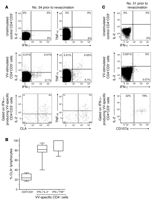
We next evaluated the expression of cutaneous lymphocyte–associated antigen (CLA) (18), a skin-homing marker on VV-specific T lymphocytes that produce IFN-γ, from 10 revaccinated individuals (Figure 6, A and B). Representative flow cytometric and cytokine profiles of VV-specific CD3+CD4+ cells are shown in Figure 6. We found that VV-specific CD3+CD4+ cells that display effector functions such as Th1 cytokine production expressed high levels of CLA antigen (mean of 98% CLAhigh expression on VV-specific CD4 cells) (Figure 6B). These results suggest the preferential homing capacities of VV-specific CD4+ lymphocytes into the skin and further reinforced the role of residual VV-specific CD4+IFN-γ+ effector/memory T cells in the control of skin lesion formation at the site of VV vaccination.
Figure 6. CLA and CD107a cytotoxic effector molecule on VV-specific CD4 cells in previously vaccinated volunteers.
(A) PBMCs prior to revaccination were thawed and stimulated for 16 hours with VV and harvested for flow cytometric assays. Multiparametric flow cytometric analyses of IL-2, IFN-γ, and/or TNF-α and gated on CD3+CD4+ as indicated using FlowJo software. Flow cytometric analyses of the basal level of cytokine production by unstimulated CD3+CD4+ cells are shown in the upper panels. We showed percentages of VV-specific CD3+CD4+ cells producing IFN-γ and/or IL-2 (middle, left) and of CD3+CD4+ cells producing IFN-γ and/or TNF-α (middle, right) for a representative individual. Flow cytometric analyses of CLA-expressing cells among VV-specific CD4+ cells producing IFN-γ (bottom). Data are representative of 10 individuals tested. (B) Box-and-whiskers representation of the frequency of VV-specific CD3+CD4+ lymphocytes expressing CLA markers is presented for 10 vaccinated volunteers. The data show the median values and the 10th, 25th, 75th, and 90th percentiles. (C) The expression of CD107a cytotoxic effector molecule was analyzed for 3 vaccinated volunteers. This latter population represents 79%–85% of VV-specific CD4+ cells producing IFN-γ and/or IL-2 in all volunteers tested. Representative plot for CD107a marker gated on CD3+CD4+ cells producing IFN-γ (C, bottom).
In addition, we have shown that VV-specific effector CD4+ cells express high levels of CD107a (cytotoxic effector molecule) (n = 3 individuals). This latter population of VV-specific CD3+CD4+CD107ahigh cells represented 79%–85%, in all volunteers tested. Representative multiparametric flow cytometric analyses are shown in Figure 6C. These results are in accordance with the previous demonstration of the persistence of long-term residual VV-specific CD4+ cells that displayed cytotoxic functions after VV vaccination (19–21). These data further emphasize the cytotoxic role of VV-specific CD4+ cells with skin-homing capacities that might be involved in the effector mechanisms of control of the skin lesion formation.
Discussion
This is the first evidence to our knowledge of the role of long-term efficacy of residual memory CD4+ effector T cells in adults that were vaccinated against smallpox more than 30 years earlier. Our data demonstrate the crucial role mediated by the residual VV-specific CD4+ memory T cells producing IFN-γ but not by anti-VV NAbs in the control of VV-induced skin lesion formation at the site of immunization in individuals preimmunized 30 years earlier. However, a large skin lesion positively correlated with high anti-VV NAb levels, and the residual effector/memory VV-specific T cells negatively influenced not only the size of the skin lesion but also the magnitude of the recall antibody response. These data represent qualitative changes in our existing knowledge and understanding of the immune response to VV.
This finding sharply contrasts with the lack of protection against VV skin lesion formation conferred by the protective arms of immunity, i.e., the NAbs and CD8+ T lymphocytes. Indeed, with global eradication of smallpox, skin lesion formation at site of immunization remains the sole available end point for vaccine “take” (22). Indeed, VV administration results in many side effects, among which virus dissemination is the most frequent. Local VV production in healthy naive individuals leads to skin lesions at the site of injection, the frequency and size of which usually decreased with revaccinations. However, the immune mechanisms controlling extension of the skin lesion remain unknown. In this study, we further characterized long-term memory to vaccinia and their potential role in the control of skin lesion that involves both virus replication and inflammatory reaction. However, the ethical committees did not permit skin biopsies, and we were also not able to analyze the virus production at the site of vaccination. Further clinical trials will be thus necessary to dissect the direct role of CD4+ T cells in the control of virus replication.
In the context of research of new pox-based recombinant vaccines, our finding suggested a novel role of the VV-specific effector/memory CD4+ T cells in controlling the size of the skin lesion formation and reveal an original feature in the immunology of pox viruses.
Immunity to VV is known to involve both T and B cells, since defects in either compartment increase the risk of complications (3, 9). Many studies have provided evidence about the modalities of the persistence of the humoral and cellular compartments more than 25 years after smallpox eradication. Antibody responses to VV were shown to decline within the first 3–5 years after vaccination (1, 23), although both VV-specific antibodies and memory B cells can persist up to 60 years after vaccination (24). A similar reduction over time of VV-specific memory T cells was observed in previously vaccinated individuals that retained a predominant CD4+ memory T cell response (15, 16). Interestingly, while the primary VV immunization induces strong humoral and T cell responses (11) involving high levels of polyfunctional CD8 responses (25, 26), the VV-specific CD4+ and CD8+ T cell contraction phases appear to differ. Indeed, the VV-specific CD8 response was shown to contract 7-fold, while the CD4 response contracts by only 2-fold (27), thus leading to the maintenance of a dominant CD4+ T cell response. Accordingly, others and we have shown the long-term cellular memory to VV predominantly involves CD4+ Th1 cells (16, 27). However, the role of these residual cells remained to be elucidated. Here, we propose that these cells were at the first line of control of skin lesion after revaccination. The cytokine-producing CD8 memory response predominated at earlier stages after antigen exposure, but levels were lower than those of cytokine-producing CD4 counterparts, suggesting differential regulation of the VV-specific memory pool. Herein, we show that solely high levels of residual IFN-γ+TNF-α+–producing effector CD4+ lymphocytes but not CD8+ effector T cells correlate with a small cutaneous skin lesion. Our investigation of the polyfunctionality of VV-specific residual T cells more than 30 years after the last immunization showed that 2 of 3 of the responding T cells displayed a single cytokine producer profile, while only 1 of 3 had a double producer profile. Among those effector T cells, only CD4+ lymphocytes that produced IFN-γ+TNF-α+ or IFN-γ+IL-2+ were involved in the control of the skin lesion but not VV-specific CD8+ effector memory cells. Importantly, these residual effector/memory CD4+ cells still expressed high levels of the skin-homing CLA marker (range, 70%–100%). However, too few VV-specific CD8 cells were detected among individuals tested to allow us to draw conclusions regarding the expression of CLA markers (approximately 40% CLA-expressing VV-specific CD8+ cells). In addition, CLA+ cells represented approximately 20% of the total number of CD4+ or CD8+ lymphocytes. These results suggest the skin-homing capacities of these residual VV-specific CD4 cells. Unfortunately, we were not authorized to perform skin biopsies in the IMMUVAR cohort, and thus further investigation on CD4 infiltration at the site of lesion was not possible.
Furthermore, other infectious models have shown that activation by antigen confers to T cells the ability to home to nonlymphoid sites. It has been proposed that activated effector/memory T cells migrate preferentially to tissues that are connected to the secondary lymphoid organs where antigen was first encountered. Accordingly, the VV vaccine is injected by an intradermal route. Thus, skin-homing properties of VV-specific T cells might be one of the key features of the VV-specific immune responses. Indeed, tissue-specific homing of effector and memory T cells to skin and small intestine requires the imprinting of specific combinations of adhesion molecules and chemokine receptors by dendritic cells in the draining lymph nodes (28, 29).
In addition to skin-homing markers, VV-specific CD4+ cells displayed the cytotoxic-effector molecule CD107a, in accordance with previous studies on the persistence of VV-specific CD4+ cytotoxic cells after VV vaccination (19–21). These results further suggest that VV-specific CD4+ cells might play a role in control of infected cells at the site of infection. The direct mechanism of control of skin lesion is still unknown.
In addition, revaccination of previously vaccinated individuals did not modify the pattern of T cell polyfunctionality regarding these 3 basic cytokine profiles. This point further suggests that revaccination boosts the residual VV-specific memory T cells.
The fact that residual NAbs do not control the size of the VV-induced skin lesion might raise concerns about the accessibility of skin tissues to antibodies. However, the scarification technique for vaccination against smallpox induces a light bleeding at the site of needle injection, and virus is deposited on the different layers of the skin. Therefore, the site of virus deposit is truly accessible to blood and to the residual plasma VV-specific NAbs. The lack of protection against skin lesion formation conferred by residual NAbs might therefore reflect either their low levels or their lack of involvement. However, we showed that large skin lesions positively correlate with a strong induction of neutralizing anti-VV Abs after revaccination.
In a recent study of clinical and immunological responses to multiple doses of MVA vaccination, the size of the skin lesion after DryVax challenge has been shown to decrease with repeated vaccinations (30, 31). Indeed, in macaque models, highly attenuated MVA vaccine induced NAbs and T cell responses that reduced skin lesions in after challenge with monkeypox virus (32, 33). However, both in humans and animal models, immunological correlates that control skin lesions were not determined in these studies.
We are gaining a better understanding of the immunological memory and its efficacy over time, which has been elegantly reviewed by Slifka et al. (34). Indeed, based on studies performed in 1958 and 1965, it has been suggested that cutaneous immunity to revaccination does not correlate with systemic protective immunity to smallpox (humoral immune responses). However, a recent publication from Simpson, Slifka, et al. (7) suggested a diminished skin reaction to VV vaccination in previously vaccinated individuals. In addition, we are currently measuring residual immune responses in the absence of circulating VV when immune responses are diminished, which is in contrast to studies performed prior to 1979. This parameter can affect the quality and persistence of memory T cells. We have applied our knowledge of cellular immune responses and have demonstrated the part played by long-term residual cellular responses rather than humoral responses on one of the measurable parameters of success in smallpox vaccination, which is the size of the skin lesion.
Finally, because variola has been eradicated, efficacy of the new generation of attenuated vaccinia virus remained to be determined. We propose that assessment of diverse parameters of immune responses including CD4+ effector T cells will be crucial in the future evaluation of the new generation of attenuated pox viruses.
Methods
Study design.
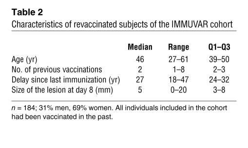
In March 2003, the French Ministry of Health implemented a program on preparedness and response to a biological attack using smallpox as a weapon. This program included the establishment of the French National Emergency Team against Smallpox IMMUVAR cohort, which is composed of 184 volunteers previously vaccinated who were revaccinated from March 2003 to November 2006 with VV vaccine and followed for the immunological responses prior to and after revaccination. Prior to vaccination, the volunteers had verification of prior vaccination, as evidenced by the presence of a scar resulting from previous smallpox vaccination or notification in a vaccination notebook. All volunteers included in the cohort were previously vaccinated in childhood. An HIV test was performed 2 weeks before vaccination. A urine pregnancy test on the day scheduled for vaccination was also performed for women of childbearing age. Pregnancy and HIV+ were exclusion criteria. Volunteers (Table 2) were revaccinated by administration of a Smallpox Elsee Lister strain (Pourquier Laboratories, batch 986, titer 108.3 PFU/ml), using bifurcated needles (15 pricks in an area of about 5 mm) as recommended on the left deltoid muscle. Blood samples and sera were collected and monitored for humoral immune response at different time points. Written informed consent was obtained from all volunteers. After revaccination, vaccination success was measured by the development of a skin lesion at the inoculation site 7–9 days after vaccination as described previously (17). The lesion was measured as the diameter (in mm) of the pustule 8 days after revaccination. All investigations were conducted after receipt of approval of the French institutional review board of the French Ministry of Health (Paris, France) and according to the Declaration of Helsinki.
Table 2 .
Characteristics of revaccinated subjects of the IMMUVAR cohort
NAb responses.
The neutralization titers were assessed at different time points after revaccination as previously described (35). Serial 2-fold dilutions of heat-inactivated serum were added to an equal volume of VV suspension (35 PFU/100 μl) and incubated for 1 hour at 37°C. Then, Vero cells in 24-well culture plates in quadruplicate were inoculated with 0.2 ml of each diluted serum/virus mixture. After 48 hours of incubation, cells were fixed and stained with 0.2% crystal violet, 4.5% formaldehyde, and 7.5% ethanol in PBS, and plaques were counted under a microscope. The neutralization titer was calculated as the mean reciprocal dilution of serum that caused a 50% VV plaque reduction compared with the number of VV plaques with negative control. A serum sample taken from a young unvaccinated individual was used as negative control. All unvaccinated individuals had an NT50 score less than 10. Individuals with NT50 values greater than 10 were considered responders. The neutralization titer was expressed in NT50 scores (for responders) and in IU/ml (for all individuals) using the European Directorate for the Quality of Medicines and Health Care (EDQM). Human VV Ig reference standard was available from the EDQM (35).
ELISpot assays.
The ELISpot assay was run as previously described (16). Briefly, fresh PBMCs were infected for 1 hour with the live VV Copenhagen wild-type strain (1 PFU/cell) (Transgène Laboratories) added to 96-well ELISpot plates (Millipore) coated with anti–human IFN-α antibody (Diaclone). Negative controls were uninfected cells with medium alone. Positive controls were uninfected PBMCs stimulated with purified PHA (1 μg/ml; Gibco, Invitrogen). Ten samples from unvaccinated individuals were used as negative control.
Cells were cultured in triplicate wells at 1 × 10⁵ PBMCs at 37°C for 18 hours. Spots were detected with an automated microscope (Zeiss). Spots were counted as positive if there were at least 50 vaccinia-specific SFU/million PBMCs after subtraction of the background obtained with unstimulated cells. This threshold for positive responder was established by taking into account: (a) the variability of the background obtained with cells alone (3 ± 5 SFU/million PBMCs; range, 0–75); and (b) the SFU range in the 10 naive unvaccinated individuals used as negative controls: median, 1.5 SFU/million PBMCs (range, 0–20 SFU/million PBMCs). The threshold for the positive response was established as previously described (16) and was set at 50 SFU/million PBMCs and yielded 100% negative results for the naive unvaccinated unexposed donors.
Proliferation assays.
The T cell proliferation assay was performed as previously described (16). Briefly, triplicate wells containing cells (105) were cultured in RPMI medium supplemented with 10% AB human serum with 0.1 PFU/cell VV Copenhagen strain for 7 days in the [3H]thymidine assays. [3H]thymidine was added during the last 18 hours of culture, and cells were harvested and counted on a Microbeta-plate beta-counter (Beckman). Ten naive unvaccinated individuals served as negative controls. Positive responses were defined by a positive stimulation index of 3 and a minimum [3H]thymidine incorporation of 3,000 counts/min.
Flow cytometric analyses.
Frozen PBMCs were available from only a few volunteers (n = 23) for further analysis of VV-specific T cells and were thawed in RPMI (Life Technologies) containing 5% FCS (Seromed), 2 mmol/l l-glutamine (Gibco BRL, Life Technologies), and antibiotics (1,000 IU/ml penicillin sodium, 1 mg/ml streptomycin sulfate, and 250 ng/ml amphotericin B). Data from all PBMCs tested are shown. Cells were stimulated for 12 hours at 37°C with 1 PFU of VV Copenhagen strain. Brefeldin A (5 μg/ml) (Sigma-Aldrich) was added to the well 4 hours before harvesting in order to detect intracellular cytokines. Then cells were stained in PBS once for 10 minutes at room temperature with CCR7-allophycocyanin first, and Abs CD4–APC-Cy7, CD3–PerCp-Cy5.5, CD8–Pacific Blue (BD Biosciences — Immunocytometry Systems or BD Biosciences — Pharmingen), and CD45RA-ECD (Beckman Coulter) were then added for 20 minutes at 4°C. Then 100 μl of Fix and Perm Medium A (Caltag) was added to each sample for 10 minutes at room temperature. Washed cells were resuspended with 100 μl Fix and Perm Medium B (Caltag) and incubated with intracellular Abs against TNF-α–PE CY7, IL-2–FITC, and IFN-γ–Alexa Fluor 700 (BD Biosciences — Immunocytometry Systems or BD Biosciences — Pharmingen) for 20 minutes at room temperature. The antibody panel used was as follows: CD4-AmCyan/CD8–Pacific Blue/CD45RA-ECD/IL-2–FITC/IFN-γ–Alexa Fluor 700/TNF-α–PE-C7 or CD4-AmCyan/CD8–Pacific Blue/CLA-PE/IL-2–FITC/IFN-γ–Alexa Fluor 700/TNF-α–PE-C7 or CD4-AmCyan/CD8–Pacific Blue/CD107a-PE/IL-2–FITC/IFN-γ–Alexa Fluor 700/TNF-α–PE-C7. Flow cytometric analysis was done using LSR2 flow cytometers (BD Biosciences — Immunocytometry Systems). FCS files were then exported and analyzed using FlowJo software (Tree Star). The lymphocyte gate was set based on forward and side scatter for further analysis. Non-T cells were excluded by gating on CD3+ cells. CD8+ and CD4+ cells were then selected according to these markers. Lymphocytes were thus identified based on their scatter patterns and on their expression of CD4 or CD8. The CD4+ T cells or CD8+ T cells were then gated for cells positive for the respective cytokines. Boolean combination gating was then performed to determine the frequencies of expression profiles corresponding to the 7 different combinations of cytokines by using the Flowjo software. Background cytokine responses detected in negative controls were subtracted from those detected in stimulated samples for every specific combination.
Statistics.
Prism 2.01 (GraphPad Software) was used for data handling, analysis, and graphic representation. The quantitative variables (i.e., age, size of the pustule, immunological data) are not normally distributed. Nonparametric tests using Wilcoxon matched pairs test and Spearman rank correlation test were used as indicated between quantitative variables. Statistical significance was set at P < 0.05.
Acknowledgments
We acknowledge members of the IMMUVAR study group for helpful discussions, E. Lefranc and M. Legendre for technical assistance, and D. Duffy and V. Abadie for helpful advice. This work was supported by the French Biotox Research Program and the French Ministry of Health (R05043DD), the French Department of Defense (DGA and Military Health Service), and association ARMI. B. Combadière is a recipient of Young Investigator Awards from the Agence Nationale de Recherche (ANR-JC05_46455) and Interface program INSERM/AP-HP.
Footnotes
Conflict of interest: The authors have declared that no conflict of interest exists.
Citation for this article: J Clin Invest. 2010;120(5):1636–1644. doi:10.1172/JCI38506.
References
- 1.Amanna IJ, Slifka MK, Crotty S. Immunity and immunological memory following smallpox vaccination. Immunol Rev. 2006;211:320–337. doi: 10.1111/j.0105-2896.2006.00392.x. [DOI] [PubMed] [Google Scholar]
- 2.Amanna IJ, Carlson NE, Slifka MK. Duration of humoral immunity to common viral and vaccine antigens. N Engl J Med. 2007;357(19):1903–1915. doi: 10.1056/NEJMoa066092. [DOI] [PubMed] [Google Scholar]
- 3.Puissant B, Combadiere B. Keeping the memory of smallpox virus. Cell Mol Life Sci. 2006;63(19–20):2249–2259. doi: 10.1007/s00018-006-6313-2. [DOI] [PMC free article] [PubMed] [Google Scholar]
- 4.Slifka MK. The future of smallpox vaccination: is MVA the key? Med Immunol . 2005;4(1):2. doi: 10.1186/1476-9433-4-2. [DOI] [PMC free article] [PubMed] [Google Scholar]
- 5. Jenner E.An inquiry into the causes and effects of the variolae vaccinae, a diseases discovered in some of the western countries of England, particularly Gloucestershire . London, UK: printed for the author by Sampson Low and sold by Law and Murray and Highley; 1798. http://pyramid.spd.louisville.edu/~eri/fos/jenner.html . Accessed February 2, 2010.
- 6.Frey SE, Newman FK, Yan L, Lottenbach KR, Belshe RB. Response to smallpox vaccine in persons immunized in the distant past. JAMA. 2003;289(24):3295–3299. doi: 10.1001/jama.289.24.3295. [DOI] [PubMed] [Google Scholar]
- 7.Simpson EL, Hercher M, Hammarlund EK, Lewis MW, Slifka MK, Hanifin JM. Cutaneous responses to vaccinia in individuals with previous smallpox vaccination. J Am Acad Dermatol. 2007;57(3):442–444. doi: 10.1016/j.jaad.2007.04.022. [DOI] [PubMed] [Google Scholar]
- 8.Lane JM, Ruben FL, Neff JM, Millar JD. Complications of smallpox vaccination, 1968. N Engl J Med. 1969;281(22):1201–1208. doi: 10.1056/NEJM196911272812201. [DOI] [PubMed] [Google Scholar]
- 9.Kempe CH. Studies smallpox and complications of smallpox vaccination. Pediatrics. 1960;26:176–189. [PubMed] [Google Scholar]
- 10.Frey SE, et al. Clinical responses to undiluted and diluted smallpox vaccine. N Engl J Med. 2002;346(17):1265–1274. doi: 10.1056/NEJMoa020534. [DOI] [PubMed] [Google Scholar]
- 11.Frey SE, et al. Dose-related effects of smallpox vaccine. N Engl J Med. 2002;346(17):1275–1280. doi: 10.1056/NEJMoa013431. [DOI] [PubMed] [Google Scholar]
- 12.Crotty S, Ahmed R. Immunological memory in humans. Semin Immunol. 2004;16(3):197–203. doi: 10.1016/j.smim.2004.02.008. [DOI] [PubMed] [Google Scholar]
- 13.Murali-Krishna K, Altman JD, Suresh M, Sourdive D, Zajac A, Ahmed R. In vivo dynamics of anti-viral CD8 T cell responses to different epitopes. An evaluation of bystander activation in primary and secondary responses to viral infection. Adv Exp Med Biol. 1998;452:123–142. doi: 10.1007/978-1-4615-5355-7_14. [DOI] [PubMed] [Google Scholar]
- 14.Slifka MK, Ahmed R. B cell responses and immune memory. Dev Biol Stand. 1998;95:105–115. [PubMed] [Google Scholar]
- 15.Hammarlund E, et al. 2003. Duration of antiviral immunity after smallpox vaccination. Nat Med. 2003;9(9):1115–1116. doi: 10.1038/nm0903-1115. [DOI] [PubMed] [Google Scholar]
- 16.Combadiere B, et al. Distinct time effects of vaccination on long-term proliferative and IFN-gamma-producing T cell memory to smallpox in humans. . J Exp Med. 2004;199(11):1585–1593. doi: 10.1084/jem.20032083. [DOI] [PMC free article] [PubMed] [Google Scholar]
- 17.Bossi P, et al. Demographic and clinical factors associated with response to smallpox vaccine in preimmunized volunteers. PLoS ONE. 2008;3(12):e4087. doi: 10.1371/journal.pone.0004087. [DOI] [PMC free article] [PubMed] [Google Scholar]
- 18.Fuhlbrigge RC, Kieffer JD, Armerding D, Kupper TS. Cutaneous lymphocyte antigen is a specialized form of PSGL-1 expressed on skin-homing T cells. Nature. 1997;389(6654):978–981. doi: 10.1038/40166. [DOI] [PubMed] [Google Scholar]
- 19.Mitra-Kaushik S, Cruz J, Stern LJ, Ennis FA, Terajima M. Human cytotoxic CD4+ T cells recognize HLA-DR1-restricted epitopes on vaccinia virus proteins A24R and D1R conserved among poxviruses. J Immunol. 2007;179(2):1303–1312. doi: 10.4049/jimmunol.179.2.1303. [DOI] [PubMed] [Google Scholar]
- 20.Demkowicz WE, Jr, Littaua RA, Wang J, Ennis FA. Human cytotoxic T-cell memory: long-lived responses to vaccinia virus. J Virol. 1996;70(4):2627–2631. doi: 10.1128/jvi.70.4.2627-2631.1996. [DOI] [PMC free article] [PubMed] [Google Scholar]
- 21.Littaua RA, Oldstone MB, Takeda A, Ennis FA. A CD4+ cytotoxic T-lymphocyte clone to a conserved epitope on human immunodeficiency virus type 1 p24: cytotoxic activity and secretion of interleukin-2 and interleukin-6. J Virol. 1992;66(1):608–611. doi: 10.1128/jvi.66.1.608-611.1992. [DOI] [PMC free article] [PubMed] [Google Scholar]
- 22.Fenner F. Risks and benefits of vaccinia vaccine use in the worldwide smallpox eradication campaign. Res Virol. 1989;140(5):465–466; discussion 487–491. doi: 10.1016/s0923-2516(89)80126-8. [DOI] [PubMed] [Google Scholar]
- 23.Hsieh SM, et al. Clinical and immunological responses to undiluted and diluted smallpox vaccine with vaccinia virus of Lister strain. Vaccine. 2006;24(4):510–515. doi: 10.1016/j.vaccine.2005.07.082. [DOI] [PubMed] [Google Scholar]
- 24.Crotty S, Felgner P, Davies H, Glidewell J, Villarreal L, Ahmed R. Cutting edge: long-term B cell memory in humans after smallpox vaccination. . J Immunol. 2003;171(10):4969–4973. doi: 10.4049/jimmunol.171.10.4969. [DOI] [PubMed] [Google Scholar]
- 25.Ennis FA, Cruz J, Demkowicz WE, Jr, Rothman AL, McClain DJ. Primary induction of human CD8+ cytotoxic T lymphocytes and interferon-gamma-producing T cells after smallpox vaccination. . J Infect Dis. 2002;185(11):1657–1659. doi: 10.1086/340517. [DOI] [PubMed] [Google Scholar]
- 26.Precopio ML, et al. Immunization with vaccinia virus induces polyfunctional and phenotypically distinctive CD8(+) T cell responses. J Exp Med. 2007;204(6):1405–1416. doi: 10.1084/jem.20062363. [DOI] [PMC free article] [PubMed] [Google Scholar]
- 27.Amara RR, Nigam P, Sharma S, Liu J, Bostik V. Long-lived poxvirus immunity, robust CD4 help, and better persistence of CD4 than CD8 T cells. . J Virol. 2004;78(8):3811–3816. doi: 10.1128/JVI.78.8.3811-3816.2004. [DOI] [PMC free article] [PubMed] [Google Scholar]
- 28.Mora JR, Cheng G, Picarella D, Briskin M, Buchanan N, von Andrian UH. Reciprocal and dynamic control of CD8 T cell homing by dendritic cells from skin- and gut-associated lymphoid tissues. . J Exp Med. 2005;201(2):303–316. doi: 10.1084/jem.20041645. [DOI] [PMC free article] [PubMed] [Google Scholar]
- 29.Edele F, et al. Cutting edge: instructive role of peripheral tissue cells in the imprinting of T cell homing receptor patterns. J Immunol. 2008;181(6):3745–3749. doi: 10.4049/jimmunol.181.6.3745. [DOI] [PubMed] [Google Scholar]
- 30.Frey SE, et al. Clinical and immunologic responses to multiple doses of IMVAMUNE (Modified Vaccinia Ankara) followed by Dryvax challenge. Vaccine. 2007;25(51):8562–8573. doi: 10.1016/j.vaccine.2007.10.017. [DOI] [PMC free article] [PubMed] [Google Scholar]
- 31.Parrino J, et al. Safety, immunogenicity and efficacy of modified vaccinia Ankara (MVA) against Dryvax challenge in vaccinia-naive and vaccinia-immune individuals. Vaccine. 2007;25(8):1513–1525. doi: 10.1016/j.vaccine.2006.10.047. [DOI] [PMC free article] [PubMed] [Google Scholar]
- 32.Bikoff EK, Otten GR, Robertson EJ. Defective assembly of class I major histocompatibility complex molecules in an embryonic cell line. Eur J Immunol. 1991;21(9):1997–2004. doi: 10.1002/eji.1830210905. [DOI] [PubMed] [Google Scholar]
- 33.Edghill-Smith Y, et al. Smallpox vaccine-induced antibodies are necessary and sufficient for protection against monkeypox virus. Nat Med. 2005;11(7):740–747. doi: 10.1038/nm1261. [DOI] [PubMed] [Google Scholar]
- 34.Slifka MK. Immunological memory to viral infection. Curr Opin Immunol. 2004;16(4):443–450. doi: 10.1016/j.coi.2004.05.013. [DOI] [PubMed] [Google Scholar]
- 35.Leparc-Goffart I, Poirier B, Garin D, Tissier MH, Fuchs F, Crance JM. Standardization of a neutralizing anti-vaccinia antibodies titration method: an essential step for titration of vaccinia immunoglobulins and smallpox vaccines evaluation. J Clin Virol. 2005;32(1):47–52. doi: 10.1016/j.jcv.2004.07.005. [DOI] [PubMed] [Google Scholar]


