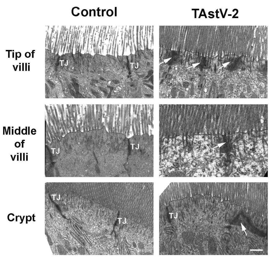
Fig 2. TAstV-2 infection mediated changes to the apical ultrastructure of epithelial cells.
Jejunum from control and TAstV-2 infected animals were collected at 4 dpi and analyzed by transmission electronic microscopy at the tip of villi, middle of villi, and the crypts. Normal tight junctions (TJ) are demarcated in the control micrographs, while electron dense aggregations (arrows) were observed in the TAstV-2-infected jejunum. White bar represents 0.5 µM

