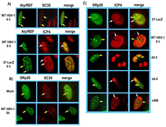
Fig. 3.
SRp20 remains associated with splicing speckles and is not recruited to viral replication compartments. A) RSF cells were transfected with pEGFP-Aly/REF and 24 h later were were infected with HSV-1 KOS or 27-LacZ, at an MOI of 10. Cells were fixed at the times indicated and immunostaining was performed with anti-ICP4 monoclonal antibody or anti-SC35 hybridoma supernatant. GFP fluorescence was visualized directly. B) RSF cells were transfected with pEGFP-SRp20 and 24 h later were either mock infected or were infected with HSV-1 KOS at an MOI of 10. At 8 h after infection, cells were fixed and stained with anti-SC35 hybridoma supernatant. GFP fluorescence was visualized directly. C) RSF cells were transfected with pEGFP-SRp20 and 24 h later were infected with 27-LacZ, HSV-1 KOS, or mutants d2–3, d4–5 or n406, all at an MOI of 10 for 8 h. Cells were stained with anti-ICP4 antibody and GFP fluorescence was visualized directly. Images were captured with Zeiss Axiovert S100 microscope at 100X. Yellow arrows point to speckled structures. White arrows point to viral replication compartments.

