Abstract
S-Nitrosothiols (RSNOs) represent an important class of post-translational modifications that preserve and amplify the actions of nitric oxide and regulate enzyme activity. Several regulatory proteins are now verified targets of cellular S-nitrosation and the direct detection of S-nitrosated residues in proteins has become essential to better understand RSNO-mediated signaling. Current RSNO detection depends on indirect assays that limit their overall specificity and reliability. Herein, we report the reaction of S-nitrosated cysteine, glutathione, and a mutated C165S alkyl hydroperoxide reductase with the water-soluble phosphine, tris(4,6-dimethyl-3-sulfonatophenyl)phosphine trisodium salt hydrate (TXPTS). A combination of NMR and MS techniques reveals these reactions produce covalent S-alkylphosphonium ion adducts (with S-P+ connectivity), TXPTS oxide and a TXPTS-derived aza-ylide. Mechanistically, this reaction may proceed through an S-substituted aza-ylide or the direct displacement of nitroxyl from the RSNO group. This work provides a new means for detecting and quantifying S-nitrosated species in solution and suggests that phosphines may be useful tools for understanding the complex physiological roles of S-nitrosation and its implications in cell signaling and homeostasis.
Keywords: S-nitrosothiol, triarylphosphine, aza-ylide, S-alkylphosphonium ion, protein labeling, chemical ligation, nitrosative stress
Reactive oxygen and nitrogen species (ROS/RNS) affect cellular signaling processes at concentrations far below those required to inflict oxidative damage (1–4). The over-oxidation and metabolism of nitric oxide (NO), an endogenous cell signaling agent, produces several RNS including nitrogen dioxide and nitrite, which can generate competent nitrosation agents (5, 6). Nitrosation of cysteine sites yields S-nitrosothiols (RSNOs) that preserve, modulate, and amplify the actions of NO. This selective and reversible process represents an important post-translational modification in cell signaling and regulation (3, 6–8). Several proteins have been identified as targets of cellular S-nitrosation including glyceraldehyde-3-phosphate dehydrogenase (GAPDH), papain, hemoglobin (Hb), and several caspases (5, 9–12). Inflammatory stimuli, including RNS, increase overall cellular RSNO formation, which further mediates the inflammatory process (9, 13–17). Direct detection and specific identification of S-nitrosated residues in proteins is essential to better understand RSNO-mediated signaling. Current techniques, such as the biotin switch assay, Saville assay, and chemiluminescence-based methods, are indirect methods of detection (18–23). Also, variability in these assays has challenged their accuracy and driven the search for alternative detection systems (24, 25). Deciphering the role and importance of RSNOs in biology requires direct and specific methods of quantification. We report a new strategy to covalently label biological RSNOs using the water-soluble phosphine tris(4,6-dimethyl-3-sulfonatophenyl) phosphine trisodium salt hydrate (TXPTS, Scheme 1) (26).
Scheme 1.
Reactions of triarylphosphines with S-nitrosothiols in organic and aqueous systems
Earlier work showed the reaction of trityl-S-nitrosothiol with triphenylphosphine yields an S-substituted aza-ylide (1, Scheme 1) and provided inspiration for developing an SNO-specific label (27). Recently, Xian revealed that the treatment of various organic SNOs with derivatized triarylphosphines provides products arising from similar S-substituted aza-ylides (28–30). In the presence of properly situated electrophiles on the triarylphosphine, the S-substituted aza-ylide intermediates undergo ligation to form stable sulfenamides or disulfide iminophosphoranes (28–30). Treatment of S-nitrosocysteine ester derivatives with various phosphines yields the corresponding dehydroalanines through intramolecular elimination via a similar S-substituted aza-ylide (28). These results encouraged our use of the water-soluble phosphine TXPTS as a potential RSNO trap. We report the reactions of TXPTS with S-nitrosocysteine (Cys-SNO), S-nitrosoglutathione (GSNO), and a mutated peroxiredoxin, S-nitrosated C165S alkyl hydroperoxide reductase (C165S AhpC-SNO) yield the covalent S-alkylphosphonium salt (2, Scheme 1).
RESULTS AND DISCUSSION
Haake’s observation that the reaction of trityl S-nitrosthiol and triphenylphosphine gives an S-substituted aza-ylide (1, Scheme 1) reveals that organic S-nitrosthiols can act in a similar manner as the organic azide in Staudinger-type reductions/ligations (27, 31). Based upon this work, we hypothesized that protein and other biologically relevant S-nitrosothiols could potentially be labeled as structurally unique S-substituted aza-ylides through their reaction with the water soluble triarylphosphine (TXPTS). Recent work by Xian shows that a variety of S-nitrosothiols react with triaryl and trialkylphopshines to give S-substiuted aza-ylides, which undergo further reactions to yield a variety of different products depending on both RSNO and phosphine structure (29, 30). In the presence of an electrophilic trap, these S-substituted aza-ylide intermediates ligate to generate sulfenamides or rearrange to disulfide iminophosphoranes (29, 30). In the absence of an electrophilic trap, the intermediate S-substituted aza-ylide eliminates to form dehydroalanine-containing products (28). This work reveals rich reaction chemistry between S-nitrosothiols and phosphines that may form the basis of new labeling methods, but in general, these processes have been limited to protected versions of cysteine and glutathione in organic or organic/buffer mixtures. Using a combination of NMR and MS-based techniques, we sought to critically evaluate the reaction between TXPTS and several physiologically relevant S-nitrosothiols. We report that the reaction between S-nitrosothiols of cysteine, glutathione and a mutant of the bacterial peroxiredoxin, AhpC with the water soluble triarylphosphine (TXPTS) yield a unique set of products that include TXPTS oxide (3), the stable aza-ylide of TXPTS (4) and an S-alkylphosphonium adduct (RS-P+ connection, 5–6, Schemes 2 and 3).
Scheme 2.
Reaction of TXPTS with S-nitrosoglutathione at pH 7.
Scheme 3.
Reaction of TXPTS with S-nitrosocysteine at pH 7.
The reaction of triphenylphosphine with trityl S-nitrosothiol
Treatment of trityl S-nitrosothiol with two equivalents of triphenylphosphine in benzene yields the S-substituted aza-ylide (1, 92% yield, Scheme 1), that corroborates previous results and provides the first X-ray crystallographic and 31P NMR characterization of this compound (Supporting Information).
The Reaction of TXPTS and S-Nitrosoglutathione (GSNO)
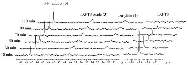
The reaction of TXPTS and S-nitrosoglutathione (GSNO, a commercially available or readily prepared stable pink solid), offers an opportunity to evaluate the formation of S-substituted aza-ylides in water or buffer. Ultraviolet-visible spectroscopy shows the rapid loss of the S-nitrosothiol functional group in GSNO upon addition of TXPTS as evidenced by the decrease in absorbance at 545 nm (Supporting Information). 31P NMR Spectroscopy provides detailed information regarding this reaction. A mixture of products results when freshly prepared GSNO (1 equivalent) and TXPTS (2 equivalents) are combined in buffer (50 mM HEPES, 1mM DTPA, pH 7.1) in the dark. The peak for phosphine (δ = −28.8 ppm) decreases with time and three new peaks at δ = 34.5, 39.7 and 47.2 ppm emerge over 30 min (Figure 1). No other phosphorus-derived species are observed on this time scale and control experiments show that TXPTS does not react with GSH and only to a small extent (~3%) with GSSG to form the same product with the resonance at 47.2 ppm as judged by 31P NMR (Supporting Information). Comparison to known standards allows identification of the peak at 39.7 ppm as TXPTS oxide (3, Scheme 2) and the peak at 34.5 ppm as the TXPTS-derived ylide (4, Scheme 2). Monitoring this reaction mixture over time shows the immediate formation of the species at 47.2 ppm, while 3 and 4 emerge more gradually over the course of 2 h. (Figure 2). Integration of the 31P NMR spectrum after 1 h indicates the formation of these three products in roughly a 1:1:1 ratio, which gives an estimated yield of 67% for 5 (based on starting GSNO). The use of 15N-labeled GSNO results in an apparent coupling (1JN-P = 23 Hz) in the peak at 34.5 ppm, which corresponds to 4, but not in any of the other peaks. This observed splitting varies over time suggesting some dynamic changes of 4 under these conditions (possibly water addition to the ylide). Treatment of this reaction mixture with dithiothreitol regenerates TXPTS (δ = −28.8 ppm) at the expense of the species at 47.2 ppm with no change in the peaks that correspond to 3 and 4 (Supporting Information). The addition of a sodium hydroxide solution to the reaction mixture results in the disappearance of the peaks at 34.5 ppm and 47.2 ppm and an increase in phosphine oxide (3, 39.7 ppm). The 13C and 1H NMR spectra of this reaction show complicated mixtures that do not allow for the clear identification of the phosphorus-containing species responsible for the resonance at 47.5 ppm or definitive estimates of the total product yields. Gas chromatographic headspace analysis and chemiluminescence nitric oxide detection reveal that no nitrous oxide (evidence for HNO) and only a trace amount of nitric oxide form during the reaction of TXPTS and GSNO.
Figure 1.
31P NMR time course experiment of the GSNO and TXPTS reaction. 15N-GSNO (1 eq.) and TXPTS (2 eq.) were incubated in a HEPES/DTPA buffered solution (pH 7.1) with 10 % D2O and the reaction was monitored using 31P NMR with 1 scan every 20 min. The peaks at 47.2, 39.7, and 34.5 ppm correspond to 5, 3, and 4, respectively and TXPTS starting material at −28.8 ppm. All peaks were referenced to a 3% H3PO4 (δ = 0 ppm) external standard which was omitted for clarity.
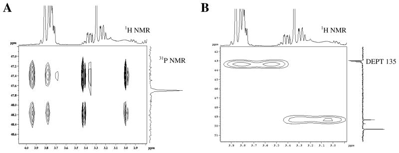
Figure 2.
2-D NMR of the GSNO and TXPTS reaction. a) 1H-31P COSY of the GSNO/TXPTS reaction mixture in D2O. b) 1H-13C HMQC of the GSNO/TXPTS reaction mixture in D2O
Liquid chromatography-mass spectrometry (LC-MS) further confirms the identity of these products and provides insight into the structure of the compound responsible for the peak at 47.2 ppm in the 31P NMR spectrum. Analysis of the GSNO/TXPTS reaction mixture validates the formation of 3 and 4 given their comparison to known standards (Supporting Information). The observed m/z of the final compound (892.1 m/z) suggests an adduct of GSNO and TXPTS with the loss of NO as opposed to the initially predicted -S-N=PR3 motif leading to the proposal of an S-alkylphosphonium product (5, Scheme 2) (27–30). The use of 15N-labeled GSNO results in the formation of 15N-labeled 4 indicating the transfer of the –S-N=O nitrogen atom to the phosphorus atom of TXPTS (Supporting Information). Similar LC-MS experiments do not support the formation of GSH or GSSG during this reaction.
A series of 2-D NMR experiments confirm the proposed S-P+ bonding motif of 5. A 1H-31P COSY indicates that the 31P signal of 5 (47.2 ppm) correlates to three protons in the 1H NMR spectrum (3.85, 3.42, and 3.01 ppm, presumably the –CH and diastereotopic –CH2 of the cysteine side chain of 5), in the reaction mixture (Figure 2A). Using this obtained 1H correlation, the 1H-13C HMQC of the same reaction mixture reveals the proximity of the P atom of the S-P+ moiety to nearby -CH2 and -CH carbons. Coupling constant analysis shows splitting of the -CH2 carbon (2JP-C = 14.6 Hz, Figure 2B), providing evidence for attachment of the phosphine through an S-P+ linkage.
The Reaction of TXPTS and S-nitroso-L-cysteine (Cys-SNO)
Similar 31P NMR experiments show the formation of three phosphorus-derived products from the mixture of freshly prepared Cys-SNO (1 equivalent) with TXPTS (2 equivalents) in buffer (50 mM HEPES, 1 mM DTPA, pH 7.1) in the dark that correspond to TXPTS oxide (3, δ = 39.7 ppm) and the TXPTS-derived ylide (4, δ = 34.5) and a presumed cysteine-derived S-alkylphosphonium adduct, (6, Scheme 3, δ = 46.1 ppm) after 30 min (Supporting Information). No other phosphorus-derived species were observed and control experiments show that TXPTS does not react with cysteine but partially reacts with cystine to form the same product with a resonance at 46.1 ppm (~15%, Supporting Information), indicating that TXPTS may react with certain disulfides. Integration of the 31P NMR spectrum after 1 h indicates the formation of compounds 3:4:6 in roughly a 3:1:1 ratio which suggests a more rapid hydrolysis of 6 compared to 5. Analogous to the GSNO reaction, treatment of this mixture with dithiothreitol regenerates TXPTS (δ = −28.8 ppm) at the expense of the species at 46.1 ppm with no change in the peaks that correspond to 3 and 4 (Supporting Information). LC-MS analysis of the Cys-SNO/TXPTS mixture also confirms the presence of 3 and 4 as well as the expected mass for the S-P+ adduct 6 (Supporting Information).
The commercial availability of (3-13C) Cys provides the opportunity to further confirm the structure of the S-P+ adduct (6) in solution. Treatment of (3-13C) Cys-SNO with TXPTS followed by 31P NMR spectroscopy showed through-bond coupling of the 31P atom to the 13C with an observed 2JC-P coupling = 3.7 Hz, which confirms the connectivity and covalent nature of the S-alkylphosphonium adduct (6, Supporting Information). Importantly, LC-MS analysis revealed a 1 amu increase in mass for the S-P+ species (6), further validating the covalent attachment of TXPTS in the adduct (Supporting Information). The 13C NMR spectrum of the reaction between TXPTS and (3-13C) Cys-SNO shows the presence of four peaks at 42.1, 42.4, 60.8 and 62.7 ppm (Supporting Information). The resonance at 42.1 ppm is a doublet (2JP-C = 8.6 Hz) indicating coupling and further corroborating the 31P NMR results. Comparison to standards reveals that the peak at 60.8 ppm corresponds to the methylene carbon of L-serine. The other two resonances (42.4 and 62.7 ppm) currently remain unidentified but appear during the preparation of Cys-SNO (Supporting Information). This spectrum also indicates the absence of cysteine, cystine and dehydroalanine (no resonances between 100–150 ppm) in the reaction mixture, in contrast to recent work that shows dehydroalanine formation in the reaction of Cys-SNO derivatives with phosphines (28).
While providing strong evidence of S-alkylphosphonium ion formation, the identification of other non-phosphorus containing products (by NMR) and our inability to completely account for the GSNO and Cys-SNO reveal the complexity of these reactions. Both NMR and LC-MS examination of these reaction mixtures do not show the presence of disulfides (GSSG/cystine), the glutathione-derived sulfenamide (GSNH2) and only small amounts of thiol (GSH/cysteine), eliminating these likely products. The identification of serine from the reaction of Cys-SNO, which may form from the hydrolysis of 6, demonstrates the complexity of these reactions. These results indicate the likelihood of other processes during S-alkylphosphonium ion formation and breakdown that may include reactions of sulfenamide intermediates, other rearrangements/dehydrations of intermediate aza-ylides and various reactions of the S-alkylphosphonium ions (5 and 6).
Literature values for S-alkylphosphonium ion 31P NMR shifts are roughly 40–50 ppm, consistent with the observed resonances for 5 and 6 (32–34). These SP+ ions also represent known intermediates in phosphine-mediated disulfide reductions. In general, phosphines, including tris(2-carboxyethyl)phosphine (TCEP), the reagent of choice for protein disulfide reduction, rapidly reduce disulfides to the S-alkylphosphonium intermediate that hydrolyzes to give free thiol and phosphine oxide (35–38). Control experiments show that TXPTS reacts with GSSG and cystine to a limited extent to yield the S-P+ intermediates (5 and 6). The unique structure of TXPTS appears to slow the hydrolysis of these SP+ species, allowing them to be isolated. The steric effects of the functionalized phenyl rings may hinder nucleophilic attack by water, and furthermore, the electron donating ability of the ortho and para methyl substituents on TXPTS likely increases the electron-density at phosphorous, reducing electrophilicity and retarding attack by water or other nucleophiles. Once formed, compounds 5 and 6 react with thiols to yield the phosphine and hydrolyze in base to the give the phosphine oxide, consistent with known reactivity of S-alkylphosphonium ions.
The Reaction of TXPTS with AhpC-SNO
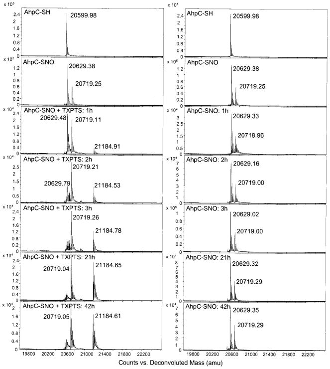
Given the reproducible formation of S-alkylthiophosphonium adducts from the reactions of TXPTS with GSNO and Cys-SNO, we examined the reaction of the S-nitrosated peroxiredoxin mutant, C165S alkyl hydroperoxide reductase C (AhpC-SNO) with TXPTS. This mutant was an ideal candidate for examining the interaction of a protein SNO with TXPTS since the remaining active site cysteine (Cys46) is susceptible to oxidative modifications, including sulfenic acid and S-nitrosothiol formation (39). Incubation of freshly prepared AhpC-SNO with 25-fold excess TXPTS in buffer (50 mM HEPES, 1 mM DTPA) at 25°C forms an S-alkylphosphonium (SP+-type) adduct over several hours (Figure 3) as shown by electrospray ionizationtime of flight (ESI-TOF) MS experiments. As seen in Figure 3, both AhpC-SNO (exact mass = 20629.38) and the S-thiolation product with cysteine (AhpC-S-S-Cys, exact mass = 20719.11) form upon mixture of AhpC (reduced) and Cys-SNO. Upon incubation of this sample with TXPTS, a time-dependent decrease of AhpC-SNO occurs with the emergence of the corresponding AhpC-SP+ adduct (exact mass = 21184.69) was formed in roughly 83 % yield after 3 hr as estimated by the relative intensities (Figure 3). Based on the m/z difference of 555.1 amu and the lack of reaction between AhpC (reduced) and TXPTS, the phosphine (exact mass = 652 as the tri-sodium salt) appears to be covalently bound to the protein in an S-P+ fashion. Direct treatment of the free thiol form of C165S AhpC (AhpC-SH) with TXPTS does not form this product by ESI-TOF MS (Supporting Information). Figure 3 also indicates that TXPTS does not react with the AhpC-S-S-Cys mixed disulfide over 42 h. The AhpC-derived S-P+ species demonstrates excellent stability over several days at room temperature and under denaturing conditions (40% acetonitrile, Supporting Information).
Figure 3.
ESI-TOF-MS data showing TXPTS covalently labeling the S-nitrosated mutated peroxiredoxin (C165S AhpC-SNO). The exact mass of the covalent adduct is 21184.69, MW of the AhpC-SNO is 20629.55. The MW 20719.14 corresponds to the mixed disulfide that forms between AhpC-SNO and Cys-SNO (AhpC-S-S-Cys). (left) C165S AhpC-SNO with 25-fold excess TXPTS over 42 hours (right) control showing C165S AhpC-SNO in the absence of TXPTS under the same conditions
To the best of our knowledge, these results describe the first covalent labeling of an S-nitrosated protein in buffer and demonstrate the potential of phosphines as protein SNO labels. While TXPTS rapidly reacts with small molecule RSNOs, Figure 3 shows that the reaction with AhpC-SNO occurs much more slowly. Steric differences in the environment of the –SNO group likely dictate the rates of these reactions and thus rates may vary among proteins with different thiol accessibility; the cysteine of AhpC has been shown to react relatively sluggishly with alkylating agents (40). Evaluation of the rate and selectivity of this reaction with protein SNOs and other oxidized sulfur species is warranted and will be required for the development of this reagent as a labeling tool.
Evaluating the Mechanism of SNO-Derived Adduct Formation
Scheme 4 depicts a proposed mechanism for the formation of S-substituted aza-ylides from an S-nitrosothiol with a triarylphosphine. This reaction could yield products through phosphorous-addition to either nitrogen or oxygen, and both could exist as a three-membered ring (7, Scheme 4) (27, 30). Addition of a second phosphine to 7 (or one of the initial addition products) would give the phosphine oxide and S-substituted aza-ylide in equal proportions (as observed for 1, Scheme 1) (27).
Scheme 4.
Proposed mechanism for the formation of the S-N=P type aza-ylide (1)
The described spectroscopic and LC-MS studies show that the reaction of GSNO or Cys-SNO with TXPTS in buffer rapidly consumes the S-nitrosothiol. While 31P NMR provides evidence of TXPTS phosphine oxide (3) formation as expected, 31P NMR chemical shift comparison to 1, the lack of observed splitting in the 31P NMR spectrum in the reaction using 15N-GSNO and LC-MS work do not support formation of the expected S-substituted aza-ylide. Hydrolysis of any S-substituted aza-ylide product to the corresponding sulfenamide (RSNH2, 8, Scheme 4) and 3 would explain the absence of the S-substituted aza-ylide, but the 31P NMR identification of two other phosphorus-containing species clearly indicates a different mechanism; direct formation of the S-substituted aza-ylide followed by hydrolysis would produce phosphine oxide (3) as the only phosphorus containing product. This work identifies the two other phosphorus-containing products as the aza-ylide (4) and the corresponding S-alkylphosphionium salts (5–6, Schemes 2 and 3). Aza-ylide (4), which has been characterized during the reaction of TXPTS and nitroxyl (HNO), demonstrates considerable water stability, presumably due to stabilization of the phosphonium ion by the electron-donating ortho and para methyl groups of TXPTS (41). The reaction of TXPTS with 15N-labled GSNO results in 15N incorporation in 4 and reveals the formal 6 e− reduction of the S-nitrosothiol nitrogen atom to ammonia (consistent with the absence of NO/HNO formation). This reaction finds some precedence in the GSNO reductase (aka alcohol dehydrogenase) mediated reduction of GSNO to ammonia (42, 43).
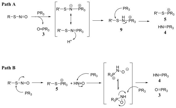
Scheme 5 shows two potential mechanisms that account for the observed phosphorus-containing products. Path A proceeds through a mechanism that includes an S-substituted aza-ylide (-S-N=P), presumably formed through a mechanism as seen in Scheme 4. In aqueous conditions, protonation of this S-substituted aza-ylide gives a new intermediate (9) that rapidly reacts with another equivalent of phosphine to simultaneously yield equal amounts of the S-alkylphosphonium ion (5 or 6) and the aza-ylide (4). This mechanism utilizes 3 equivalents of phosphine and predicts a 1:1:1 ratio of 3:4:5, as experimentally observed for the GSNO reaction. While 2 equivalents of phosphine were used in these experiments, the inherent instability of GSNO provided for excess phosphine during the reaction. The lack of experimental evidence of an S-subsituted aza-ylide (1) and previous work showing that TXPTS reacts with HNO to form equal amounts of 3 and 4 suggest a more direct mechanism for observed product formation (41). Path B (Scheme 5) depicts the direct nucleophilic attack of TXPTS on the sulfur atom of an S-nitrosothiol to form the S-alkylphosphonium ion (5, for GSNO) with release of HNO. The reaction of 2 equivalents of TXPTS with nascent HNO would yield 3 and 4. This mechanism also requires three equivalents of phosphine and predicts a 1:1:1 ratio of 3:4:5. The 31P NMR time-course experiment (Figure 2) supports Path B with the rapid initial formation of 5 followed by the emergence of 3 and 4 while Path A would initially produce 3 followed by 4 and 5. Given the rapid reaction of TXPTS and HNO, the failure to detect N2O (the HNO dimerization/dehydration product) does not eliminate Path B. While both mechanisms account for the observed products and ratios, further experiments, particularly a detailed kinetic analysis of the constituent reactions of this sequence, are required to delineate the mechanisms.
Scheme 5.
Two proposed mechanisms for the formation of an SNO-derived adduct from TXPTS.
In summary, the triarylphosphine TXPTS reacts directly with S-nitrosothiol residues to produce stable S-alkylphosphonium ions (5–6, or AhpC-SP+) as well as TXPTS oxide (3) and the TXPTS-derived aza-ylide (4), This work details the first reaction of triarylphosphines with RSNOs in water. Mechanistically, this reaction may proceed through an S-substituted aza-ylide or through the direct displacement of HNO from the RSNO group. This reaction, which is amenable to MS detection and 31P NMR spectroscopy, provides a new method for identifying S-nitrosated species in solution. This unique reactivity suggests that phosphines may be useful tools for understanding the complex physiological roles of biological S-nitrosation and its implications in cell signaling and homeostasis.
METHODS
Tris(4,6-dimethyl-3-sulfonatophenyl)phosphine trisodium salt hydrate (TXPTS) was purchased from Strem Chemicals. Glutathione (reduced and oxidized), L-cysteine, L-cystine, 1,4-dithiothreitol, sodium nitrite, sodium hydroxide, triphenylphosphine, triphenylmethane thiol, butyl nitrite, hydrogen peroxide solution (10 M), deuterium oxide, and deuterated chloroform were purchased from Sigma Aldrich. Hydrochloric acid solution (1 N) was purchased from TCI. (3-13C) L-Cysteine and 15N-sodium nitrite were purchased from Cambridge Isotope Laboratory. All reagents were used directly from suppliers without further purification.
Preparation of S-substituted aza-ylide (1)
A solution of trityl thionitrite in CHCl3 (0.1905 g, 0.62 mmol, (λmax, (CHCl3) 335, 545 nm)(44) was added to a solution of triphenylphosphine (0.33 g, 1.25 mmol) in anhydrous benzene (5 mL) at room temperature. After 30 minutes, the solids were filtered to give 1 as a bright yellow solid (27). Recrystalization in CH3CN/toluene afforded 1 as bright yellow crystals. 31P NMR (121 MHz, CDCl3) δ 17.3.
Preparation of S-nitrosocysteine (Cys-SNO)
A solution of L-cysteine (0.06 g, 0.34 mmol) in 0.75 N HCl (4 mL) with 15 mM EDTA was added to a solution of sodium nitrite (0.035g, 0.5 mmol) in H2O (5 mL) with 0.5 mM EDTA at 4°C. After 10 minutes, the reaction turned deep red and was neutralized with 1 N NaOH (2.5 mL) to afford a solution of L-Cys-SNO (H2O, ε335nm 503 M−1cm−1). (3-13C) L-Cys-SNO was made using the same procedure. (15N)-S-Nitrosocysteine was prepared using 15N-sodium nitrite.
Preparation of S-nitrosoglutathione (GSNO) (45)
Sodium nitrite (0.345 g, 5 mmol) was added to an ice-cold solution of glutathione (1.53 g, 5 mmol) in 2N HCl (5 mL). After 40 minutes at 4°C, the red solution was treated with acetone (10 mL) and stirred for an additional 10 minutes. The resulting pink solid was filtered off and washed successively with ice-cold water (5 mL), acetone (3 mL), and ether (3 mL) to afford S-nitrosoglutathione (1.29 g, 76%) (H2O, ε335nm 922 M−1cm−1, ε545nm 15.9 M−1cm−1). (15N)-S-Nitrosoglutathione was prepared using 15N-sodium nitrite.
Preparation of S-nitroso (C165S) AhpC
The mutant form of Salmonella typhimurium AhpC, C165S, was overexpressed and purified from Escherichia coli as previously described.(40, 46) A solution of reduced protein (0.5 mg/mL, 500 μL) in HEPES (50 mM), DTPA (1 mM) buffer (pH 7.1) was incubated with freshly prepared S-nitrosocysteine (1 mM) at 25°C for 60 min in the dark. Removal of S-nitrosocysteine was performed using BioRad™ spin columns with Bio-Gel P6™ equilibrated in HEPES (50 mM), DTPA (1 mM) buffer (pH 7.1). The formation of C165S AhpC-SNO (as well as a lesser amount of C165S AhpC/Cys mixed disulfide) was monitored by ESI-TOF MS as described below.
Labeling of AhpC-SNO using TXPTS
A solution of TXPTS (2 μL, 250 mM) was added to a solution of freshly prepared AhpC-SNO (200 μL, 100 μM) in HEPES (50 mM), DTPA (1 mM) buffer. Aliquots (50 μL) were taken at different time points and diluted with ammonium bicarbonate buffer (50 μL, 50 mM, pH 7.5), then TXPTS was removed using BioRad spin columns packed with Bio-Gel P6 (BioRad) and samples were equilibrated with ammonium bicarbonate buffer (50 mM, pH 7.5). The samples were analyzed by ESI-TOF MS as described below.
NMR Analysis
1-D Analyses: 1H NMR spectra were recorded on Bruker Avance DPX-300 and DRX-500 instruments at 300.13 and 500.13 MHz, respectively. 13C and DEPT NMR spectra were recorded on a Bruker DRX-500 instrument operating at 125.76 MHz. 31P NMR spectra were recorded on Bruker DPX-300 and DRX-500 instruments operating at 121.49 MHz and 202.46 MHz respectively and 31P chemical shifts are relative to 3% H3PO4 (δ = 0 ppm) contained in a concentric internal capillary (Wilmad). 2-D Analyses: The 1H-31P gradient selected COSY spectrum was acquired with 2048 complex points in t2, 256 points in t1 and 40 transients with a pulse repetition delay of 2 s. A sweep width of 10 ppm in 1H (centered at 4.5 ppm) and 100 ppm in 31P (centered at 10 ppm) was used. Data sets were multiplied with 90° phase shifted squared-sinebell apodization function and zero-filled to 512 × 512 data points before Fourier transformation. Chemical shifts were referenced to the residual H2O signal and the observed SP+ chemical shift (31P NMR shift of 47.7 ppm). The 2-D HMQC spectrum was collected with 256 complex points in t2, 256 points in t1 and 32 transients with a pulse repetition delay of 1.5s. A sweep width of 7.78 ppm in 1H (centered at 5 ppm) and 250 ppm in 13C (centered at 110 ppm) was used. Data sets were multiplied with 90° phase shifted squared-sinebell apodization function and zero-filled to 1024 × 1024 data points before Fourier transformation.
LC-MS Analysis
LC-MS experiments were performed on an Agilent Technologies 1100 LC/MSD Trap instrument. Separations were achieved using an Agilent Zorbax Rapid Resolution SB-C18 reverse phase column (2.1 × 30 mm/3.5μm) at 25°C with solvent A = 0.1% ammonium formate in water and solvent B = HPLC-grade acetonitrile. Flow rate: 0.4 mL/min. Gradient for conditioning of column: 100% B to 15% B over 90 minutes. Gradient for GSNO + TXPTS analyses: 1% B for 1 min, ramp to 3% B over 2 min, ramp to 5% B over 3 min, and hold for 1 min, for a total run time of 7 min. The post-run time was 3 min at 5% B. Gradient for Cys-SNO + TXPTS analyses: 3% B for 3 min, ramp to 10% B for 3 min, and lower to 5% B for 1 min giving a total run time of 7 min. The post-run time was 3 min at 5% B. The ion trap mass spectrometer was equipped with an atmospheric pressure electrospray ionization source, and was operated in smart mode. Nebulization was achieved with a N2 pressure of 50 psi, and solvent evaporation assisted by a flow of He drying gas (11 L/min, 325°C). Mass spectra were obtained in positive ion mode, and target masses (m/z) were set at 603 (TXPTS oxide, 3), 602 (aza-ylide, 4), and 892 (S-alkylphosphonium adduct, 5). Retention times and masses of extracted ions were compared to standards on the same instrument. TXPTS oxide (3) was prepared by incubating H2O2 and TXPTS in a 1:1 mole ratio in H2O for 5 min. Aza-ylide (4) was prepared by incubating hydroxylamine-O-sulfonic acid and TXPTS in a 1:1 mole ratio in H2O for 1h. as previously reported (41).
ESI-TOF-MS Analysis
All ESI-TOF MS data was collected on an Agilent MSD TOF system. The operating conditions for MS analysis were as follows: positive ion mode, capillary voltage (VCap) 3500 V, nebulizer gas 30 psig, drying gas 5.0 L/min; fragmentor 140 V; gas temperature 325°C. The samples were injected into the ion source using a syringe pump (KD Scientific) and a 250 μL Hamilton syringe connected to the ion probe with a 50 μm ID fused silica capillary. The injection flow rate was 10 μL/min. The averaged MS spectra were deconvoluted using the Agilent MassHunter Workstation Software vs B.01.03.
Supplementary Material
Acknowledgments
This work was supported by the National Institutes of Health (R01 HL062198, SBK), (R01 CA136810, CMF) and (R33 CA126659, LBP) and Wake Forest University. The NMR spectrometers used in this work were purchased with partial support from NSF (CHE-9708077) and the North Carolina Biotechnology Center (9703-IDG-1007). We thank Dr. C. S. Day (Wake Forest University) for assistance with X-ray crystallography.
Footnotes
Supporting Information available: Experimental details for the synthesis of Cys-SNO, GSNO and AhpC-SNO. 31P and 13C NMR spectra, UV/vis data and LC-MS results for various reactions. ESI-TOF-MS controls and x-ray structural data for 1. This material is available free of charge via the Internet at http://pubs.acs.org.
References
- 1.Davies MJ, Fu S, Wang H, Dean RT. Stable markers of oxidant damage to proteins and their application in the study of human disease. Free Radic Biol Med. 1999;27:1151–1163. doi: 10.1016/s0891-5849(99)00206-3. [DOI] [PubMed] [Google Scholar]
- 2.Poole LB, Karplus PA, Claiborne A. Protein sulfenic acids in redox signaling. Annu Rev Pharmacol Toxicol. 2004;44:325–347. doi: 10.1146/annurev.pharmtox.44.101802.121735. [DOI] [PubMed] [Google Scholar]
- 3.Deem S, Kim SS, Min JH, Eveland R, Moulding J, Martyr S, Wang X, Swenson ER, Gladwin MT. Pulmonary vascular effects of red blood cells containing S-nitrosated hemoglobin. Am J Physiol Heart Circ Physiol. 2004;287:H2561–2568. doi: 10.1152/ajpheart.00310.2004. [DOI] [PubMed] [Google Scholar]
- 4.Kobayashi-Miura M, Shioji K, Hoshino Y, Masutani H, Nakamura H, Yodoi J. Oxygen sensing and redox signaling: the role of thioredoxin in embryonic development and cardiac diseases. Am J Physiol Heart Circ Physiol. 2007;292:H2040–2050. doi: 10.1152/ajpheart.01316.2006. [DOI] [PubMed] [Google Scholar]
- 5.Haendeler J, Weiland U, Zeiher AM, Dimmeler S. Effects of redox-related congeners of NO on apoptosis and caspase-3 activity. Nitric Oxide. 1997;1:282–293. doi: 10.1006/niox.1997.0134. [DOI] [PubMed] [Google Scholar]
- 6.Hess DT, Matsumoto A, Kim SO, Marshall HE, Stamler JS. Protein S-nitrosylation: purview and parameters. Nat Rev Mol Cell Biol. 2005;6:150–166. doi: 10.1038/nrm1569. [DOI] [PubMed] [Google Scholar]
- 7.Hogg N. The Biochemistry and Physiology of S-Nitrosothiols. Annu Rev Pharmacol Toxicol. 2002;42:585–600. doi: 10.1146/annurev.pharmtox.42.092501.104328. [DOI] [PubMed] [Google Scholar]
- 8.Janssen-Heininger YM, Mossman BT, Heintz NH, Forman HJ, Kalyanaraman B, Finkel T, Stamler JS, Rhee SG, van der Vliet A. Redox-based regulation of signal transduction: Principles, pitfalls, and promises. Free Radic Biol Med. 2008;45:1–17. doi: 10.1016/j.freeradbiomed.2008.03.011. [DOI] [PMC free article] [PubMed] [Google Scholar]
- 9.Crawford JH, Chacko BK, Pruitt HM, Piknova B, Hogg N, Patel RP. Transduction of NO-bioactivity by the red blood cell in sepsis: novel mechanisms of vasodilation during acute inflammatory disease. Blood. 2004;104:1375–1382. doi: 10.1182/blood-2004-03-0880. [DOI] [PubMed] [Google Scholar]
- 10.Martinez-Ruiz A, Lamas S. Detection and identification of S-nitrosylated proteins in endothelial cells. Methods Enzymol. 2005;396:131–139. doi: 10.1016/S0076-6879(05)96013-8. [DOI] [PubMed] [Google Scholar]
- 11.Padgett CM, Whorton AR. S-nitrosoglutathione reversibly inhibits GAPDH by S-nitrosylation. Am J Physiol. 1995;269:C739–749. doi: 10.1152/ajpcell.1995.269.3.C739. [DOI] [PubMed] [Google Scholar]
- 12.Xian M, Chen X, Liu Z, Wang K, Wang PG. Inhibition of papain by S-nitrosothiols. Formation of mixed disulfides. J Biol Chem. 2000;275:20467–20473. doi: 10.1074/jbc.M001054200. [DOI] [PubMed] [Google Scholar]
- 13.Stamler JS. S-nitrosothiols in the blood: roles, amounts, and methods of analysis. Circ Res. 2004;94:414–417. doi: 10.1161/01.RES.0000122071.55721.BC. [DOI] [PubMed] [Google Scholar]
- 14.Jourd’heuil D, Gray L, Grisham MB. S-nitrosothiol formation in blood of lipopolysaccharide-treated rats. Biochem Biophys Res Commun. 2000;273:22–26. doi: 10.1006/bbrc.2000.2892. [DOI] [PubMed] [Google Scholar]
- 15.Xu J-W, Ikeda K, Yamori Y. Inhibitory effect of polyphenol cyanidin on TNF-[alpha]-induced apoptosis through multiple signaling pathways in endothelial cells. Atherosclerosis. 2007;193:299–308. doi: 10.1016/j.atherosclerosis.2006.09.006. [DOI] [PubMed] [Google Scholar]
- 16.Marshall HE, Stamler JS. Inhibition of NF-kappa B by S-nitrosylation. Biochemistry. 2001;40:1688–1693. doi: 10.1021/bi002239y. [DOI] [PubMed] [Google Scholar]
- 17.Stamler JS, Hausladen A. Oxidative modifications in nitrosative stress. Nat Struct Biol. 1998;5:247–249. doi: 10.1038/nsb0498-247. [DOI] [PubMed] [Google Scholar]
- 18.Forrester MT, Foster MW, Stamler JS. Assessment and application of the biotin switch technique for examining protein S-nitrosylation under conditions of pharmacologically induced oxidative stress. J Biol Chem. 2007;282:13977–13983. doi: 10.1074/jbc.M609684200. [DOI] [PubMed] [Google Scholar]
- 19.Jaffrey SR, Snyder SH. The biotin switch method for the detection of S-nitrosylated proteins. Sci STKE. 2001;2001:PL1. doi: 10.1126/stke.2001.86.pl1. [DOI] [PubMed] [Google Scholar]
- 20.Doctor A, Gaston B, Kim-Shapiro DB. Detecting physiologic fluctuations in the S-nitrosohemoglobin micropopulation: Triiodide versus 3C. Blood. 2006;108:3225–3226. doi: 10.1182/blood-2006-05-026047. author reply 3226–3227. [DOI] [PubMed] [Google Scholar]
- 21.Gow A, Doctor A, Mannick J, Gaston B. S-Nitrosothiol measurements in biological systems. J Chromatogr B Analyt Technol Biomed Life Sci. 2007;851:140–151. doi: 10.1016/j.jchromb.2007.01.052. [DOI] [PMC free article] [PubMed] [Google Scholar]
- 22.Xu X, Cho M, Spencer NY, Patel N, Huang Z, Shields H, King SB, Gladwin MT, Hogg N, Kim-Shapiro DB. Measurements of nitric oxide on the heme iron and beta-93 thiol of human hemoglobin during cycles of oxygenation and deoxygenation. Proc Natl Acad Sci U S A. 2003;100:11303–11308. doi: 10.1073/pnas.2033883100. [DOI] [PMC free article] [PubMed] [Google Scholar]
- 23.Park JK, Kostka P. Fluorometric detection of biological S-nitrosothiols. Anal Biochem. 1997;249:61–66. doi: 10.1006/abio.1997.2159. [DOI] [PubMed] [Google Scholar]
- 24.Giustarini D, Dalle-Donne I, Colombo R, Milzani A, Rossi R. Is ascorbate able to reduce disulfide bridges? A cautionary note. Nitric Oxide. 2008;19:252–258. doi: 10.1016/j.niox.2008.07.003. [DOI] [PubMed] [Google Scholar]
- 25.Landino LM, Koumas MT, Mason CE, Alston JA. Ascorbic acid reduction of microtubule protein disulfides and its relevance to protein S-nitrosylation assays. Biochem Biophys Res Commun. 2006;340:347–352. doi: 10.1016/j.bbrc.2005.12.013. [DOI] [PubMed] [Google Scholar]
- 26.Moore LR, Shaughnessy KH. Efficient aqueous-phase Heck and Suzuki couplings of aryl bromides using tri(4,6-dimethyl-3-sulfonatophenyl)phosphine trisodium salt (TXPTS) Org Lett. 2004;6:225–228. doi: 10.1021/ol0360288. [DOI] [PubMed] [Google Scholar]
- 27.Haake M. Zur desoxygenierung von tritylthionitrit. Tet Lett. 1972;33 [Google Scholar]
- 28.Wang H, Zhang J, Xian M. Facile formation of dehydroalanine from S-nitrosocysteines. J Am Chem Soc. 2009;131:13238–13239. doi: 10.1021/ja905558w. [DOI] [PubMed] [Google Scholar]
- 29.Zhang J, Wang H, Xian M. Exploration of the “traceless” reductive ligation of S-nitrosothiols. Org Lett. 2009;11:477–480. doi: 10.1021/ol802663q. [DOI] [PubMed] [Google Scholar]
- 30.Wang H, Xian M. Fast reductive ligation of S-nitrosothiols. Angew Chem Int Ed Engl. 2008;47:6598–6601. doi: 10.1002/anie.200801654. [DOI] [PubMed] [Google Scholar]
- 31.Saxon E, Bertozzi CR. Cell surface engineering by a modified Staudinger reaction. Science. 2000;287:2007–2010. doi: 10.1126/science.287.5460.2007. [DOI] [PubMed] [Google Scholar]
- 32.Langer R, Shi W, Rothenberger A. Copper-mediated cleavage of disulfides by tertiary phosphines: a new route to As-S anions. Dalton Transactions. 2006;37:4435–4437. doi: 10.1039/b610228j. [DOI] [PubMed] [Google Scholar]
- 33.Valeriy EP, Alexei VB, Yuriy GS. Comparison of the reactivity of N-(p-toluenesulfonyl)-sulfinimidoyl fluorides and chlorides toward triphenylphosphine. Heteroatom Chemistry. 2008;19:66–71. [Google Scholar]
- 34.Omelanczuk J, Mikolajczyk M. Optically active trivalent phosphorus compounds. 2. Reactivity of alkylthio- and alkylselenophosphonium salts. The first stereospecific synthesis of a chiral phosphinite. Journal of the American Chemical Society. 1979;101:7292–7295. [Google Scholar]
- 35.Cline DJ, Redding SE, Brohawn SG, Psathas JN, Schneider JP, Thorpe C. New water-soluble phosphines as reductants of peptide and protein disulfide bonds: reactivity and membrane permeability. Biochemistry. 2004;43:15195–15203. doi: 10.1021/bi048329a. [DOI] [PubMed] [Google Scholar]
- 36.Han JC, Han GY. A procedure for quantitative determination of tris(2-carboxyethyl)phosphine, an odorless reducing agent more stable and effective than dithiothreitol. Anal Biochem. 1994;220:5–10. doi: 10.1006/abio.1994.1290. [DOI] [PubMed] [Google Scholar]
- 37.Overman LE, Matzinger D, O’Connor EM, Overman JD. Nucleophilic cleavage of the sulfur-sulfur bond by phosphorus nucleophiles. Kinetic study of the reduction of aryl disulfides with triphenylphosphine and water. Journal of the American Chemical Society. 1974;96:6081–6089. [Google Scholar]
- 38.Houk J, Singh R, Whitesides GM. Measurement of thiol-disulfide interchange reactions and thiol pKa values. Methods Enzymol. 1987;143:129–140. doi: 10.1016/0076-6879(87)43023-1. [DOI] [PubMed] [Google Scholar]
- 39.Poole LB, Ellis HR. Identification of cysteine sulfenic acid in AhpC of alkyl hydroperoxide reductase. Methods Enzymol. 2002;348:122–136. doi: 10.1016/s0076-6879(02)48632-6. [DOI] [PubMed] [Google Scholar]
- 40.Nelson KJ, Parsonage D, Hall A, Karplus PA, Poole LB. Cysteine pKa Values for the Bacterial Peroxiredoxin AhpC. Biochemistry. 2008 doi: 10.1021/bi801718d. [DOI] [PMC free article] [PubMed] [Google Scholar]
- 41.Reisz JA, Klorig EB, Wright MW, King SB. Reductive Phosphine-Mediated Ligation of Nitroxyl (HNO) Organic Letters. 2009;11:2719–2721. doi: 10.1021/ol900914s. [DOI] [PubMed] [Google Scholar]
- 42.Liu L, Hausladen A, Zeng M, Que L, Heitman J, Stamler JS. A metabolic enzyme for S-nitrosothiol conserved from bacteria to humans. Nature. 2001;410:490–494. doi: 10.1038/35068596. [DOI] [PubMed] [Google Scholar]
- 43.Jensen DE, Belka GK, Du Bois GC. S-Nitrosoglutathione is a substrate for rat alcohol dehydrogenase class III isoenzyme. Biochem J. 1998;331(Pt 2):659–668. doi: 10.1042/bj3310659. [DOI] [PMC free article] [PubMed] [Google Scholar]
- 44.Lecher H, Siefken W. Nitrosyl derivatives of bivalent sulfur. II. Nitrosyl ethyl mercaptide. II. Chem Ber. 1926;59:2594–2601. [Google Scholar]
- 45.Hart TW. Some observations concerning the S-nitroso and S-phenylsulphonyl derivatives of L-cysteine and glutathione. Tet Lett. 1985;26:2013–2016. [Google Scholar]
- 46.Poole LB, Ellis HR. Flavin-dependent alkyl hydroperoxide reductase from Salmonella typhimurium. 1. Purification and enzymatic activities of overexpressed AhpF and AhpC proteins. Biochemistry. 1996;35:56–64. doi: 10.1021/bi951887s. [DOI] [PubMed] [Google Scholar]
Associated Data
This section collects any data citations, data availability statements, or supplementary materials included in this article.


