Abstract
A simple common path optical coherence tomography using a fibre optic bundle as a probe is demonstrated experimentally. The mechanical lateral scans are accomplished outside the specimen, proximal entrance of the fibre bundle, which eliminated the need for moving parts in the distal end of the probe. This feature allows the probe to be made submillimetre in size and easily integrated into surgical tools for intraoperative imaging. The axial and lateral resolutions of the system, and preliminary images of phantom samples, are reported.
Introduction
Current optical coherence tomography (OCT) adapts various kinds of miniaturised scanning optic probes that can be used in minimally invasive imaging for diagnosing or micro-sensitive image-guided surgery purposes [1-3]. There also have been great efforts to avoid the use of mechanically moving probes for the lateral scans either in high-resolution microscopy [4-6] or in conventional OCT systems to obtain 2-D or 3-D images [7, 8]. Some of the previous works that introduced fibre bundle conduits (either rigid or flexible), were taking images using full-field (or wide beam projection) imaging with a 2-D image sensor. Others implemented a remote scanning with the conventional Michelson interferometer type OCT system with directly coupling the beam into the bundle [9, 10]. However, because there are large variations of effective index, core size and dispersion properties between fibre bundle pixels, obtaining such an OCT image using a fibre bundle can be difficult and complex. Since common path (CP) OCT obtains the reference at the distal end of the probe, it can overcome the difference between the optical properties between the fibre bundle pixels [11, 12]. In this work, we explored an approach for a scanningless probe using common path OCT that can be used for possible applications in endoscopic diagnostic imaging and microsurgery applications.
Experiment and results
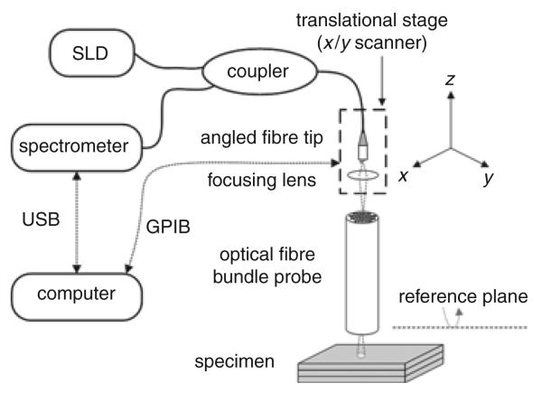
The schematic view of a CP-OCT system with an optical fibre bundle probe is illustrated in Fig. 1. The CP-OCT system comprises a low coherence superluminescence diode (SLD), a 2 × 1 fibre directional coupler, and a spectrometer. We used a broadband 0.8 μm light source with 5 mW output and a high-resolution spectrometer to obtain signal spectra which were subsequently processed for depth imaging. The fibre coupler was used to route the SLD output to an X/Y scanner which was used to scan the beam across the proximal entrance of the fibre bundle. We used 76 mm-long 10 000 and 50 000 pixels fibre optic bundles with a core diameter of ~10 μm. One of the outputs from the fibre coupler was placed at the proximal input of the fibre bundle and was attached to the X/Y scanner for the transverse 2-D scanning. A microscope objective was used to couple the light from the coupler into each fibre pixel. The numerical aperture (NA) and magnification of the objective was 0.25 and 10x, respectively.
Fig. 1.
Experimental setup for FD CP-OCT system with fibre bundle conduit probe
The reference plane for the CP-OCT occurs at the distal end of the fibre bundle where a strong Fresnel reflection occurs at the glass–air interface. The computer controls both the spectrometer (connected by USB cable) and the X/Y scanner (connected by GPIB cable) for data collecting and lateral scanning, respectively. We used a US Air Force resolution target, glass slides, and multilayer polymer tapes to evaluate the probe performance.
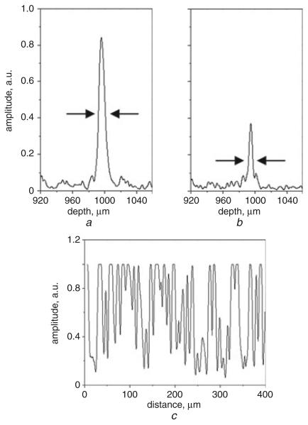
The axial and lateral responses of selected pixel fibres are shown in Fig. 2. The signal amplitude from a silvered plane (mirror) placed 1 mm away from the tip to avoid detector saturation varies from pixel to pixel both in the axial (Figs. 2a and b) and in the lateral (Fig. 2c) direction. The full width half maximum (FWHM) of the resolved envelope is ~9 μm, which is the source coherence length limited axial resolution for fibre pixels having different amplitudes as in Figs. 2a and b. The irregularity of the fibre bundle is presented with missing and/or damaged fibre pixels from the response in Fig. 2c which is laterally scanned for 400 μm along the fibre bundle with 1 μm steps.
Fig. 2.
Measured different pixel responses from mirror
a Fibre pixel 1 (axial response)
b Fibre pixel 2 (axial response)
c Consecutive fibre pixels (lateral response)
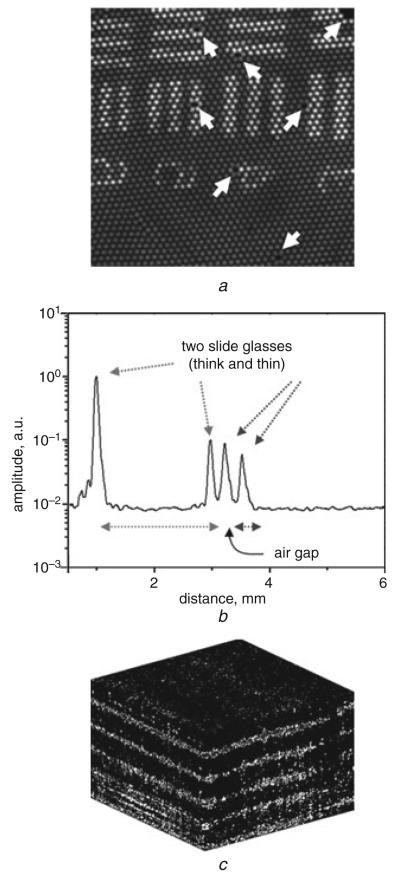
From an en face OCT image of the US Air Force test chart in Fig. 3a, we clearly observe the pixilation effect by each fibre core in which the individual pixel limits the lateral resolution of the image. We can also see several partially or totally damaged and/or missing pixels marked with arrows. Fig. 3b shows an A-scan image of two glass slides where the glass interfaces can be clearly seen. A 3-D cross-sectional image of a multilayer polymer tape is shown in Fig. 3c where the boundaries between different layers can be clearly seen. Factors that degrade image quality are the impact of discrete pixilation of fibres, uneven optical characteristics between each core (as compared in Fig. 2), and the coupling loss due to the mismatches in NAs between the fibre bundle (0.55) and the objective (0.25) in the setup (Fig. 1).
Fig. 3.
Obtained OCT images
a En face image of US Air Force target (2 × 2 mm)
b A-scan image of two glass slides
c 3-D image of polymer layers (0.6 × 0.6 × 0.4 mm)
Conclusion
We have experimentally demonstrated a fibre bundle optic probe based common path OCT system that can be used to obtain scanningless OCT imaging at the distal end of the probe for image-guided microsurgery and diagnostic imaging. We were able to achieve axial and lateral resolutions limited by the coherence length of the source and the size of the fibre pixel of the bundle imager, respectively.
Acknowledgment
This work was supported in part by NIH grant BRP 1R01 EB 007969-01.
Contributor Information
C.G. Song, School of Electronics & Information Engineering, Chonbuk National University, Deokjin-Dong, Jeonju, Jeonbuk 561-756, South Korea
J.U. Kang, Department of Electrical and Computer Engineering, Johns Hopkins University, 3400 N. Charles St., Baltimore, MD 21218, USA
References
- 1.Munce NR, Mariampillai A, Standish BA, Pop M, Anderson KJ, Liu GY, Luk T, Courtney BK, Wright GA, Vitkin IA, Yang VXD. Electrostatic forward-viewing scanning probe for Doppler optical coherence tomography using a dissipative polymer catheter. Opt. Lett. 2008;33:657–659. doi: 10.1364/ol.33.000657. [DOI] [PubMed] [Google Scholar]
- 2.Jung W, McCormick DT, Zhang J, Wang L, Tien NC, Chen Z. Three-dimensional endoscopic optical coherence tomography by use of a two-axis microelectromechanical scanning mirror. Appl. Phys. Lett. 2006;88:163901. [Google Scholar]
- 3.Jafri MS, Farhang S, Tang RS, Desai N, Fishman PS, Rohwer RG, Tang C-M, Schmitt JM. Optical coherence tomography in the diagnosis and treatment of neurological disorders. J. Biomed. Opt. 2005;10:051603. doi: 10.1117/1.2116967. [DOI] [PubMed] [Google Scholar]
- 4.Oron D, Tal E, Silberberg Y. Scanningless depth-resolved microscopy. Opt. Express. 2005;13:1468–1476. doi: 10.1364/opex.13.001468. [DOI] [PubMed] [Google Scholar]
- 5.Spitz J-A, Yasukuni R, Sandeau N, Takano M, Vachon J-J, Meallet-Renault R, Pansu RB. Scanning-less wide-field single-photon counting device for fluorescence intensity, lifetime and time-resolved anisotropy imaging microscopy. J. Microscopy. 2008;229:104–114. doi: 10.1111/j.1365-2818.2007.01873.x. [DOI] [PubMed] [Google Scholar]
- 6.Chen X, Reichenbach KL, Xu C. Experimental and theoretical analysis of core-to-core coupling on fibre bundle imaging. Opt. Express. 2008;16:21598–21607. doi: 10.1364/oe.16.021598. [DOI] [PubMed] [Google Scholar]
- 7.Oh WY, Bouma BE, Iftimia N, Yelin R, Tearney GJ. Spectrally-modulated full-field optical coherence microscopy for ultrahigh-resolution endoscopic imaging. Opt. Express. 2006;14:8675–8684. doi: 10.1364/oe.14.008675. [DOI] [PMC free article] [PubMed] [Google Scholar]
- 8.Ford HD, Tatam RP. Fibre imaging bundles for full-field optical coherence tomography. Meas. Sci. Technol. 2007;18:2949–2957. [Google Scholar]
- 9.Pyhtila JW, Boyer JD, Chalut KJ, Wax A. Fourier-domain angle-resolved low coherence interferometry through an endoscopic fibre bundle for light-scattering spectroscopy. Opt. Lett. 2006;31:772–774. doi: 10.1364/ol.31.000772. [DOI] [PubMed] [Google Scholar]
- 10.Xie T, Mukai D, Guo S, Brenner M, Chen Z. Fiber-optic-bundle-based optical coherence tomography. Opt. Lett. 2005;30:1803–1805. doi: 10.1364/ol.30.001803. [DOI] [PubMed] [Google Scholar]
- 11.Tan KM, Mazilu M, Chow TH, Lee WM, Taguchi K, Ng BK, Sibbett W, Herrington CS, Brown CTA, Dholakia K. In-fibre common-path optical coherence tomography using a conical-tip fibre. Opt. Express. 2009;17:2375–2384. doi: 10.1364/oe.17.002375. [DOI] [PubMed] [Google Scholar]
- 12.Vakhtin AB, Kane DJ, Wood WR, Peterson KA. Common-path interferometer for frequency-domain optical coherence tomography. Appl. Opt. 2003;42:6953–6958. doi: 10.1364/ao.42.006953. [DOI] [PubMed] [Google Scholar]


