Abstract
The development of attention in the infant can be characterized by changes in overall arousal (attentiveness) and by changes in attention's effect on specific cognitive processes (e.g., stimulus orienting, spatial selection, recognition memory). These attention systems can be identified using behavioral and psychophysiological methods. The development of infant attention is thought to be closely related to changes in the neural systems underlying attention control. The recent application of cortical source analysis of event-related potentials (ERP) and structural magnetic resonance imaging (MRI) has led to the identification of some of these the neural systems.
Keywords: development, attention, infancy, cognitive neuroscience, psychophysiology
Research on the early development of attention began in the 1960s with elegant demonstrations that infants could direct their fixation for long periods of time to interesting visual stimuli. When presented with two stimuli, infants often would look longer at one stimulus than the other, and a catalogue of visual preferences was developed for everything from pictures of faces, checkerboard patterns, and geometric shapes to landscape scenes and realistic 3-D objects. The duration of fixation toward these stimuli became the standard measure of infant visual attention. The differential preference for interesting stimuli was understood as an example of infant selective attention. It was noticed very early in this research that the patterns of looking behavior changed significantly over the first 2 years of postnatal life. With increased age, infants paid attention to patterns for longer periods of time and seemed to attend to increasing amounts of visual complexity. These early studies established some basic characteristics of infant attention and introduced many of the tools that are still used today. Many of these techniques were subsequently applied to the study of infant cognitive development in many domains and have been instrumental in establishing the extensive literature on infant cognitive development that exists today (see Colombo, 2002).
These early observations of infants' attention were based on infants' visual behavior, such as where an infant was looking and for how long it looked at a visual stimulus. However, often neural mechanisms were proposed to explain the development of attention. For example, neonatal and early visual functioning were attributed to subcortical structures that were relatively mature at birth and that were known to support such visual behavior in adults and juvenile nonhuman primates. One example is the relatively mature form of the superior colliculus (a subcortical brain structure) at birth in human infants. This brain structure is involved in moving the head and eyes to a sound in the periphery. All parents know that an infant will move its head toward a rattle shaken at the side of its head. The later onset of more mature attention patterns and more selective attention was attributed to developmental advances in brain areas that supported these more complex cognitive skills. For example, the age-related changes in infants' preferences for complex patterns and realistic visual scenes over simple displays was attributed to the increasing specificity of synaptic connections in the primary visual cortex and the corresponding increase in visual acuity. However, a deficit in these neuroscientific explanations of the development of infant attention was a lack of tools for measuring brain structure and brain activity in human infant participants. Only recently has it been possible to link developmental changes in infant attention directly to the neural systems hypothesized to be responsible for this development.
Varieties of Infant Attention
One aspect of attention that shows developmental change in the first 2 years of life is the arousal component that has been called sustained attention (Richards, 2008). The onset of a new stimulus (e.g., a pattern on a computer monitor, a person entering a room, a sound) will result in the infant moving its head and eyes toward that stimulus (stimulus orienting). The infant will keep looking at that stimulus for some period of time. In addition to looking at the stimulus, body changes (posture, facial expression, limb movements), physiological changes (breathing, heart rate), and brain changes (electroencephalogram or EEG) indicate the infant is attentive toward the new stimulus. For example, a large deceleration in heart rate accompanies the onset of a stimulus that elicits orienting and fixation. The heart rate stays below the prestimulus level as long as the infant is attentive. Sustained attention refers to this pattern of fixation, body changes, and physiological reactions. Often these “body signals” diminish although the infant will continue to look toward the stimulus. This period has been called attention termination, and represents a time when the infant is no longer attentive even though he or she continues to look toward the stimulus. Several studies have shown that infants are differentially responsive to stimuli when they are in sustained attention versus attention termination. In sustained attention they are better able to resist peripheral distracters and show superior memory encoding and enhanced identification of briefly presented stimuli. Sustained attention is likely controlled by neurotransmitter systems in the brain that control arousal or attentiveness (Richards, 2008).
Studying Brain Activity Related to Infant Attention
Attention has been studied in infants in a variety of ways (see reviews in Richards, 2008, 2010). How can we study the brain activity related to this attention? One way that has been used is the “modified oddball” procedure. This procedure involves presenting a series of brief visual stimuli frequently (“standard”), infrequently (“oddball”), or as novel stimuli (“novel”). Adults and children easily recognize the oddball and novel stimuli and can identify them in later memory tasks. To do this, a rudimentary form of recognition memory must occur, and attention is elicited by the infrequent and novel stimuli. This procedure is done with infants by first making sure they are familiar with the standard and oddball stimuli by showing them for 20 to 30 seconds. Then one of the familiar stimuli serves as the standard stimulus (“frequent familiar”), one of the familiar stimuli serves as the oddball stimulus (“infrequent familiar”), and a set of stimuli that have never been seen by the infant are the novel stimuli (“infrequent novel”). This modified procedure insures that the infant is equally familiar with the standard and oddball stimulus before the presentation.
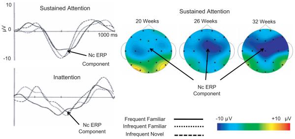
This procedure often is used in psychophysiological studies. Psychophysiology uses noninvasive physiological recordings to make inferences about psychological activity. The EEG is recorded simultaneously with the presentation of the brief visual stimuli. The EEG signal is averaged at the point of time at which the visual stimulus is presented, and the resulting average is called an event-related potential (ERP). The EEG is a measure of electrical activity recorded on the scalp that is generated by electrical activity of the brain. Thus any changes in brain activity closely linked to the psychological processes occurring in response to the brief visual stimuli will affect the ERP response. An ERP component termed the Nc (for negative central) typically occurs in response to these briefly presented stimuli. Figure 1 (left panels) shows the Nc response at the Cz electrode recording site for infants from 4.5 to 7.5 months of age. This ERP component is maximal over frontal and central scalp areas for all three stimulus types and tends to be larger for novel stimuli.
Fig. 1.
The Nc (negative-central) ERP (event-related potential) component in the infant oddball procedure. The oddball procedure presents familiar and novel stimuli to the infant briefly, and the ERP is recorded at the onset of the stimuli. The Nc is shown on the Cz electrode recording, separately for attentive and inattentive periods and the frequent familiar, infrequent familiar, and infrequent novel stimuli (left panels). Scalp topographical potential maps (upper right panels) show the Nc component occurring during sustained attention for infants at three ages.
The role of attention on brain activity in the modified oddball procedure is illustrated in a study of infants' brain responses to briefly presented visual stimuli (Richards, 2003). Infants at ages 4.5, 6.0, and 7.5 months were presented with a Sesame Street movie called “Follow that Bird” that elicited periods of sustained attention and attention termination. The stimuli in the modified-oddball procedure were then briefly presented, overlaid on the attention-eliciting movie. The brief stimuli were presented either when the infant was attentive (sustained attention) or inattentive (preattention or attention termination). Figure 1 (left panels) shows the Nc response for the participants separately during attention and inattention and for the three stimulus types. There was a larger Nc during sustained attention, and this was true for all three familiarity-type stimuli. Figure 1 (right panels) shows scalp topographical potential maps during sustained attention. There was an increase in the amplitude of the Nc response from 4.5 to 6 to 7.5 months, but only for the brief stimuli presented during sustained attention. The Nc is believed to signal the initial orienting of attention to interesting stimuli. These findings show that this orienting is enhanced during the sustained-attention phase—that is, when the brain is aroused and the neurotransmitter systems controlling arousal are active. The brain areas responding to these brief stimuli show more coherent, more organized, and larger-amplitude neural activity. We assume that there are changes in the neurotransmitter systems controlling attention and that these changes in the neurotransmitter systems over this age range affect the extent to which the specific brain area will respond.
New Tools for Measuring the Neural Basis of Development in Infant Attention
Nearly all neuroscientific explanations of attention development have relied either on measurement of brain function in animal models or on measurements of overt behavior putatively linked to brain areas (marker tasks). More recently, the field of cognitive neuroscience has been advanced significantly with the availability of tools that measure brain activity directly when adults are engaging in cognitive tasks. The first wave of cognitive neuroimaging studies started with PET (positron emission tomography), an invasive method that required an infusion of radioactive material. More recently, noninvasive neuroimaging tools have been developed for use with infants. One approach is to apply near-infrared optical spectroscopy (NIRS, or optical topography, OT). The NIRS measures blood flow in cortical areas near the surface of the cortex with infrared detector/emitter pairs placed on the surface of the head. Areas of the brain that are active during a psychological task will receive increased blood flow during and following that task, so that the blood-flow changes are a measure of which areas of the brain are involved in the psychological processes for that task. NIRS is currently being developed for use with infants in psychological tasks (see Aslin & Mehler, 2005). A second technique is cortical source analysis of scalp electrical currents (EEG, ERP). The source analysis technique identifies putative sources in the cortex that could create the topographical distribution of the surface-recorded EEG, with EEG being measured with high-density scalp electrode configurations (see Reynolds & Richards, 2009 for methods and assumptions of this technique). The activity of these cortical sources can be tracked over time with adequate resolution to fully capture the brain electrical activity (e.g., milliseconds temporal resolution for EEG). This activity can be averaged across trials to identify the brain response related to experimental events or psychological processes. Both techniques are useful for infant participants because they allow the measurement of brain activity in individuals while they participate in psychological tasks. Both use measurement tools “outside the head” that are direct measures of brain activity generated “inside the head” and give information about where the activity is generated.
We will describe briefly our current attempts to refine the cortical source analysis technique for infant participants (Reynolds & Richards, 2009; Richards, 2010). The EEG is electrical activity occurring on the scalp that is generated by neural activity occurring in the brain. Cortical source analysis tests hypotheses about which cortical areas that generate the observed topographical distribution of current flow on the scalp (i.e., the observed EEG). This quantitative technique requires a model of the electrical properties of the head to calculate how current would flow from the cortical sources to the scalp. The simulated topographical distribution of the scalp current is compared to the observed current flow on the scalp. The modeling process is iterated until the model that provides the best fit with the observed data is determined.
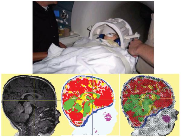
Cortical source analysis procedures have been used almost exclusively in adult participants, and the parameters used in current models are based upon adult heads. Adult heads are different than infant heads in many ways. The size of the head, the relative thickness of the skull, the topological placement of the cortical areas relative to external skull landmarks, and changes in myelination and brain structure are all differences that could affect the assumptions of this technique. One way to get around this issue is to use cortical source analysis with models of infant heads rather than models of adult heads. This is currently being accomplished by doing structural MRI scans of individual infants and producing a head model based on that participant. Figure 2 shows an infant being placed into a scanner. The MRI is done when the infant is sleeping and structural scans are obtained. The MRI volumes are used to segment the head into tissue types (cerebrospinal fluid, white matter, gray matter, skull, scalp, muscle, eyes) and quantitative models that describe the electrical properties of each element inside the head are produced. Figure 2 (bottom panels) shows the MRI for the infant being scanned, the segmented head model, and the high-resolution 3-D wireframe model representing the electrical model of this infant's head. This infant participated in studies of the recognition of brief visual stimuli, spatial cueing, and hidden objects. The EEG and ERP recorded in the psychophysiological study were analyzed with cortical source analysis, and the ongoing activity in the cortex that corresponded to the infant's attention toward the stimuli in the psychological task was identified.
Fig. 2.
Structural magnetic resonance imaging (MRIs) for cortical source analysis of psychophysiological EEG (electroencephalogram) and ERP (event-related potentials). The top panel shows a 7.5-month-old (32 weeks) infant being placed into a Siemens 3T Trio Scanner. The procedure results in a MRI volume for the child's head, (bottom left), a MRI volume that segments the head materials (bottom center), and a high-resolution 3-D wireframe model representing the electrical model of the head used in computer programs for source analysis (bottom right).
Neural Basis of Infant Attention and Recognition Memory
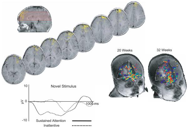
The potential of the cortical source analysis procedure is illustrated in recent studies of visual attention and recognition memory. The cortical sources of the Nc ERP component were investigated in two recent studies using the modified-oddball procedure. In the first, cortical source analysis was used to hypothesize the locations of cortical areas that were likely sources of the current recorded on the scalp (Reynolds & Richards, 2005). Figure 3 shows the location of these dipoles. The dipole locations represent the modeled sources of the Nc ERP component. The dipoles occurred in a wide range of areas in the frontal and prefrontal cortical areas, probably a result of the broad distribution of the Nc ERP component on the scalp. However, the dipoles seemed to be centered in the anterior cingulate and basomedial/basolateral prefrontal cortex. The time course of the activity of these cortical sources was examined. Figure 3b shows the averaged activity of the dipole sources when the novel stimulus was presented and the infant was either attentive or inattentive. The onset of the activity of these dipoles when the infants were attentive was nearly immediate (around 200 milliseconds) and was sustained throughout the period of time corresponding to the time course of the Nc ERP component. The activity of the dipole sources during inattentive periods showed a peak only about the same time as the peak of the Nc component. This finding suggests that the brain areas that generate the scalp-recorded Nc ERP component are shorter in onset latency and are enhanced during sustained attention. This implies that this brain area acts in a more organized and efficient manner during sustained attention. Given that sustained attention reflects the ongoing activity of the neurotransmitter systems involved in arousal, this finding implies that arousal leads to increased efficiency of neural transmission in these brain areas. The functional association of the activity of these cortical sources with attention implies that these brain areas are closely involved in the initial orienting of attention to novel stimuli.
Fig. 3.
Cortical sources for the Nc (negative-central) ERP (event-related potential) in infants. The sources for the Nc ERP component are shown and occur over a wide range of ages in the prefrontal cortex (top left). The bottom left diagram shows the activity of the brain areas to a novel stimulus presented during sustained attention or when the infant is inattentive. The right-hand images show common brain locations for the oddball procedure and a paired-comparison procedure for infants at 20 weeks (4.5 months) and 32 weeks (7.5 months) of age.
A second study (Reynolds, Courage, & Richards, 2009) shows that the cortical areas controlling attention to these stimuli may be similar to cortical areas controlling recognition memory. The paired-comparison/recognition-memory procedure is commonly used to measure infant recognition memory. Presented simultaneously with a familiar and a novel visual stimulus, an infant will tend to look more toward the novel stimulus. The amount of the novelty preference is often used as an index of an infant's visual recognition memory. In this study, the modified-oddball presentations of the briefly presented visual stimuli were interspersed with paired-comparison presentations of the novel and familiar stimuli. There were three notable findings. First, we found that the magnitude of novelty preference shown during paired-comparison trials was closely related to the amplitude of the Nc ERP component that had been demonstrated following individual ERP presentations of each stimulus. Second, in this study the ERP during the paired-comparison procedure was measured. An ERP component that was highly similar to the Nc ERP found in traditional ERP studies (latency, amplitude, relation to experimental events, scalp topography) was found. The Nc recorded during the paired-comparison presentations was positively correlated with the amount of time spent looking at one stimulus or another. The third interesting finding was the age differences in the distribution of the cortical sources for the Nc ERP. The cortical sources for the Nc ERP component found in the modified-oddball and paired-comparison presentations shared common locations, primarily in the basal prefrontal cortex area. Figure 3 (bottom right panels) shows the locations of the common cortical sources for the oddball and paired-comparison procedures for 4.5- and 7.5-month-old infants. The sources for the youngest age were scattered over a wide lateral area of prefrontal cortex whereas the sources for the oldest age were concentrated near the basomedial prefrontal cortex.
The age changes for the Nc shown in Figure 1 may be due to the brain areas becoming more organized, less widely scattered, and more efficient. This would imply that the brain systems controlling attention are less efficient or less localized in the younger infants. There are also significant structural and neurophysiological changes that are occurring across this age range. We should be able to distinguish the structural changes with neuroimaging procedures (voxel-based white-matter counts or diffusion tensor imaging) from the functional changes that occur in this task. This will enable us to test specific hypotheses about the neural basis of the development of the Nc ERP response and attention-linked recognition memory processing. It is also possible that some of the changes in the Nc ERP component across this age range are due to the electrical properties of the head at different ages. This would not detract from the close functional relationship between the Nc ERP component and the psychological processes in this task.
These findings show that brain activity in the paired-comparison procedure shows a rapid orienting to novel stimuli. This is similar to the Nc component found in the modified-oddball procedure. This implies that this novelty detection may be the basis for neural control of the “novelty preference” phenomenon The common cortical sources for brain activity in the paired-comparison and modified-oddball presentations, and the close link between brain activity in the modified-oddball presentations and the novelty-preference measure of recognition memory, imply that attention and recognition memory are closely associated in infant participants. Novel objects elicit a large neural orienting response and the paired-comparison procedure takes advantage of the novelty-preference behavior that is driven by this attention response. These results continue to show the importance of sustained attention in infant cognitive processes, as these recognition memory results are shown primarily when the infant is in the sustained-attention phase (see similar findings in Richards, 2008). The paired-comparison procedure was first developed in the 1960s by the pioneers of research on infant cognition. The conclusions about infant attention development drawn from that early work were based primarily on infant looking. The cognitive processes that are active in this task can now be examined with tools that enable the identification of the brain areas that control behavior. This will help us provide testable neuroscientific explanations for how attention develops in the early months of life.
Recommended Reading
- Aslin RN, Mehler J. A review of studies and methods for doing near-infrared spectroscopy (NIRS) in infant participants. 2005 See References. [Google Scholar]
- Colombo J. A cognitive neuroscience interpretation of infant attention. 2002 See References. [Google Scholar]
- Reynolds GD, Richards JE. A discussion of issues and methods for doing cortical source analysis of infant ERP. 2009 See References. [Google Scholar]
- Richards JE. A review of current research on infant attention. 2008 See References. [Google Scholar]
- Richards JE. Issues and methods for studying the role of brain development in infant attention development. 2010 See References. [Google Scholar]
Acknowledgments
Funding
Preparation of this article and the work described herein was supported by a MERIT Award, HD18942 from the National Institutes of Health (NIH). The work reported in this paper also was supported by NIH Grant R03-HD05600 awarded to Greg Reynolds and by Natural Sciences and Research Council of Canada, OPG0093057, to Mary L. Courage.
References
- Aslin RN, Mehler J. Near-infrared spectroscopy for functional studies of brain activity in human infants: promise, prospects, and challenges. Journal of Biomedical Optics. 2005;10:011009-1–011009-3. doi: 10.1117/1.1854672. [DOI] [PubMed] [Google Scholar]
- Colombo J. Infant attention grows up: The emergence of a developmental cognitive neuroscience perspective. Current Directions in Psychological Science. 2002;11:196–200. [Google Scholar]
- Reynolds GD, Courage ML, Richards JE. Infant attention and visual preferences: Converging evidence from behavior, event-related potentials, and cortical source localization. 2009 doi: 10.1037/a0019670. Manuscript submitted for publication. [DOI] [PMC free article] [PubMed] [Google Scholar]
- Reynolds GD, Richards JE. Familiarization, attention, and recognition memory in infancy: An ERP and cortical source localization study. Developmental Psychology. 2005;41:598–615. doi: 10.1037/0012-1649.41.4.598. [DOI] [PMC free article] [PubMed] [Google Scholar]
- Reynolds GD, Richards JE. Cortical source analysis of infant cognition. Developmental Neuropsychology. 2009;3:312–329. doi: 10.1080/87565640902801890. [DOI] [PMC free article] [PubMed] [Google Scholar]
- Richards JE. Attention affects the recognition of briefly presented visual stimuli in infants: An ERP study. Developmental Science. 2003;6:312–328. doi: 10.1111/1467-7687.00287. [DOI] [PMC free article] [PubMed] [Google Scholar]
- Richards JE. Attention in young infants: A developmental psychophysiological perspective. In: Nelson CA, Luciana M, editors. Handbook of developmental cognitive neuroscience. MIT Press; Cambridge, MA: 2008. pp. 479–479. [Google Scholar]
- Richards JE. Attention in the brain and early infancy. In: Johnson SP, editor. Neoconstructivism. The new science of cognitive development. Oxford University Press; New York: 2010. pp. 3–37. [Google Scholar]


