Abstract
Growth in aerobic environments has been shown to generate reactive oxygen species (ROS) and to cause oxidative stress in most organisms. Antioxidant enzymes (i.e., superoxide dismutases and hydroperoxidases) and DNA repair mechanisms provide protection against ROS. Acid stress has been shown to be associated with the induction of Mn superoxide dismutase (MnSOD) in Lactococcus lactis and Staphylococcus aureus. However, the relationship between acid stress and oxidative stress is not well understood. In the present study, we showed that mutations in the gene coding for MnSOD (sodA) increased the toxicity of lactic acid at pH 3.5 in Streptococcus thermophilus. The inclusion of the iron chelators 2,2′-dipyridyl (DIP), diethienetriamine-pentaacetic acid (DTPA), and O-phenanthroline (O-Phe) provided partial protection against 330 mM lactic acid at pH 3.5. The results suggested that acid stress triggers an iron-mediated oxidative stress that can be ameliorated by MnSOD and iron chelators. These findings were further validated in Escherichia coli strains lacking both MnSOD and iron SOD (FeSOD) but expressing a heterologous MnSOD from S. thermophilus. We also found that, in E. coli, FeSOD did not provide the same protection afforded by MnSOD and that hydroperoxidases are equally important in protecting the cells against acid stress. These findings may explain the ability of some microorganisms to survive better in acidified environments, as in acid foods, during fermentation and accumulation of lactic acid or during passage through the low pH of the stomach.
Superoxide dismutases (SODs; EC 1.15.1.1) are metalloenzymes that catalyze the conversion of the superoxide anion to hydrogen peroxide and dioxygen (41). Four types of SOD have been characterized, which differ in their metal cofactors (i.e., copper and zinc [Cu/ZnSOD], manganese [MnSOD], iron [FeSOD], or nickel [NiSOD]) (30, 65). These enzymes are found across a broad range of organisms, and each organism can use one or more types of the enzyme to meet their antioxidant needs (30). For example, Escherichia coli possesses three isoforms: MnSOD, FeSOD, and Cu/ZnSOD (9, 34, 64).
Lactic acid bacteria (LAB) are acid-tolerant organisms that require sugar as a source of carbon and energy, generating mainly lactate as a final fermentation product. In particular, the homofermentative organism Streptococcus thermophilus, either alone or together with other species, is extensively employed in the production of yoghurt and other dairy products in which acidification guarantees preservation. LAB are constantly faced with environmental conditions that can affect their growth and viability. Two of the major threats are acid stress caused by organic acids generated during the fermentation process and oxidative stress caused by the generation of reactive oxygen species (ROS) during growth in the presence of oxygen.
The majority of the LAB possess an inducible acid tolerance response (ATR), also known as the acid adaptive response, which improves the survival of adapted cells upon exposure to a lethal acid challenge compared to that of the unadapted cells. The induction of the ATR often protects the cells not only from acid challenge, but also from other stresses (18, 24, 61). Regarding the oxidative stress, LAB are classified as catalase (hydroperoxidases) negative and microaerophilic (7). They lack a functional electron transport chain, but they can grow in the presence of molecular oxygen. However, they contain enzymes that use oxygen, such as pyruvate oxidases (17, 51, 52), NADH oxidases that produce H2O2, and NADH peroxidases able to break down peroxides (17, 26, 33, 37, 56, 57). Consequently, they generate ROS during their growth in aerobic environments. To offset the harmful effects of ROS, most organisms have evolved protective mechanisms that utilize antioxidant enzymes, such as superoxide dismutases and hydroperoxidases (i.e., catalases and peroxidases or KatE and KatG), which scavenge superoxide radicals and hydrogen peroxide, respectively, and thus prevent the formation of HO. via Fenton chemistry (21).
In most Streptococcus and Lactococcus spp., elimination of ROS conforms to this general antioxidant defense system since both genera possess MnSOD (44, 48); however, they lack hydroperoxidases. Instead of using SOD, Lactobacillus plantarum developed an alternative nonenzymatic defense system that involves the accumulation of high intracellular concentrations of manganese ions, which can scavenge O2− (4). Previous work has shown that S. thermophilus possesses only one type of SOD, the Mn-containing enzyme (MnSOD) (3, 15, 44). The gene encoding MnSOD (sodA) from S. thermophilus AO54 has been characterized, cloned, and heterologously expressed in other bacteria (3, 13, 14). Unlike most sodA genes, the S. thermophilus sodA gene is constitutively expressed and is not induced by oxygen or the redox cycling compound paraquat (15). This antioxidant enzyme (MnSOD) was shown to be essential for the aerobic growth of S. thermophilus (3). Consequently, the activity of MnSOD was found to increase in a growth-dependent fashion, increasing 3- to 4-fold upon entry into stationary phase (15), which may be related to regulation of manganese transport (32).
Stress responses are complicated processes that involve the synthesis of a variety of proteins (8, 60). Only a few of the putative acid resistance proteins have been characterized (8, 18, 61). Here we present evidence showing that MnSOD and hydroperoxidases provide protection against acid stress. A plausible mechanism is proposed to explain the relationship between acid stress and oxidative stress and how antioxidant enzymes confer a survival advantage against both types of stress.
MATERIALS AND METHODS
Bacterial strains and media.
The bacterial strains and plasmids used in this study are listed in Table 1. E. coli strains were grown at 37°C in Luria-Bertani (LB) medium (47) supplemented with the appropriate antibiotics. The antibiotics used were chloramphenicol (20 μg/ml), kanamycin (50 μg/ml), ampicillin (100 μg/ml), and erythromycin (200 μg/ml).
TABLE 1.
Bacterial strains and plasmids used in this study
| Strain or plasmid | Relevant characteristic(s) | Source or reference |
|---|---|---|
| Strains | ||
| Escherichia coli K-12 | ||
| NC906a | OX362A; E. coli K-12 with ΔsodA Φ(sodB-kan)1-Δ2 Kmr | 55 |
| NC906/pSKSODA | As NC906, but contains pBluescript II KS(+) with 1.2-kb sodA from S. thermophilus AO54 | 3, 13 |
| UM2 | F−leuB6 proC83 purE42 trpE28 his-208 argG77 ilvA681 met-160 thi-1 ara-14 lacY1 galK2 xyl-5 mtl-1 azi-6 rpsL109 tonA23 tsx-67 supE44 malA38 xthA katE2 katG15 | 38 |
| UM2A | As UM2, but Φ(sodA-lacZ)49 Cmr Lac+ | 49 |
| UM2B | As UM2, but Φ(sodB-kan)1-Δ2 Kmr Lac− | 49 |
| UM2AB | As UM2, but Φ(sodA-lacZ)49 Cmr Φ(sodB-kan)1-Δ2 Kmr Lac+ | 49 |
| Streptococcus thermophilus | ||
| AO54 | Wild-type industrial strain | 43 |
| KO2-4 | As AO54, but ΔsodA | 3 |
| Plasmids | ||
| pSodA | 1.2-kb sodA fragment from S. thermophilus cloned into pTRK563 | 13 |
| pSKSODA | 1.2-kb sodA fragment from S. thermophilus cloned into EcoRI pBluescript II KS(+) | 3, 13 |
North Carolina State University culture collection.
S. thermophilus AO54 (43) was grown at 37°C in Difco Lactobacilli MRS broth (19). When required, erythromycin (2 μg/ml) was added to S. thermophilus cultures. Solid media for plating were prepared by adding 1.5% agar to the appropriate liquid media.
Bacterial transformations.
E. coli strains were transformed via electroporation using a Bio-Rad Gene Pulser (Bio-Rad, Richmond, CA) according to the manufacturer's instructions.
Sources of chemicals and enzymes.
dl-Lactic acid (89%), 2,2′-dipyridyl (DIP), diethylenetriamine-pentaacetic acid (DTPA), O-phenanthroline (O-Phe), and all antibiotics used were purchased from Sigma (St. Louis, MO). All other chemicals and the bacteriological media were purchased from Fisher Scientific (Pittsburgh, PA).
Acid tolerance studies.
To prepare exponentially growing cells of S. thermophilus or E. coli, overnight cultures were used to inoculate appropriate media to an initial optical density at 600 nm (OD600) of 0.05. Cells were allowed to grow at 37°C, and changes in OD600 were monitored over time until the cultures reached an OD600 of 0.2 to 0.4. The cells were then harvested by centrifugation, washed, and resuspended in the same medium preacidified with dl-lactic acid (330 mM, pH 3.5).
In the acid preadaptation experiments, cells were incubated for 30 min in the appropriate media preacidified with dl-lactic acid (33 mM, pH 5.5) before they were subsequently challenged with dl-lactic acid (330 mM, pH 3.5) as described above. Aliquots of the cell culture were withdrawn at various time intervals (0, 30, 60, 90, and 120 min), diluted in the nonacidified media (pH 6.5 to ∼7.0), and spread on LB or MRS agar plates. Plates were incubated at 37°C and counted (CFU/ml) after 24 h for E. coli or 48 h for S. thermophilus.
Effect of iron chelators.
Increasing concentrations of the iron chelators (2, 2′-dipyridyl, diethylenetriamine-pentaacetic acid, and O-phenanthroline) were added to exponentially growing S. thermophilus cells during acid challenge in MRS medium containing 330 mM dl-lactic acid or HCl, (pH 3.5) at 37°C. At different time intervals, 10-μl aliquots were removed, spotted on solid medium, and incubated at 37°C.
Metal analyses.
Overnight culture of S. thermophilus AO54 was used to inoculate MRS medium to an initial OD600 of 0.05. Cells were grown to an OD600 of 1 before they were harvested by centrifugation, washed in sterile medium, and subsequently challenged in 20 ml of MRS medium preacidified with dl-lactic acid (330 mM, pH 3.5) or HCl (330 mM, pH 3.5) for 30 min. At the end of the acid challenge, cells were harvested, washed with deionized distilled water, ashed, and dissolved by boiling in 1 ml of 10% nitric acid. Inductively coupled plasma-mass spectrometry was subsequently performed.
Reproducibility.
All results presented are the means of triplicate values. Two independent replicate assays were performed, and the variations were less than 10%. Statistical analysis and graphical representations were performed using OriginLab Corporation software (Northampton, MA).
RESULTS
Can MnSOD protect S. thermophilus against acid stress?
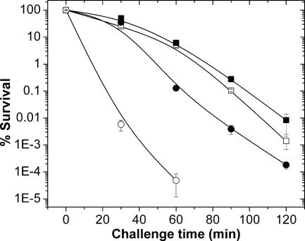
Acid stress has been suggested to lead to oxidative stress (16, 48); however, a conclusive relationship between the two stresses is lacking. In this study, we hypothesized that antioxidant enzymes like superoxide dismutases (SODs) and hydroperoxidases must have a role in protecting the cells against acid stress. Thus, we evaluated the physiological role of MnSOD in protecting S. thermophilus against acid stress. We used both acid-adapted and nonadapted cells from exponentially growing cultures of the wild-type (WT) S. thermophilus strain AO54, which contains the MnSOD gene (sodA), and its isogenic ΔsodA mutant strain (KO2-4), which lacks MnSOD (Fig. 1). Nonadapted cells were directly exposed to dl-lactic acid (330 mM, pH 3.5), while adapted cells were first incubated in a nonlethal concentration of dl-lactic acid (33 mM and pH 5.5) to trigger the acid tolerance response (ATR) prior to exposure to dl-lactic acid (330 mM, pH 3.5). By using both adapted and nonadapted cells, we were able to differentiate between resistance to acid stress and the ATR response.
FIG. 1.
Response of S. thermophilus to lactic acid stress. Unadapted (open symbols) and adapted (closed symbols) cells of exponentially growing S. thermophilus parent strain A054 (□, ▪) and its isogenic ΔsodA mutant strain, KO2-4 (○, •), preexposed or not for 30 min to 33 mM dl-lactic acid (pH 5.5), were challenged in MRS medium containing 330 mM dl-lactic acid (pH 3.5). At specific time intervals, samples were diluted and plated on agar medium to monitor cell viability. The data are means of triplicate points.
Figure 1 shows that exponential-phase cells of S. thermophilus KO2-4 (ΔsodA) were more sensitive to lactic acid-acidified medium (pH 3.5) than cells of its isogenic wild-type strain. Data in Fig. 1 also show that both wild-type (WT) and ΔsodA cultures were able to mount an adaptive ATR. However, in adapted cells, the mutant was still more sensitive to dl-lactic acid (330 mM, pH 3.5) than the WT (compare solid circles and solid squares in Fig. 1).
Is lactic acid toxicity related to iron-mediated Fenton chemistry?
In this part of the study, we examined the effect of added lactate or HCl on the intracellular levels of iron in S. thermophilus AO54. As described in Materials and Methods, the cells were grown, collected, and exposed to preacidified (i.e., 330 mM dl-lactic acid or HCl, pH 3.5) MRS medium. The MRS medium used contained 10.56 μM total iron (i.e., 0.59 μg/ml). The concentrations of iron in cells exposed to either lactate or HCl were 3.09 ± 0.8 ng and 3.68 ± 0.4 ng iron/OD600, respectively, while the iron content in untreated cells was 4.02 ± 0.9 ng of iron/OD600. It is clear that total intracellular iron content of acid-exposed cells was not significantly different from that of the untreated control cells. These results, however, do not differentiate between free and bound iron in the cells.
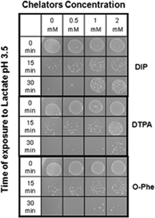
It has been shown that lactate stimulates fibroblast proliferation (62) and wound healing (59), enhances iron bioavailability in foods (45), and increases iron absorption by the human colon carcinoma cell line (Caco-2 cells) (10). Furthermore, lactic acid has been shown to chelate Fe3+ in a 1:1 ratio (29), and that lactate-iron complex can generate hydroxyl radicals (2). Therefore, we decided to verify the involvement of iron and Fenton chemistry in lactate toxicity by examining the effects of iron chelators on acid toxicity. We employed chelators that are known to be able to chelate intracellular iron: 2, 2′-dipyridyl (DIP), diethylenetriamine-pentaacetic acid (DTPA), and O-phenanthroline (O-Phe). Data in Fig. 2 indicate that the removal of intracellular free iron provided partial protection against lactate toxicity. It should be noted that the permeability and the iron binding affinity of the chelators used in this study are, most likely, not identical. Therefore, the results in Fig. 2 qualitatively demonstrate the involvement of free intracellular iron in the toxicity of lactic acid under the experimental conditions employed.
FIG. 2.
Effect of iron chelators in protecting S. thermophilus KO2-4 against lactic acid toxicity. Unadapted exponentially growing cells of S. thermophilus KO2-4 (AO54 ΔsodA) were exposed at 37°C in MRS medium containing 330 mM dl-lactic acid (pH 3.5) and in the presence of increasing concentrations of chelators (2, 2′-dipyridyl, diethylenetriamine pentaacetic acid, and O-phenanthroline). Ten-microliter aliquots were removed at 0, 15, and 30 min from the different treatments, spotted on solid medium, and incubated at 37°C as described in Materials and Methods.
Can heterologous MnSOD protect the sodA sodB mutant of E. coli against acid stress?
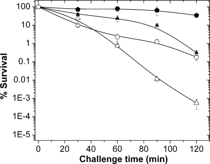
To further corroborate the contribution of MnSOD to acid resistance, we used the sodA gene from S. thermophilus to complement E. coli NC906, a strain lacking both of the endogenous Mn- and FeSOD genes (sodA sodB). Data in Fig. 3 show that the heterologous MnSOD was able to protect E. coli NC906 cells against acid stress.
FIG. 3.
Effect of heterologous MnSOD from S. thermophilus AO54 on the survival and adaptative response of the sodA sodB mutant of E. coli (NC906) exposed to lactic acid stress. Exponentially growing cells of E. coli NC906 (▵, ▴) and NC906/pSODA (○, ) were preexposed to 33 mM dl-lactic acid (pH 5.5) (closed symbols) or not exposed (open symbols). After 30 min of treatment, cells (preexposed or not exposed) were resuspended in MRS medium containing 330 mM dl-lactic acid (pH 3.5). At specific time intervals, samples were diluted and plated on LB agar medium to monitor cell viability. The data are means of triplicate points.
Can FeSOD protect E. coli against acid stress?
The Mn- and Fe-SODs of E. coli are highly homologous, and the levels of coordination of the metals in the active sites are nearly identical (22). Both enzymes are equally important in protecting E. coli against oxygen toxicity (25, 30, 49). Therefore, it is expected that FeSOD would have the same role as MnSOD in protecting E. coli against acid stress. For this part of the study, we used an E. coli strain (UM2) deficient in katG and katE (38), since previous studies have found the accumulation of weak acids induces catalase expression (50). Thus, we compared the roles of FeSOD and MnSOD in acid stress using isogenic UM2 strains harboring mutations in sodA, sodB, or both (49) (Table 1).
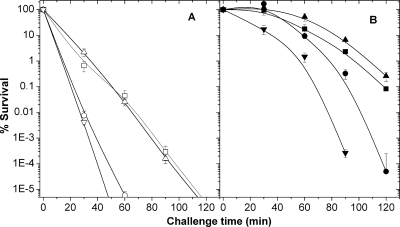
Figure 4 shows that strains lacking sodA (UM2A) or both sodA and sodB (UM2AB) were more sensitive to acid stress than the parent strain (UM2). In contrast, the loss of sodB (UM2B) did not result in a greater sensitivity to lactic acid than that seen in the SOD-proficient strain (UM2). However, cells lacking both sodA and sodB (UM2AB) were more sensitive to lactic acid than cells lacking only sodA (UM2A). These data demonstrated that FeSOD is not as efficient as MnSOD in protecting the cells against acid stress.
FIG. 4.
Roles of MnSOD and FeSOD in the absence of hydroperoxidases (KatG− and KatE−) on the survival and adaptative response of exponentially growing E. coli cells exposed to lactic acid stress. (A) Cells were not preadapted. (B) Cells were preadapted by exposure to 33 mM dl-lactic acid (pH 5.5) for 30 min. The unadapted and adapted cells were resuspended in MRS medium containing 330 mM dl-lactic acid (pH 3.5). At specific time intervals, samples were diluted and plated on LB agar medium to monitor cell viability. ▪, parent Kat− strain (UM2); •, SodA− Kat− (UM2A); ▴, SodB− Kat− (UM2B); and ▾, SodA− SodB− Kat− (UM2AB). The data are means of triplicate points.
Can hydroperoxidases protect E. coli against acid stress?
Data in Fig. 3 and 4B (compare the lines for preadapted NC906 and UM2) show that deficiency of SODs (NC906) or hydroperoxidases (UM2) resulted in equal losses in viable counts after 120 min of exposure to 330 mM lactic acid at pH 3.5 (i.e., a loss of 2.7 versus 3.0 logs, respectively). The data clearly suggest that both SODs and hydroperoxidases are important in protecting the cells against acid stress.
DISCUSSION
The ability of microorganisms to adapt and survive high-acid/low-pH conditions is essential for their viability in acid foods and/or during passage through the acidic environment of the stomach. This adaptive ability is essential for both beneficial and pathogenic organisms. Exposure to mild acidic conditions triggers an adaptive response also called the acid tolerance response (ATR), in which the cells adjust the expression of several genes required for survival in the hostile high-acid environment. Proteins whose expression is increased during ATR include F1Fo-ATPase proton pumps, membrane proteins, DNA and protein repair enzymes, etc. Indeed, the role of acid pH in inducing the proton-translocation F1Fo-ATPase operon has been demonstrated in both Gram-positive and Gram-negative organisms (24, 36, 39).
MnSOD has been shown to be induced under low-pH conditions in Lactococcus lactis (48), Streptococcus mutans (53), and Staphylococcus aureus (16). Additionally, accumulation of weak acids in the culture medium has been shown to induce the catalase genes in E. coli (50) and to induce both catalase and superoxide dismutase in Listeria monocytogenes (20). Alignment of amino acid sequences of MnSODs from different prokaryotes and eukaryotes shows a high degree of homology, a highly conserved active center (22), and a conserved catalytic function(s) (i.e., to disproportionate O2.− to H2O2 and O2) (41). The role of MnSOD in protecting cells against oxidative stress is widely understood and accepted (25). However, its role in protecting the cells against acid stress has yet to be elucidated.
In this study, we tested the hypothesis that MnSODs also protect cells against acid stress and that free iron plays an important role in cell death during acid exposure. Our data showed that exponential-phase cells from sodA mutant strains of S. thermophilus and E. coli (i.e., KO2-4 and NC906) were less tolerant to pH 3.5 than their isogenic counterparts expressing sodA (Fig. 1 and 3). Furthermore, the data showed that MnSOD, regardless of the origin of the sodA gene, has a role in protecting E. coli against acid stress (Fig. 3 and 4). We also demonstrated that the addition of iron chelators (Fig. 2), and the presence of hydroperoxidases (KatG and KatE) (Fig. 3 and 4) provided significant protection against acid toxicity. Taken together, these findings strongly suggest that acid toxicity is mediated by the greater availability of free iron that can react with the partially reduced oxygen species (O2− and H2O2) to cause the generation of the damaging hydroxyl radical (HO.).
It was interesting and unexpected to discover that FeSOD was not as effective as MnSOD in protecting E. coli against acid stress (Fig. 4). Thus, the loss of sodA (UM2A) in acid-preadapted cells (Fig. 4B) resulted in a 7-log reduction in cell viability after 120 min of acid challenge, while the loss of sodB (UM2B) resulted in ∼2.9 logs of reduction, which is similar to that seen in the SOD-competent cells (i.e., 3-log reduction in UM2). This finding is best explained by the fact that iron-containing SODs are inactivated by H2O2 (6, 11, 12). The finding that FeSOD was less efficient than MnSOD in protecting E. coli against acid stress also enforces the conclusion that acid stress is mediated by iron-catalyzed Fenton chemistry. Furthermore, since hydroperoxidases remove H2O2, it is not surprising that UM2 cells lacking of both KatG and KatE enzymes were as sensitive to lactic acid challenge as those cells lacking SODs (NC906) (Fig. 3 and 4). Indeed, a recent report has shown that overexpression of catalase reduces lactic acid-induced oxidative stress in Saccharomyces cerevisiae (1). Furthermore, the coinduction of catalase and superoxide dismutase by the accumulation of weak acids (20, 50) supports the notion that acid stress and oxidative stress are related phenomena.
Hydrogen peroxide and superoxide radical are normally generated during growth in aerobic and microaerobic environments. The deleterious effects of H2O2 on growth and cell survival have been shown to be dependent on the availability of free intracellular soluble iron, [Fe2+] (23, 35). Lactic acid has been found to increase the dissociation of catalytic iron from proteins (46), provoking the reaction between ferrous iron and H2O2 to generate the highly reactive hydroxyl radical (HO.) via the Fenton chemistry (21) or via the Haber-Weiss reaction (27, 40, 54). Furthermore, the Fenton reaction has been shown to be optimum at acidic pHs (∼pH 3.0) (5). In addition, in vitro studies have shown that the lactic acid-iron complex enhances the generation of HO. (2). The data presented here and in the literature indicate that the addition of lactic acid increases the availability of “free intracellular iron,” which can then participate in the generation of HO. that reacts indiscriminately with most of the biological molecules and kill the cell.
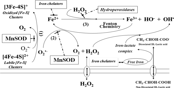
Previous studies have shown that the endogenous SOD levels control the iron-dependent HO. formation when cells are exposed to hydrogen peroxide (13, 14, 42) or such formation is due to an iron overload as in the Fur mutant of E. coli (58). The continuous generation of HO. requires a continuous supply of Fe2+, which can be provided by the labile iron-sulfur [4Fe-4S]2+ clusters or via the reduction of Fe3+ by O2− (13, 15, 31). Previously, we demonstrated that MnSOD provides protection against H2O2 (3, 13), likely by interfering with the generation of HO. Figure 5 is a schematic presentation showing how MnSOD, iron chelation, or hydroperoxidases could protect the cells from both organic acid (e.g., lactic acid) and ROS stress.
FIG. 5.
Schematic presentation showing how MnSOD, iron chelators, or hydroperoxidases could protect cells against oxidative stress mediated by lactic acid. Reaction 1 shows the oxidation of labile iron-sulfur clusters by O2−; reaction 2 shows the regeneration of Fe(II) from Fe(III) by the O2− (the sum of reactions 2 and 3 is also known as the Haber-Weiss reaction); reaction 3 shows the generation of HO. by Fenton chemistry. Protective molecules and/or mechanisms are shown in boxes: MnSOD inhibits reactions 1, 2, and 3; hydroperoxidases also inhibit reaction 3; iron chelators inhibit reaction 3. Lactic acid provides protons and forms an iron-lactate complex that can enhance the generation of HO..
From this study, we conclude that the cytotoxic effects of acid stress and oxidative stress are remarkably similar: i.e., both involve the generation of hydroxyl radicals. We predict that other antioxidant enzymes (e.g., SodC, alkyl-hydroperoxide reductase) and iron-chelating proteins (e.g., Dps and Dpr) could also have a protective role against acid stress. Interestingly, the expression of Dpr in S. mutans was differentially increased by acid exposure (54), and in Salmonella, Dps was reported to be regulated by Fur (28), which was shown in an earlier study to be essential for acid resistance (63).
Acidophilic microorganisms, like S. thermophilus (which can grow under low-pH conditions), seem to rely on MnSOD and probably other antioxidants for their survival in acidic environments. Furthermore, expression of MnSOD and/or hydroperoxidases in Lactobacillus spp. that lack these antioxidant enzymes may enhance their ability to survive and resist ROS and acid stress.
Acknowledgments
Partial support was provided by the North Carolina Agricultural Research Service.
We thank Matt Evans for technical assistance and critical reading of the manuscript.
Footnotes
Published ahead of print on 19 March 2010.
REFERENCES
- 1.Abbott, D. A., E. Suir, G. H. Duong, E. de Hulster, J. T. Pronk, and A. J. A. van Maris. 2009. Catalase overexpression reduces lactic acid induced oxidative stress in Saccharomyces cerevisiae. Appl. Environ. Microbiol. 75:2320-2325. [DOI] [PMC free article] [PubMed] [Google Scholar]
- 2.Ali, M. A., F. Yasui, S. Matsugo, and T. Konishi. 2000. The lactate-dependent enhancement of hydroxyl radical generation by the Fenton reaction. Free Radic. Res. 32:429-438. [DOI] [PubMed] [Google Scholar]
- 3.Andrus, J. M., S. W. Bowen, T. R. Klaenhammer, and H. M. Hassan. 2003. Molecular characterization and functional analysis of the manganese-containing superoxide dismutase gene (sodA) from Streptococcus thermophilus AO54. Arch. Biochem. Biophys. 420:103-113. [DOI] [PubMed] [Google Scholar]
- 4.Archibald, F. S., and I. Fridovich. 1981. Manganese and defenses against oxygen toxicity in Lactobacillus plantarum. J. Bacteriol. 145:442-451. [DOI] [PMC free article] [PubMed] [Google Scholar]
- 5.Arnold, S. M., W. J. Hickey, and R. F. Harris. 1995. Degradation of atrazine by Fentons reagent—condition optimization and product quantification. Environ. Sci. Technol. 29:2083-2089. [DOI] [PubMed] [Google Scholar]
- 6.Asada, K., K. Yoshikawa, M. A. Takahashi, Y. Maeda, and K. Enmanji. 1975. Superoxide dismutases from a blue green alga, Plectonema boryanum. J. Biol. Chem. 250:2801-2807. [PubMed] [Google Scholar]
- 7.Axelsson, L. 1998. Lactic acid bacteria: classification and physiology, p. 1-12. In S. Salminen and A. von Wright (ed.), Lactic acid bacteria: microbiology and functional aspects. Marcel Dekker, Inc., New York, NY.
- 8.Azcarate-Peril, M. A., E. Altermann, R. L. Hoover-Fitzula, R. J. Cano, and T. R. Klaenhammer. 2004. Identification and inactivation of genetic loci involved with Lactobacillus acidophilus acid tolerance. Appl. Environ. Microbiol. 70:5315-5322. [DOI] [PMC free article] [PubMed] [Google Scholar]
- 9.Benov, L. T., and I. Fridovich. 1994. Escherichia coli express a copper- and zinc-containing superoxide dismutase. J. Biol. Chem. 269:25310-25314. [PubMed] [Google Scholar]
- 10.Bergqvist, S. W., T. Andlid, and A. S. Sandberg. 2006. Lactic acid fermentation stimulated iron absorption by Caco-2 cells is associated with increased soluble iron content in carrot juice. Br. J. Nutr. 96:705-711. [PubMed] [Google Scholar]
- 11.Beyer, W. F., and I. Fridovich. 1987. Effect of hydrogen peroxide on the iron-containing superoxide-dismutase of Escherichia coli. Biochemistry 26:1251-1257. [DOI] [PubMed] [Google Scholar]
- 12.Britton, L., D. P. Malinowski, and I. Fridovich. 1978. Superoxide dismutase and oxygen metabolism in Streptococcus faecalis and comparisons with other organisms. J. Bacteriol. 134:229-236. [DOI] [PMC free article] [PubMed] [Google Scholar]
- 13.Bruno-Barcena, J. M., J. M. Andrus, S. L. Libby, T. R. Klaenhammer, and H. M. Hassan. 2004. Expression of a heterologous manganese superoxide dismutase gene in intestinal lactobacilli provides protection against hydrogen peroxide toxicity. Appl. Environ. Microbiol. 70:4702-4710. [DOI] [PMC free article] [PubMed] [Google Scholar]
- 14.Bruno-Barcena, J. M., M. A. Azcarate-Peril, T. R. Klaenhammer, and H. M. Hassan. 2005. Marker-free chromosomal integration of the manganese superoxide dismutase gene (sodA) from Streptococcus thermophilus into Lactobacillus gasseri. FEMS Microbiol. Lett. 246:91-101. [DOI] [PubMed] [Google Scholar]
- 15.Chang, S. K., and H. M. Hassan. 1997. Characterization of superoxide dismutase in Streptococcus thermophilus. Appl. Environ. Microbiol. 63:3732-3735. [DOI] [PMC free article] [PubMed] [Google Scholar]
- 16.Clements, M. O., S. P. Watson, and S. J. Foster. 1999. Characterization of the major superoxide dismutase of Staphylococcus aureus and its role in starvation survival, stress resistance, and pathogenicity. J. Bacteriol. 181:3898-3903. [DOI] [PMC free article] [PubMed] [Google Scholar]
- 17.Condon, S. 1987. Responses of lactic acid bacteria to oxygen. FEMS Microbiol. Rev. 46:269-280. [Google Scholar]
- 18.Cotter, P. D., and C. Hill. 2003. Surviving the acid test: responses of gram-positive bacteria to low pH. Microbiol. Mol. Biol. Rev. 67:429-453. [DOI] [PMC free article] [PubMed] [Google Scholar]
- 19.de Man, J. C., M. Rogosa, and M. E. Sharpe. 1960. A medium for the cultivation of lactobacilli. J. Appl. Bacteriol. 23:130-135. [Google Scholar]
- 20.Dimmig, L. K., E. R. Myers, and S. E. Martin. 1994. Catalase, superoxide-dismutase and listeriolysin-O production by Listeria monocytogenes in broth containing acetic and hydrochloric acids. J. Food Prot. 57:626-628. [DOI] [PubMed] [Google Scholar]
- 21.Fenton, H. J. H. 1894. Oxidation of tartaric acid in the presence of iron. J. Chem. Soc. Trans. 65:899-910. [Google Scholar]
- 22.Fink, R. C., and J. G. Scandalios. 2002. Molecular evolution and structure-function relationships of the superoxide dismutase gene families in angiosperms and their relationship to other eukaryotic and prokaryotic superoxide dismutases. Arch. Biochem. Biophys. 399:19-36. [DOI] [PubMed] [Google Scholar]
- 23.Flint, D. H., J. F. Tuminello, and M. H. Emptage. 1993. The inactivation of Fe-S clusters containing hydro-lyases by superoxide. J. Biol. Chem. 268:22369-22376. [PubMed] [Google Scholar]
- 24.Foster, J. W. 1999. When protons attack: microbial strategies of acid adaptation. Curr. Opin. Microbiol. 2:170-174. [DOI] [PubMed] [Google Scholar]
- 25.Fridovich, I. 1995. Superoxide radical and superoxide dismutases. Annu. Rev. Biochem. 64:97-112. [DOI] [PubMed] [Google Scholar]
- 26.Gotz, F., B. Sedewitz, and E. F. Elstner. 1980. Oxygen utilization by Lactobacillus plantarum. I. Oxygen consuming reactions. Arch. Microbiol. 125:209-214. [DOI] [PubMed] [Google Scholar]
- 27.Haber, F., and J. Weiss. 1934. The catalytic decomposition of hydrogen peroxide by iron salts. Proc. R. Soc. 147:332-351. [Google Scholar]
- 28.Hall, H. K., and J. W. Foster. 1996. The role of Fur in the acid tolerance response of Salmonella typhimurium is physiologically and genetically separable from its role in iron acquisition. J. Bacteriol. 178:5683-5691. [DOI] [PMC free article] [PubMed] [Google Scholar]
- 29.Hamada, Y. Z., B. Carlson, and J. Dangberg. 2005. Interaction of malate and lactate with chromium(III) and iron(III) in aqueous solutions. Synthesis and reactivity in inorganic metal. Org. Nano-Met. Chem. 35:515-522. [Google Scholar]
- 30.Hassan, H. M. 1997. Cytotoxicity of oxyradicals and the evolution of superoxide dismutases, p. 25-51. In D. Massaro and L. Clerch (ed.), Oxygen, gene expression and cellular function. Marcel Dekker, New York, NY.
- 31.Imlay, J. A. 2006. Iron-sulphur clusters and the problem with oxygen. Mol. Microbiol. 59:1073-1082. [DOI] [PubMed] [Google Scholar]
- 32.Jakubovics, N. S., A. W. Smith, and H. F. Jenkinson. 2002. Oxidative stress tolerance is manganese (Mn2+) regulated in Streptococcus gordonii. Microbiology 148:3255-3263. [DOI] [PubMed] [Google Scholar]
- 33.Kanbe, C., and K. Uchida. 1985. Oxygen consumption by Pediococcus halophilus. Agric. Biol. Chem. 49:2931-2937. [Google Scholar]
- 34.Keele, B. B., Jr., J. M. McCord, and I. Fridovich. 1970. Superoxide dismutase from Escherichia coli B. A new manganese-containing enzyme. J. Biol. Chem. 245:6176-6181. [PubMed] [Google Scholar]
- 35.Keyer, K., and J. A. Imlay. 1996. Superoxide accelerates DNA damage by elevating free iron levels. Proc. Natl. Acad. Sci. U. S. A. 93:13635-13640. [DOI] [PMC free article] [PubMed] [Google Scholar]
- 36.Kullen, M. J., and T. R. Klaenhammer. 1999. Identification of the pH-inducible, proton-translocating F1F0-ATPase (atpBEFHAGDC) operon of Lactobacillus acidophilus by differential display: gene structure, cloning and characterization. Mol. Microbiol. 33:1152-1161. [DOI] [PubMed] [Google Scholar]
- 37.Lloyd, G. T., A. J. Hillier, I. Barlow, and G. R. Jago. 1978. Aerobic formation of acetate from pyruvate by Lactobacillus bulgaricus. Aust. J. Biol. Sci. 31:565-571. [Google Scholar]
- 38.Loewen, P. C., B. L. Triggs, C. S. George, and B. E. Hrabarchuk. 1985. Genetic mapping of katG, a locus that affects synthesis of the bifunctional catalase-peroxidase hydroperoxidase I in Escherchia coli. J. Bacteriol. 162:661-667. [DOI] [PMC free article] [PubMed] [Google Scholar]
- 39.Martin-Galiano, A. J., M. J. Ferrandiz, and A. G. de la Campa. 2001. The promoter of the operon encoding the F0F1 ATPase of Streptococcus pneumoniae is inducible by pH. Mol. Microbiol. 41:1327-1338. [DOI] [PubMed] [Google Scholar]
- 40.McCord, J. M., and E. D. Day, Jr. 1978. Superoxide-dependent production of hydroxyl radical catalyzed by iron-EDTA complex. FEBS Lett. 86:139-142. [DOI] [PubMed] [Google Scholar]
- 41.McCord, J. M., and I. Fridovich. 1969. Superoxide dismutase. An enzymatic function for erythrocuprein (hemocuprein). J. Biol. Chem. 244:6049-6055. [PubMed] [Google Scholar]
- 42.McCormick, M. L., G. R. Buettner, and B. E. Britigan. 1998. Endogenous superoxide dismutase levels regulate iron-dependent hydroxyl radical formation in Escherichia coli exposed to hydrogen peroxide. J. Bacteriol. 180:622-625. [DOI] [PMC free article] [PubMed] [Google Scholar]
- 43.Mercenier, A., C. Robert, D. A. Romero, P. Slos, and Y. Lemoine. 1987. Transformation of Streptococcus thermophilus spheroplasts, p. 234-237. In J. J. Ferretti and R. Curtiss III (ed.), Streptococcal genetics. American Society for Microbiology, Washington, DC.
- 44.Poyart, C., P. Berche, and P. Tryeu-Cout. 1995. Characterization of superoxide dismutase genes from Gram-positive bacteria by polymerase chain reaction using degenerate primers. FEMS Microbiol. Lett. 131:41-45. [DOI] [PubMed] [Google Scholar]
- 45.Proulx, A. K., and M. B. Reddy. 2007. Fermentation and lactic acid addition enhance iron bioavailability of maize. J. Agric. Food Chem. 55:2749-2754. [DOI] [PubMed] [Google Scholar]
- 46.Rehncrona, S., H. N. Hauge, and B. K. Siesjo. 1989. Enhancement of iron-catalyzed free-radical formation by acidosis in brain homogenates—difference in effect by lactic-acid and CO2. J. Cereb. Blood Flow Metab. 9:65-70. [DOI] [PubMed] [Google Scholar]
- 47.Sambrook, J., E. F. Fritsch, and T. Maniatis. 1989. Molecular cloning: a laboratory manual, 2nd ed. Cold Spring Harbor Laboratory Press, Cold Spring Harbor, NY.
- 48.Sanders, J. W., K. J. Leenhousts, A. J. Haandrikman, G. Venema, and J. Kook. 1995. Stress response in Lactococcus lactis: cloning, expression analysis and mutation of the lactococcal superoxide dismutase gene. J. Bacteriol. 177:5254-5260. [DOI] [PMC free article] [PubMed] [Google Scholar]
- 49.Schellhorn, H. E., and H. M. Hassan. 1988. Response of hydroperoxidase and superoxide dismutase deficient mutants of Escherichia coli K-12 to oxidative stress. Can. J. Microbiol. 34:1171-1176. [DOI] [PubMed] [Google Scholar]
- 50.Schellhorn, H. E., and V. L. Stones. 1992. Regulation of katF and katE in Escherichia coli K-12 by weak acids. J. Bacteriol. 174:4769-4776. [DOI] [PMC free article] [PubMed] [Google Scholar]
- 51.Sedewitz, B., K. H. Schleifer, and F. Gotz. 1984. Physiological role of pyruvate oxidase in the aerobic metabolism of Lactobacillus plantarum. J. Bacteriol. 160:462-465. [DOI] [PMC free article] [PubMed] [Google Scholar]
- 52.Sedewitz, B., K. H. Schleifer, and F. Gotz. 1984. Purification and biochemical characterization of pyruvate oxidase from Lactobacillus plantarum. J. Bacteriol. 160:273-278. [DOI] [PMC free article] [PubMed] [Google Scholar]
- 53.Senadheera, D., K. Krastel, R. Mair, A. Persadmehr, J. Abranches, R. A. Burne, and D. G. Cvitkovitch. 2009. Inactivation of VicK affects acid production and acid survival of Streptococcus mutans. J. Bacteriol. 191:6415-6424. [DOI] [PMC free article] [PubMed] [Google Scholar]
- 54.Starke, P. E., and J. L. Farber. 1985. Ferric iron and superoxide anions are required for the killing of cultured hepatocytes by hydrogen peroxide. Fed. Proc. 44:1275. [PubMed] [Google Scholar]
- 55.Steinman, H. M. 1992. Construction of an Escherichia coli K12 strain deleted for manganese and iron superoxide-dismutase genes and its use in cloning the iron superoxide-dismutase gene of Legionella pneumophila. Mol. Gen. Genet. 232:427-430. [DOI] [PubMed] [Google Scholar]
- 56.Strittmatter, C. F. 1959. Electron transport to oxygen in lactobacilli. J. Exp. Biol. 234:2789. [PubMed] [Google Scholar]
- 57.Teraguchi, S., J. Ono, I. Kiyosawa, and S. Okonogi. 1987. Oxygen-uptake activity and aerobic metabolism of Streptococcus thermophilus STH450. J. Dairy Sci. 70:514-523. [DOI] [PubMed] [Google Scholar]
- 58.Touati, D., M. Jacques, B. Tardat, L. Bouchard, and S. Despied. 1995. Lethal oxidative damage and mutagenesis are generated by iron in Δfur mutants of Escherichia coli: protective role of superoxide dismutase. J. Bacteriol. 177:2305-2314. [DOI] [PMC free article] [PubMed] [Google Scholar]
- 59.Trabold, O., S. Wagner, C. Wicke, H. Scheuenstuhl, M. Z. Hussain, N. Rosen, A. Seremetiev, H. D. Becker, and T. K. Hunt. 2003. Lactate and oxygen constitute a fundamental regulatory mechanism in wound healing. Wound Repair Regen. 11:504-509. [DOI] [PubMed] [Google Scholar]
- 60.VanBogelen, R. A., P. M. Kelley, and F. C. Neidhardt. 1987. Differential induction of heat-shock, SOS, and oxidation stress regulons and accumulation of nucleotides in Escherichia coli. J. Bacteriol. 169:26-32. [DOI] [PMC free article] [PubMed] [Google Scholar]
- 61.van de Guchte, M., P. Serror, C. Chervaux, T. Smokvina, S. D. Ehrlich, and E. Maguin. 2002. Stress responses in lactic acid bacteria. Antonie Van Leeuwenhoek 82:187-216. [PubMed] [Google Scholar]
- 62.Wagner, S., M. Z. Hussain, T. K. Hunt, B. Bacic, and H. D. Becker. 2004. Stimulation of fibroblast proliferation by lactate-mediated oxidants. Wound Repair Regen. 12:368-373. [DOI] [PubMed] [Google Scholar]
- 63.Yoo, A. Y., S. W. Kim, J. E. Yu, Y. H. Kim, J. Cha, J. I. Oh, S. K. Eo, J. H. Lee, and H. Y. Kang. 2007. Requirement of Fur for the full induction of dps expression in Salmonella enterica serovar typhimurium. J. Microbiol. Biotechnol. 17:1452-1459. [PubMed] [Google Scholar]
- 64.Yost, F. J., Jr., and I. Fridovich. 1973. An iron-containing superoxide dismutase from Escherichia coli. J. Biol. Chem. 248:4905-4908. [PubMed] [Google Scholar]
- 65.Youn, H. D., E. J. Kim, J. H. Roe, Y. C. Hah, and S. O. Kang. 1996. A novel nickel-containing superoxide dismutase from Streptomyces spp. Biochem. J. 318:889-896. [DOI] [PMC free article] [PubMed] [Google Scholar]


