Abstract
Accurate diagnosis of canine leishmaniasis (CanL) is essential toward a more efficient control of this zoonosis, but it remains problematic due to the high incidence of asymptomatic infections. In this study, we present data on the development of enzyme-linked immunosorbent assay (ELISA)-based techniques for the detection of antibodies against the recombinant protein Leishmania infantum cytosolic tryparedoxin peroxidase (LicTXNPx) and a comparison of the results with those employing soluble Leishmania antigens from promastigote or amastigote forms and the homologue recombinant protein L. infantum mitochondrial TXNPx (LimTXNPx). Moreover, we offer an evaluation of the diagnostic potential of rK39 for CanL in the Portuguese canine population and propose an improvement to the existing ELISA-based serological techniques by combining the LicTXNPx and rK39 antigens as a Leishmania antigen mixture (LAM). The data demonstrated that ELISAs based on soluble promastigote or amastigote antigens had generally higher levels of sensitivity for detection of antibodies in symptomatic or asymptomatic dogs than for detection of those against isolated recombinant proteins. Nevertheless, the specificities were found to be similar for all target antigens used. Importantly, the LAM-ELISA methodology improved the overall sensitivity, maintaining a high overall level of specificity. In addition, it was demonstrated that the detection of anti-LAM IgG2 can increase the accuracy of the serological diagnosis. Overall, the obtained results showed that the strategy of combining two well-defined Leishmania antigens, LicTXNPx and rK39, proved to be a sensitive and specific improvement to current serological diagnosis of CanL, being a useful tool for the detection of both clinical and subclinical forms of canine Leishmania infection.
Leishmaniasis, a disease caused by protozoan parasites of the genus Leishmania, is one of the major communicable illnesses in the world and the third most important vector-borne disease, after malaria and sleeping sickness (5). The incidence of leishmaniasis has increased in recent years due to growth and redistribution of populations, increasing poverty, and climate changes. Visceral leishmaniasis (VL) is caused by species of the Leishmania donovani complex and may result, in the absence of treatment, in a progressive and often fatal illness in dogs and humans (4, 18).
The domestic dog is considered the main reservoir for human VL in the Mediterranean basin and South America (2), where the zoonotic disease is caused by L. infantum (syn. L. chagasi). Interestingly, some evidence has been presented that suggests that dogs also contribute to transmission in northeastern Africa, where L. donovani is the causative agent of human disease (17). Although the prevalence of Leishmania infection in dogs has been reported to rarely exceed 10%, it may rise up to 35% in areas where the disease is highly endemic (11). Dogs constitute a primary source of infection, contributing, in conjunction with sand fly vectors, to the perpetuation of the infectious cycle. Although other reservoirs have been incriminated in the epidemiological chain of human VL, such as wild canids, rodents, or marsupials, zoonotic VL epidemics have been described only for areas where canine leishmaniasis (CanL) is endemic (31). In certain areas in which CanL is endemic, the importance of controlling the infected canine population is sustained by the correlation of spatial distribution between canine and human cases of VL (21), since the existence of high canine infection rates leads to increased transmission to humans due to the presence of sand fly vectors. Furthermore, the systematic culling of infected dogs in areas where CanL is endemic led to a reduction in the incidence of human and canine disease (2, 14). Therefore, the design of effective control programs to prevent human infection in countries where CanL is endemic should include an accurate evaluation of the prevalence and the incidence of CanL, with special attention paid to the unsatisfactory sensitivity associated with the detection of infection in asymptomatic infected dogs (1). Indeed, although the number of dogs currently infected has been estimated in the low millions (13), the apparent global burden is believed to be much higher, due mostly to the inaccuracy of the diagnostic tests currently used to detect asymptomatic infections.
Current control measures rely on early diagnosis and chemotherapy of patients suffering from this disease. Thus, rapid diagnostic tests capable of providing early and accurate diagnoses of leishmaniasis, especially for the visceral form, are among the highest priorities defined for leishmaniasis research (9). Ideally, all cases of leishmaniasis should be confirmed by direct parasite detection in stained microscopic preparations of skin biopsy specimens, aspirates from spleen, bone marrow, or lymph nodes. Although parasitological diagnosis still remains the gold standard to confirm Leishmania infection, it is time-consuming and requires trained technical staff for the collection of samples and microscopic evaluation. The use of molecular biology techniques that are capable of detecting DNA or RNA unique to the parasite, with a high degree of specificity and sensitivity, just a few weeks after the onset of the first clinical signs or symptoms is becoming increasingly relevant (13, 24). These techniques are frequently considered the most valid diagnosis methods. Furthermore, their use as routine diagnostic tests often requires specific and expensive equipment, limiting the feasibility of PCR diagnosis in developing countries and in field conditions. Nevertheless, considerable advances, such as quantitative nucleic acid sequence-based amplification (QT-NASBA), are being developed to circumvent these problems (37, 38).
Therefore, serological approaches to detect anti-Leishmania antibodies constitute a valuable alternative for early, rapid, and user-friendly diagnostic tests for both human and canine Leishmania infection. The current available serological techniques include the direct agglutination test (DAT) and fast agglutination screening test (FAST) (32, 33), indirect fluorescent antibody test (IFAT), immunoblotting, and enzyme-linked immunosorbent assay (ELISA) (15). Among these, ELISA is a classical method used in the detection of antibodies to Leishmania which is neither normally limited to laboratory conditions nor dependent on technical expertise. ELISA-based methods allow high-throughput screening of a large number of samples with high levels of sensitivity, with specificity depending on the antigen used (36). Total soluble antigens derived from the promastigote stage of different Leishmania species are the most commonly used antigens in ELISA. These ELISAs have demonstrated sensitivities and specificities of 80 to 100% and 85 to 95%, respectively (20). Nevertheless, the use of total soluble antigens is limited due to problems of reproducibility and manufacturing. Thus, several defined Leishmania antigens have been tested to overcome these difficulties and to improve both sensitivity and specificity. The recombinant protein rK39, which is a repetitive immunodominant epitope of a kinesin-related protein expressed predominantly in the amastigotes of viscerotropic L. chagasi (syn. L. infantum) (6), has proved to be an exceptionally strong marker of disease (16). Nevertheless, rK39 performs suboptimally in asymptomatic cases with proven infection (22). We have recently described the L. infantum cytosolic tryparedoxin peroxidase (LicTXNPx) antigen, a member of unique enzymatic Leishmania cascades for detoxification of peroxides (8), as a highly immunogenic probe during natural human or experimental canine infections (28, 35). Our previous studies indicated that anti-LicTXNPx antibodies were present in both symptomatic and asymptomatic experimental canine infections, making this antigen a good candidate marker for both clinical and subclinical Leishmania infections (35). In the present study, we evaluate the ability of the LicTXNPx antigen to detect both clinical and subclinical forms of Leishmania infection using a large cohort of naturally infected dogs living in areas of Portugal in which CanL is endemic. In addition, we provide the first evaluation of the diagnostic potential of rK39 for Leishmania infection in the Portuguese canine population. The development of a sensitive and specific diagnostic ELISA for VL may depend on the synergic accuracies of two or more defined antigens that will constitute the ultimate serological marker. Hence, we proposed a defined Leishmania antigen mixture, composed of the LicTXNPx and rK39 antigens, as an improvement to current ELISA-based serological techniques for the accurate detection of both clinical and subclinical forms of CanL.
MATERIALS AND METHODS
Dogs and samples.
This study used a panel of 250 serum samples from male and female domestic dogs of various breeds and ages. Permission was obtained from all owners to test their animals, and physical examination and clinical specimen collection were performed by expert veterinarians. Dogs were clinically classified as symptomatic based on the presence of at least two clinical signs compatible with CanL, including lymphadenopathy, alopecia, dermatitis, skin ulceration, keratoconjunctivitis, onychogryposis, lameness, epistaxis, anorexia, and weight loss. Asymptomatic and healthy dogs had no detectable clinical signs of disease. Peripheral blood for serology was collected from the cephalic vein, processed, and stored at −20°C. The DAT was used for quantitative specific detection of anti-Leishmania antibodies with a cutoff titer of 400. DAT used a standard freeze-dried antigen (KIT Biomedical Research, Amsterdam, Netherlands) and followed the protocol described by Schallig et al. (30). Bone marrow or lymph node aspirates were smeared onto slides, Giemsa stained, and microscopically examined for the presence of amastigote forms of Leishmania. Four groups of serum samples were established based on the clinical, serological, and/or parasitological data available from the dogs.
Group A (n = 83) comprised samples from natural cases of CanL, i.e., symptomatic dogs confirmed as being infected with Leishmania by means of either the DAT (n = 34), detection of amastigotes (n = 27), or both procedures (n = 22).
Group B (n = 57) consisted of sera from asymptomatic dogs living in areas of northeastern Portugal where CanL is endemic but with no history of CanL. These animals were all found seropositive for anti-Leishmania antibodies by means of DAT during field surveys for canine Leishmania infection.
Group C (n = 97) was constituted by clinically healthy dogs from areas where CanL is regarded as not endemic and that were all Leishmania seronegative (DAT titer, <1:100).
Group D (n = 13) included samples from dogs seronegative for anti-Leishmania antibodies (DAT, <1:100) but infected with other agents or affected by other confirmed pathological conditions. This group comprised animals living in areas of CanL endemicity and suffering from Ancylostoma caninum infection (n = 1), Babesia canis and Leptospira spp. mixed infection (n = 1), Leptospira spp. (n = 1), Dipylidium caninum, Taenia spp., Toxocara canis, and Sarcoptes scabiei mixed infection (n = 1), Demodex canis (n = 1), Hepatozoon canis (n = 1), Trichuris vulpis (n = 1), tick infestation (n = 1), autoimmune disorder (n = 1), lymphoma (n = 1), pneumonia (n = 1), pyodermitis (n = 1), and tumor (n = 1).
Antigens used for the enzyme-linked immunosorbent assay (ELISA).
A cloned line of L. infantum (MHOM/MA/67/ITMAP-263) was used in all experiments. This strain was frequently passaged through mice to maintain its virulence. Each subculture was initiated at 5 × 10⁵ or 1 × 10⁶ parasites/ml of medium for promastigote and axenic amastigote forms, respectively. Promastigotes were maintained at 26°C by weekly subpassage in RPMI 1640 culture medium (RPMIc; Invitrogen Life Technologies) supplemented with 10% heat-inactivated fetal calf serum (FCS), 100 U/ml penicillin, 100 mg/ml streptomycin, and 20 mM HEPES. Axenically grown amastigotes of L. infantum were obtained through the differentiation of stationary-phase promastigotes in a cell-free culture medium called MAA/20 (medium for axenically grown amastigotes) at an acidic pH and at vertebrate host temperature in 25-ml flasks (34). After the differentiation process, axenic amastigotes were maintained at 37°C with 5% CO2 by weekly subpassages. After 5 days of culture, both parasite forms were washed three times with phosphate-buffered saline (PBS), pH 7.4 (3,500 × g, 10 min, 4°C); the pellet was resuspended in PBS containing 2 mM phenylmethylsulfonyl fluoride (PMSF) protease inhibitor, and the mixture was submitted to 10 freeze-thaw cycles (a −80°C-capable freezer and a water bath at 37°C were used) and sonication (Vibra Cell; Sonics Materials Inc., Danbury, CT) for complete rupture of the parasites. The mixture was centrifuged at 13,000 × g for 30 min at 4°C to separate cellular debris from the supernatant that was taken as the parasite extract, aliquoted, and stored at −80°C. The recombinant proteins LicTXNPx and LimTXNPx, two complementary peroxiredoxins of the Leishmania antioxidant machinery (8), were purified by affinity chromatography on a Ni-NTA column (Qiagen) as described in a previous report (10). The proteins used were obtained as recombinant proteins containing a six-histidine residue at their N-termini. In addition, the recombinant Leishmania antigen K39 (6) was similarly used for antibody detection by ELISA. All antigens were analyzed on 10% polyacrylamide gels and visualized by staining with Coomassie blue. The protein content of each antigen preparation was determined by the Lowry assay.
Enzyme-linked immunosorbent assay (ELISA).
The ELISA was performed as described elsewhere (35). Briefly, 96-well flat-bottomed microtiter plates (Greiner, Frickenhausen, Germany) were coated with 10 μg/ml of soluble promastigote or amastigote Leishmania antigens or 5 μg/ml of LimTXNPx, LicTXNPx, or rK39. Anti-dog IgG, IgG1, and IgG2 conjugated to horseradish peroxidase (Bethyl Laboratories, Montgomery, TX) were used as secondary antibodies. In all experiments, a reference dog IgG (Dog Reference Serum; Bethyl Laboratories, Montgomery, TX) was used as an internal control. The working dilutions for the conjugated antibody were determined according to the titration performed for the lot (1:500, 1:3,000, and 1:5,000 for anti-dog IgG1, anti-dog IgG2, and anti-dog IgG, respectively). A492 values were read in an automatic ELISA reader (PowerWave XS; Bio-Tek, Vermont).
Statistical analysis.
Statistical analyses were performed using GraphPad Prism 5 (GraphPad Software Inc., La Jolla, CA). A receiver operating characteristic (ROC) curve was generated for each tested antigen by applying the sensitivity values in the ordinate and the complement of specificity in the abscissa. These curves were used to select the cutoff values that more effectively discriminate positive from negative samples. Differences in immunoglobulin levels between groups were analyzed using the Mann-Whitney U test, and a P value of <0.05 was considered statistically significant.
RESULTS
Determination of cutoff values.
The ELISA cutoff values for all Leishmania antigens used in the present study were defined based on the ROC curves (40). For each antigen, a ROC curve was constructed using the reactivity of sera obtained from group C dogs, clinically negative for CanL, and the reactivity of all CanL-positive sera, both symptomatic and asymptomatic, as positive values (Fig. 1). Thus, we established ELISA cutoff optical density (OD) values at 492 nm of 0.156 for soluble promastigote Leishmania antigens (SPLA), 0.125 for soluble amastigote Leishmania antigens (SALA), 0.154 for L. infantum mitochondrial tryparedoxin peroxidase (LimTXNPx), 0.146 for L. infantum cytosolic tryparedoxin peroxidase (LicTXNPx), and 0.094 for rK39.
FIG. 1.
ROC curves obtained for the ELISAs. (A) ROC curves for SALA (thick dotted line) and SPLA (thick solid line) antigens. (B) ROC curves for LimTXNPx (thick dotted line) and LicTXNPx (thick solid line) antigens and rK39 (thin dotted line). The dotted gray line represents the reference line.
The area under the curve (AUC) is used to compare the accuracies of different diagnostic antigens or tests (39). In order for a test to demonstrate excellent accuracy, the AUC should be in the region of 0.97 or above (19). Although the determined AUC values were below 0.97, the Leishmania antigens tested presented similar accuracies, since proximal values were obtained for the AUC of each ROC curve: SPLA AUC = 0.952, 95% confidence interval (CI) = 0.913 to 0.990 (Fig. 1A); SALA AUC = 0.940, 95% CI = 0.908 to 0.991 (Fig. 1A); LimTXNPx AUC = 0.886, 95% CI = 0.8348 to 0.9371 (Fig. 1B); LicTXNPx AUC = 0.903, 95% CI = 0.853 to 0.953 (Fig. 1B), and rK39 AUC = 0.945, 95% CI = 0.905 to 0.985 (Fig. 1B).
The recombinant LicTXNPx antigen detects asymptomatic CanL.
We measured the presence of anti-LicTXNPx antibodies in a cohort of 250 serum samples from male and female domestic dogs of various breeds and ages. Our panel included samples from symptomatic (group A) and asymptomatic (group B) natural cases of canine Leishmania infection, clinically healthy dogs from areas where CanL is nonendemic (group C), and animals that were Leishmania negative but presented other confirmed pathological conditions (group D). For comparison, the same cohort was tested for the presence of antibodies against SPLA, SALA, or LimTXNPx. The antigen concentration and serum dilution used in all ELISA assays were previously optimized (10, 28, 35). Therefore, we used 5 μg/ml of each recombinant antigen and 10 μg/ml of both Leishmania promastigote and amastigote soluble antigens as optimal concentrations. A serum dilution of 1:1,500 was used in all assays, as this dilution allowed a clear separation between negative and positive sera.
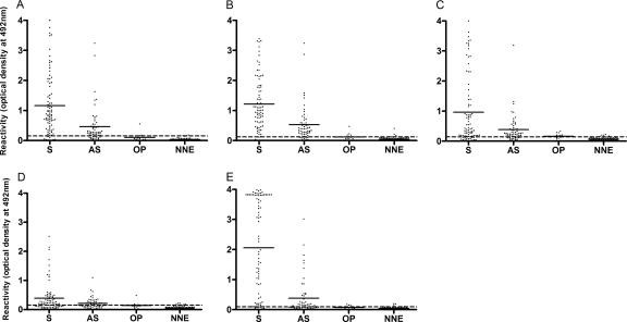
The reactivity of sera from symptomatic dogs (group A) was found to be significantly higher with SPLA (P < 0.0001), SALA (P < 0.0001), and LicTXNPx (P = 0.0105) than the reactivity of sera from asymptomatic infected dogs from group B (Fig. 2A to C). The exception was the result with LimTXNPx, with which no significant differences were found between the two groups of infected animals (P = 0.0591; Fig. 2D). The sera of symptomatic dogs were found to react more actively against SALA and SPLA antigens than against the recombinant Leishmania proteins LicTXNPx or LimTXNPx. Hence, among the 83 serum samples from symptomatic dogs in group A, 89.2% and 90.4% were reactive against SPLA and SALA, respectively, while the LicTXNPx antigen recognized 80.7% and LimTXNPx detected only 66.3% of all infections in symptomatic animals (Table 1). Indeed, the average optical density values for symptomatic dogs reached 1.22 ± 0.73 and 1.16 ± 0.78 for SALA and SPLA, respectively, while lower levels were observed for LicTXNPx and LimTXNPx (0.96 ± 0.89 and 0.39 ± 0.32, respectively) (Fig. 2). The LicTXNPx antigen displayed a high level of sensitivity in the detection of infections in asymptomatic infected dogs. LicTXNPx was capable of detecting 77.2% of the 57 tested sera, while LimTXNPx could discriminate only 49.1% (Table 1). Similarly to what we have previously described for a cohort of experimentally infected dogs (35), SALA was more sensitive than promastigote antigens in detecting infections in asymptomatic naturally infected dogs (82.4% and 73.7% for SALA and SPLA, respectively). Although LicTXNPx presented an overall lower degree of sensitivity than soluble promastigote or amastigote Leishmania antigens (78.6% for LicTXNPx, 82.9% for SPLA, and 87.1% for SALA), this antigen displayed similar capacities to detect symptomatic and asymptomatic cases of canine Leishmania infection, as demonstrated by the asymptomatic/symptomatic index of 0.96 (Table 1). Taken together, these observations support the role of LicTXNPx as a good candidate marker for subclinical CanL.
FIG. 2.
Levels of IgG antibodies against SPLA (A), SALA (B), and the L. infantum recombinant proteins LicTXNPx (C), LimTXNPx (D), and rK39 (E) in sera of symptomatic (S) and asymptomatic (AS) dogs, dogs that were Leishmania negative but presented other infections or pathological conditions (OP), and Leishmania-negative healthy dogs from areas in which CanL is not endemic (NNE). Results are expressed as the optical densities at 492 nm. Each dot represents an individual dog serum sample. Bars represent medians, and dotted lines represent cutoff values.
TABLE 1.
Sensitivity and specificity of the ELISA technique using different Leishmania antigens in the diagnosis of symptomatic and asymptomatic infected dogs
| Antigen | Sensitivitya |
Asymptomatic/symptomatic index | Specificityc | ||
|---|---|---|---|---|---|
| Symptomatic | Asymptomatic | Totalb | |||
| SPLA | 74/83 (89.2) | 42/57 (73.7) | 116/140 (82.9) | 0.83 | 100/109 (91.7) |
| SALA | 75/83 (90.4) | 47/57 (82.4) | 122/140 (87.1) | 0.91 | 100/109 (91.7) |
| LicTXNPx | 67/83 (80.7) | 44/57 (77.2) | 110/140 (78.6) | 0.96 | 97/109 (89.0) |
| LimTXNPx | 55/83 (66.3) | 28/57 (49.1) | 83/140 (59.3) | 0.74 | 99/109 (90.1) |
| rK39 | 73/83 (88.0) | 32/57 (56.1) | 105/140 (75.0) | 0.64 | 101/109 (92.7) |
| LAM | 80/83 (96.4) | 47/57 (82.4) | 127/140 (90.7) | 0.85 | 105/109 (96.3) |
True positives/(true positives + false negatives); numbers in parentheses are percentages.
Symptomatic- and asymptomatic-dog samples.
True negatives/(true negatives + false positives); numbers in parentheses are percentages.
Evaluation of rK39 reactivity in the Portuguese CanL population.
The rK39 antigen has proven to be sensitive for the ELISA diagnosis of human VL and symptomatic CanL (3, 22, 25, 29). Nevertheless, a poorer performance of rK39-ELISA was shown when asymptomatic dogs from Brazilian and Italian regions in which CanL is endemic were tested (22, 29). Hence, we evaluated the accuracy of rK39-ELISA in a canine cohort from regions of Portugal in which CanL is endemic. As shown in Fig. 2E, the serum reactivity at 492 nm of symptomatic and asymptomatic dogs was 2.07 ± 1.07 and 0.38 ± 0.20, respectively, when rK39 was used as the antigen. Among the Leishmania symptomatic infections, the rK39 antigen was more specific (88.0%) than LicTXNPx and LimTXNPx antigens (80.7% and 66.3%, respectively) (Table 1). Nevertheless, rK39 detected only 56.1% of all asymptomatic tested sera, which clearly demonstrated the lack of sensitivity in detecting asymptomatic Leishmania infections in dogs.
Leishmania antigen mixture (LAM) composition and cutoff value.
We have previously showed that the LicTXNPx antigen displayed very high levels of sensitivity in detecting experimental infections in both symptomatic and asymptomatic dogs (35). Since, as previously mentioned, rK39 antigen has been characterized as an exceptionally strong antigen recognized by sera of humans and canines with symptomatic VL (3, 22), we anticipated that a combination of these antigens could improve the performance of ELISA in the serodiagnosis of both clinical and subclinical CanL.
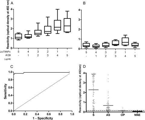
In order to determine the optimal combination of LicTXNPx and rK39 antigens that constitute the Leishmania antigen mixture (LAM) used during the study, we had previously determined the reactivity of 40 randomly chosen sera from symptomatic (Fig. 3A) and asymptomatic (Fig. 3B) dogs against several defined combinations of both antigens. As shown in Fig. 3A and B, we defined the combination of 4 μg/ml of rK39 and 1 μg/ml of LicTXNPx as the optimal LAM composition. This combination achieved the higher optical reactivity for either symptomatic or asymptomatic dog sera without significant reactivity against negative-control sera (data not shown). The reactivity cutoff was established at 0.149, as determined by the ROC curve (Fig. 3C). Importantly, LAM was clearly demonstrated to increase the overall accuracy of the ELISA method, as evaluated by the AUC value of 0.984 (95% CI, 0.961 to 1.007) (19).
FIG. 3.
Leishmania antigen mixture (LAM) characterization and evaluation. The reactivities of representative symptomatic (A) and asymptomatic (B) sera to different defined combinations of LicTXNPx and rk39 antigens (ranging from 0 to 5 μg/ml) were tested. Results are expressed as the optical densities at 492 nm. Values in box plots reflect the median and the interquartile range. The boxes represent 95% of the values. (C) A ROC curve was generated for the LAM-ELISA. (D) The levels of IgG antibodies against LAM were measured in sera of symptomatic (S) and asymptomatic (AS) dogs, dogs that were Leishmania negative but presented other pathologies (OP), and Leishmania-negative healthy dogs from areas in which CanL is not endemic (NNE). Results are expressed as the optical densities at 492 nm. Each dot represents an individual dog serum sample. Bars represent medians, and dotted lines represent cutoff values.
LAM antigen increases overall sensitivity and specificity.
The sensitivity of LAM-ELISA in the diagnosis of symptomatic and asymptomatic dogs was determined using the described cohort of dogs (Fig. 3D). Of 140 sera from symptomatic and asymptomatic Leishmania-infected dogs (groups A and B, respectively), 90.7% reacted with LAM (Table 1). Among all the antigens tested, LAM presented the highest level of sensitivity in both symptomatic (96.4%) and asymptomatic (82.4%) dogs (Table 1). These results clearly demonstrated that LAM antigen improved the overall performance of the ELISA method, detecting with high levels of specificity and sensitivity both clinical and subclinical forms of CanL.
The cross-reactivity of the antigens used in the ELISA-based methods was assessed using sera from Leishmania-negative animals affected by other confirmed pathological conditions (group D) which are frequently found in areas in which leishmaniasis is endemic. In addition, we included sera recovered from non-Leishmania-infected dogs living in areas in which the disease is not endemic (group C). In general, we have found a low level of cross-reactivity. The overall specificity level of the ELISA technique was high, varying from 89.0% with LicTXNPx, 90.1% with LimTXNPx, and 92.7% with rK39 to 91.7% for SALA and SPLA. The LAM-ELISA test presented the highest result, reaching 96.3% specificity (Table 1). Moreover, considering a specificity of 99%, the LAM antigen was still capable of detecting 87.1% of positive sera (Table 2).
TABLE 2.
Sensitivity of the ELISA technique using different Leishmania antigens for a specificity of 99% in the diagnosis of symptomatic and asymptomatic infected dogs
| Antigen | Sensitivitya |
||
|---|---|---|---|
| Symptomatic | Asymptomatic | Totalb | |
| SPLA | 72/83 (86.8) | 38/57 (66.7) | 110/140 (78.5) |
| SALA | 74/83 (89.2) | 43/57 (75.4) | 117/140 (83.6) |
| LicTXNPx | 54/83 (65.1) | 28/57 (49.1) | 82/140(58.6) |
| LimTXNPx | 43/83 (51.8) | 22/57 (38,6) | 65/140 (46.4) |
| rK39 | 65/83 (78.3) | 19/57 (33.3) | 84/140 (60.0) |
| LAM | 80/83 (96.4) | 42/57 (73.7) | 122/140 (87.1) |
True positives/(true positives + false negatives); numbers in parentheses are percentages.
Total results for symptomatic- and asymptomatic-dog samples.
Detection of the IgG2 subclass against LAM antigen increases the sensitivity of LAM-ELISA method.
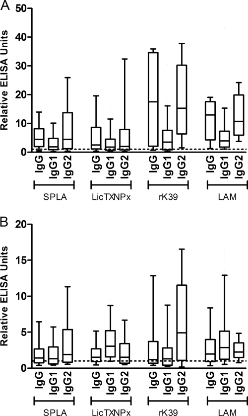
To further characterize the immune response, we have determined the reactivity of IgG1 and IgG2 to each of the antigens previously tested. We used the ROC curves to define the ELISA cutoff values for each IgG subclass against all Leishmania antigens (data not shown). We have found a significant positive correlation between the levels of IgG2 and IgG antibodies against SPLA, LicTXNPx, rK39, or LAM in both symptomatic (Fig. 4A) and asymptomatic (Fig. 4B) dogs (P < 0.001 for all antigens; data not shown). No correlation was found between IgG1 and IgG reactivities with SPLA, rK39, or LAM, which suggests that IgG2 is the predominant subclass during symptomatic and asymptomatic CanL. Interestingly, we detected higher levels of anti-LicTXNPx IgG1 antibodies in the sera of asymptomatic dogs, which improved the sensitivity obtained with total IgG (sensitivity of 93.0% for IgG1 and 77.2% for IgG in sera from asymptomatic dogs). Generally, there was a loss of sensitivity and specificity when each of the IgG subclasses were used to determine the levels of anti-SPLA or anti-rK39 in both groups of infected animals (Table 3). However, we observed an increase in sensitivity when detecting anti-LAM IgG2 (97.1%) in comparison to that when detecting total anti-LAM IgG antibodies (90.7%), maintaining the specificity of the test (Table 3). Overall, these results confirm the sensitivity and specificity improvements using LAM antigen and suggest the determination of anti-LAM IgG2 antibodies as a potential alternative to further increase the accuracy of the serological diagnosis of canine Leishmania infection and CanL.
FIG. 4.
Levels of IgG, IgG1, and IgG2 antibodies against SPLA, LicTXNPx, rK39, and LAM in the sera of symptomatic (A) and asymptomatic (B) infected dogs. For each antigen, immunoglobulin levels (y axis) are represented as relative ELISA units (optical density at 492 nm of analyzed samples/optimal cutoff value). Values in box plots reflect the median and the interquartile range. The boxes represent 95% of the values. Dotted lines represent cutoff values.
TABLE 3.
Isotype sensitivity and specificity of the ELISA technique using different Leishmania antigens in the diagnosis of symptomatic and asymptomatic infected dogs
| Antigen | Sensitivitya |
Specificityc | ||
|---|---|---|---|---|
| Symptomatic | Asymptomatic | Totalb | ||
| SPLA | ||||
| IgG1 | 59/83 (71.1) | 45/57 (78.9) | 104/140 (74.2) | 86/109 (78.9) |
| IgG2 | 69/83 (83.1) | 40/57 (71.2) | 109/140 (77.9) | 103/109 (94.4) |
| IgG | 74/83 (89.2) | 42/57 (73.7) | 116/140 (82.9) | 100/109 (91.7) |
| LicTXNPx | ||||
| IgG1 | 56/83 (67.5) | 53/57 (93,0) | 109/140 (77,9) | 83/109 (76.1) |
| IgG2 | 58/83 (69.9) | 35/57 (61.4) | 93/140 (66.4) | 102/109 (93.6) |
| IgG | 67/83 (80.7) | 44/57 (77.2) | 110/140 (78.6) | 97/109 (89.0) |
| rK39 | ||||
| IgG1 | 62/83 (74.7) | 35/57 (61.4) | 97/140 (69.3) | 102/109 (93.6) |
| IgG2 | 73/83 (88.0) | 45/57 (78.9) | 118/140 (84.3) | 96/109 (88,1) |
| IgG | 73/83 (88.0) | 32/57 (56.1) | 105/140 (75.0) | 101/109 (92.7) |
| LAM | ||||
| IgG1 | 77/83 (92.8) | 50/57 (87.8) | 127/140 (90.7) | 99/109 (90.1) |
| IgG2 | 83/83 (100) | 53/57 (93.0) | 136/140 (97.1) | 105/109 (96.3) |
| IgG | 80/83 (96.4) | 47/57 (82.4) | 127/140 (90.7) | 104/109 (95.4) |
True positives/(true positives + false negatives); numbers in parentheses are percentages.
Symptomatic- and asymptomatic-dog samples.
True negatives/(true negatives + false positives); numbers in parentheses are percentages.
DISCUSSION
The traditional characterization of VL as a rural disease has been progressively replaced by its definition as an emerging semiurban or urban disease that potentially affects not only the canine population but also children and human adults. In areas in which CanL is endemic, its increasing incidence poses a considerable health risk for both human and canine populations. In this context, the development of a specific and efficient diagnostic method, which can detect symptomatic and especially asymptomatic cases of canine Leishmania infection, constitutes a primordial, pivotal, and urgent step toward the control of zoonotic leishmaniasis. In the current study, the diagnostic potential of the rK39 and LicTXNPx antigens in comparison with SPLA, SALA, and LimTXNPx was assessed in cohorts of Leishmania-positive and -negative dogs. Animals found to be positive for Leishmania infection were further characterized as symptomatic or asymptomatic to separately determine the diagnostic potential in each of these groups. Among the Leishmania-negative dogs, a population of dogs with confirmed infection by other agents or affected by other pathological conditions was also included, which allowed the assessment of potential cross-reactivities. The assessment of new diagnostic methods has been hampered by the lack of a proper gold standard against which alternative methods could be compared. Therefore, in the absence of such a standard, SPLA and SALA were used as internal controls of our ELISA-based method. Moreover, LimTXNPx was also used, not for its potential diagnostic value but instead as a control of the purification process to demonstrate that the diagnostic potential is antigen specific.
In the absence of a gold standard to integrate the ELISA results, they were analyzed using ROC curves to determine the theoretical cutoff values for the different antigens (40). All Leishmania antigens used in the ELISA technique enabled good predictive values for the study cohort, with AUCs ranging from 0.886 to 0.984 (Fig. 1). The LimTXNPx protein was found to be the least accurate of the antigens tested, with an AUC of 0.886, whereas all the others antigens displayed AUCs of >0.900. This fact allowed the comparison of the performances of the different coating antigens with acceptable confidence (Table 1). In the present study, the specificity level of all tested proteins was found to be higher than 89%. It was determined that all tested antigens presented higher levels of sensitivity for symptomatic than asymptomatic cohorts, which was expected and is, in fact, a trademark of CanL (1). Similarly to results of studies conducted in several canine populations (22, 25, 29), the rK39 reached a sensitivity of 88% when detecting infections in symptomatic dogs. The rK39 antigen enabled predictive values on the same level as the ones obtained from SPLA and SALA, 89.2% and 90.4%, respectively, which is a good indication for the possible use of rK39 as a tool for the detection of infections in symptomatic dogs in Portugal. As previously suggested with the sera recovered from experimental CanL (35), the recombinant LicTXNPx protein was confirmed to have the ability to function as a diagnostic marker for naturally infected symptomatic dogs. In this group, although LicTXNPx reached similar levels of specificity, its level of sensitivity was lower than that registered for rK39. In contrast, LicTXNPx showed a better performance in the detection of infections in asymptomatic infected dogs, following the indication of good asymptomatic detection in an experimental canine model of infection (35). In fact, the symptomatic/asymptomatic index was the highest among all the coating antigens tested, enabling a marginal but better overall performance than rK39 in detecting infections in the overall infected population (sensitivities of 78.8% and 75.0% for LicTXNPx and rK39, respectively). Although rK39 demonstrated a superior performance in the symptomatic group, the improvement in overall performance with LicTXNPx was strictly due to its ability to detect asymptomatic infections. Indeed, LicTXNPx was 21% more sensitive than rK39 in detecting asymptomatic canine Leishmania infection.
In comparison to assays established with total promastigote or amastigote preparations, tests based on a single antigen can be disadvantageous due to reduced sensitivity. Therefore, in order to obtain a better performance from both rK39 and LicTXNPx proteins, we devised an approach with the conjunction of both proteins to examine the complementarity of immunogenicity. The use of synergic defined antigens as final serological markers with higher levels of sensitivity and specificity has been proposed for the development of CanL diagnostic tests (16, 22). Preliminary studies enabled us to define LAM as 80% rK39 and 20% LicTXNPx (Fig. 3A and 3B). The use of LAM permitted a general improvement in the test sensitivity. Hence, LAM was 8.4% more sensitive then rK39 antigen for the symptomatic population and 5.2% more sensitive than LicTXNPx antigen for the asymptomatic population, with an overall improvement of 12.1% in the detection of all infections in infected dogs (Table 1). Indeed, LicTXNPx detected 5 of 10 symptomatic sera considered negative when rK39 was used as the target antigen. Similarly, of the 25 asymptomatic sera negative for rK39, 20 recognized the LicTXNPx antigen, which can explain the increased sensitivity of LAM. The diagnostic potential of LAM was proved to be superior to those of both SPLA and SALA, which utilization is limited due to problems of reproducibility and manufacturing. Furthermore, as determined be the ROC curve, the AUC value for the LAM was the highest among the antigens tested (0.984) and well above the defined 0.97 threshold level for tests with excellent accuracy (19). To further access the LAM potential in assessing clinical leishmaniasis in dogs, we reanalyzed the results using a stringent 99% specificity cutoff (Table 2). In general, we observed a loss of sensitivity in all tested antigens. The only exception was LAM, which presented a similar level of sensitivity in the symptomatic group of animals, although a 9.7% decrease in sensitivity was observed for the asymptomatic group. Nonetheless, the use of this stringent cutoff clearly demonstrated the best overall performance of LAM in the detection of infections in both groups of infected dogs. Indeed, the use of LAM in our cohort translates to a concrete improvement of 8.6% over results with SPLA in the detection of infections in infected dogs, without raising the above-mentioned problems found with total Leishmania extracts. Moreover, we obtained an overall improvement of 27.1% and 28.5% compared to results with isolated rK39 and LicTXNPx antigens, respectively.
To further characterize the response associated with each of the antigens and to investigate the possible improvements in sensitivity and specificity, we determined the levels of IgG subclasses (IgG1 and IgG2) in the sera of symptomatic and asymptomatic dogs that recognize specifically SPLA, rK39, LicTXNPx, and LAM (Fig. 4). Several studies have determined the levels of anti-Leishmania IgG subclasses (IgG1 and IgG2) in the sera of symptomatic and asymptomatic dogs (23, 26, 27), although contradictory reports introduce the possible lack of specificity of the commercial reagents used to detect the canine IgG1 and IgG2 subclasses (12). Nonetheless, a positive correlation between IgG2 and IgG for all antigens tested in both symptomatic and asymptomatic cohorts was observed (data not shown), confirming that IgG2 is the main contributor to the humoral response detected (7). In fact, for SPLA the data obtained with IgG2 were very similar to those obtained for total IgG, with only marginal gains or losses of sensitivity and specificity (Table 3). However, this trend was not found when recombinant proteins were used. The detection of IgG2 anti-LicTXNPx provided a 10.8% loss of sensitivity for the symptomatic population and a 13.2% loss for the asymptomatic population. Interestingly, the IgG2 anti-rK39 levels enabled a 22.8% improvement in the detection of the asymptomatic population, which resulted in a 9.3% improvement in the detection of all infected dogs. Also noteworthy was the excellent sensitivity obtained in the asymptomatic cohort using IgG1 anti-LicTXNPx. Indeed, these data confirm our previous observations in experimental canine infections, where dogs were infected through the intradermal route, mimicking the asymptomatic infection (35). We speculate that the superior diagnostic performance of total IgG anti-LAM originates from the combined optimal sensitivity of IgG2 anti-rK39 in asymptomatic infections and IgG1 anti-LicTXNPx in asymptomatic animals. Indeed, LAM antigen reacted very strongly, with the highest level of sensitivity for both subclasses, with the exception of IgG1 anti-LicTXNPx in asymptomatic animals. Therefore, the use of IgG2 anti-LAM, despite an approximately 1% loss in specificity, translated into an overall 6.4% increase in sensitivity (97.1%) for the infected population compared to the use of total IgG (90.7%). Finally, a sensitivity of 100% for symptomatic and 93% for asymptomatic samples for the IgG2 isotope anti-LAM was the best-performing combination among all the antigens and subclasses tested.
In conclusion, the present study confirmed the good performance of LicTXNPx antigen in detecting asymptomatic Leishmania infections and the high level of specificity of rK39 antigen in the diagnosis of symptomatic dogs. Indeed, we confirmed the potential of rK39 as a diagnostic marker for CanL in Portugal due to its elevated sensitivity in our study cohort. The complementarity observed between the two antigens enabled the improvement of a diagnostic method using a mixture of both recombinant proteins that was defined as LAM. The LAM-ELISA achieved the highest score in both symptomatic and asymptomatic dogs among all antigens used. Moreover, the determination of IgG2 anti-LAM was demonstrated to enable a superior and specific detection of CanL, providing the best all-around sensitivity, with 100 and 93% for symptomatic and asymptomatic dogs, respectively, and a specificity of 95.4%. Further studies using large cohorts of CanL-negative dogs from areas in which CanL is endemic and animals that were submitted to treatment against leishmaniasis will permit a full assessment of the LAM potential in the detection of active CanL and the capacity to discriminate between true infections from past and present contacts with the parasite.
Acknowledgments
This work was supported by Fundação para a Ciência e Tecnologia (FCT), Programa Operacional Ciência e Inovação 2010 (POCI 2010), and FEDER project code PTDC/CVT/65047/2006. N.S. and R.S. were supported by fellowships from FCT and FEDER (codes SFRH/BD/37352/2007 and SFRH/BPD/41476/2007, respectively).
Footnotes
Published ahead of print on 17 February 2010.
REFERENCES
- 1.Alvar, J., C. Canavate, R. Molina, J. Moreno, and J. Nieto. 2004. Canine leishmaniasis. Adv. Parasitol. 57:1-88. [DOI] [PubMed] [Google Scholar]
- 2.Ashford, R. W. 1996. Leishmaniasis reservoirs and their significance in control. Clin. Dermatol. 14:523-532. [DOI] [PubMed] [Google Scholar]
- 3.Badaro, R., D. Benson, M. C. Eulalio, M. Freire, S. Cunha, E. M. Netto, D. Pedral-Sampaio, C. Madureira, J. M. Burns, R. L. Houghton, J. R. David, and S. G. Reed. 1996. rK39: a cloned antigen of Leishmania chagasi that predicts active visceral leishmaniasis. J. Infect. Dis. 173:758-761. [DOI] [PubMed] [Google Scholar]
- 4.Baneth, G., A. F. Koutinas, L. Solano-Gallego, P. Bourdeau, and L. Ferrer. 2008. Canine leishmaniosis—new concepts and insights on an expanding zoonosis: part one. Trends Parasitol. 24:324-330. [DOI] [PubMed] [Google Scholar]
- 5.Bern, C., J. H. Maguire, and J. Alvar. 2008. Complexities of assessing the disease burden attributable to leishmaniasis. PLoS Negl. Trop. Dis. 2:e313. [DOI] [PMC free article] [PubMed] [Google Scholar]
- 6.Burns, J. M., Jr., W. G. Shreffler, D. R. Benson, H. W. Ghalib, R. Badaro, and S. G. Reed. 1993. Molecular characterization of a kinesin-related antigen of Leishmania chagasi that detects specific antibody in African and American visceral leishmaniasis. Proc. Natl. Acad. Sci. U. S. A. 90:775-779. [DOI] [PMC free article] [PubMed] [Google Scholar]
- 7.Cardoso, L., H. D. Schallig, A. Cordeiro-da-Silva, M. Cabral, J. M. Alunda, and M. Rodrigues. 2007. Anti-Leishmania humoral and cellular immune responses in naturally infected symptomatic and asymptomatic dogs. Vet. Immunol. Immunopathol. 117:35-41. [DOI] [PubMed] [Google Scholar]
- 8.Castro, H., C. Sousa, M. Santos, A. Cordeiro-da-Silva, L. Flohe, and A. M. Tomas. 2002. Complementary antioxidant defense by cytoplasmic and mitochondrial peroxiredoxins in Leishmania infantum. Free Radic. Biol. Med. 33:1552-1562. [DOI] [PubMed] [Google Scholar]
- 9.Chappuis, F., S. Sundar, A. Hailu, H. Ghalib, S. Rijal, R. W. Peeling, J. Alvar, and M. Boelaert. 2007. Visceral leishmaniasis: what are the needs for diagnosis, treatment and control? Nat. Rev. Microbiol. 5:873-882. [DOI] [PubMed] [Google Scholar]
- 10.Cordeiro-da-Silva, A., L. Cardoso, N. Araujo, H. Castro, A. Tomas, M. Rodrigues, M. Cabral, B. Vergnes, D. Sereno, and A. Ouaissi. 2003. Identification of antibodies to Leishmania silent information regulatory 2 (SIR2) protein homologue during canine natural infections: pathological implications. Immunol. Lett. 86:155-162. [DOI] [PubMed] [Google Scholar]
- 11.Dantas-Torres, F. 2007. The role of dogs as reservoirs of Leishmania parasites, with emphasis on Leishmania (Leishmania) infantum and Leishmania (Viannia) braziliensis. Vet. Parasitol. 149:139-146. [DOI] [PubMed] [Google Scholar]
- 12.Day, M. J. 2007. Immunoglobulin G subclass distribution in canine leishmaniosis: a review and analysis of pitfalls in interpretation. Vet. Parasitol. 147:2-8. [DOI] [PubMed] [Google Scholar]
- 13.Dujardin, J. C., L. Campino, C. Canavate, J. P. Dedet, L. Gradoni, K. Soteriadou, A. Mazeris, Y. Ozbel, and M. Boelaert. 2008. Spread of vector-borne diseases and neglect of leishmaniasis, Europe. Emerg. Infect. Dis. 14:1013-1018. [DOI] [PMC free article] [PubMed] [Google Scholar]
- 14.Franca-Silva, J. C., R. T. da Costa, A. M. Siqueira, G. L. Machado-Coelho, C. A. da Costa, W. Mayrink, E. P. Vieira, J. S. Costa, O. Genaro, and E. Nascimento. 2003. Epidemiology of canine visceral leishmaniosis in the endemic area of Montes Claros Municipality, Minas Gerais State, Brazil. Vet. Parasitol. 111:161-173. [DOI] [PubMed] [Google Scholar]
- 15.Gomes, Y. M., M. Paiva Cavalcanti, R. A. Lira, F. G. Abath, and L. C. Alves. 2008. Diagnosis of canine visceral leishmaniasis: biotechnological advances. Vet. J. 175:45-52. [DOI] [PubMed] [Google Scholar]
- 16.Goto, Y., R. F. Howard, A. Bhatia, J. Trigo, M. Nakatani, E. M. Netto, and S. G. Reed. 2009. Distinct antigen recognition pattern during zoonotic visceral leishmaniasis in humans and dogs. Vet. Parasitol. 160:215-220. [DOI] [PMC free article] [PubMed] [Google Scholar]
- 17.Hassan, M. M., O. F. Osman, F. M. El-Raba'a, H. D. Schallig, and D. E. Elnaiem. 2009. Role of the domestic dog as a reservoir host of Leishmania donovani in eastern Sudan. Parasit. Vectors 2:26. [DOI] [PMC free article] [PubMed] [Google Scholar]
- 18.Herwaldt, B. L. 1999. Leishmaniasis. Lancet 354:1191-1199. [DOI] [PubMed] [Google Scholar]
- 19.Jones, C. M., and T. Athanasiou. 2005. Summary receiver operating characteristic curve analysis techniques in the evaluation of diagnostic tests. Ann. Thorac. Surg. 79:16-20. [DOI] [PubMed] [Google Scholar]
- 20.Noya, O., M. E. Patarroyo, F. Guzman, and B. Alarcon de Noya. 2003. Immunodiagnosis of parasitic diseases with synthetic peptides. Curr. Protein Pept. Sci. 4:299-308. [DOI] [PubMed] [Google Scholar]
- 21.Oliveira, C., M. Morais, and G. Machado-Coelho. 2008. Visceral leishmaniasis in large Brazilian cities: challenges for control. Cad. Saude Publica 24:2953-2958. [DOI] [PubMed] [Google Scholar]
- 22.Porrozzi, R., M. V. Santos da Costa, A. Teva, A. Falqueto, A. L. Ferreira, C. D. dos Santos, A. P. Fernandes, R. T. Gazzinelli, A. Campos-Neto, and G. Grimaldi, Jr. 2007. Comparative evaluation of enzyme-linked immunosorbent assays based on crude and recombinant leishmanial antigens for serodiagnosis of symptomatic and asymptomatic Leishmania infantum visceral infections in dogs. Clin. Vaccine Immunol. 14:544-548. [DOI] [PMC free article] [PubMed] [Google Scholar]
- 23.Quinnell, R. J., O. Courtenay, L. M. Garcez, P. M. Kaye, M. A. Shaw, C. Dye, and M. J. Day. 2003. IgG subclass responses in a longitudinal study of canine visceral leishmaniasis. Vet. Immunol. Immunopathol. 91:161-168. [DOI] [PubMed] [Google Scholar]
- 24.Reithinger, R., and J. C. Dujardin. 2007. Molecular diagnosis of leishmaniasis: current status and future applications. J. Clin. Microbiol. 45:21-25. [DOI] [PMC free article] [PubMed] [Google Scholar]
- 25.Rhalem, A., H. Sahibi, N. Guessous-Idrissi, S. Lasri, A. Natami, M. Riyad, and B. Berrag. 1999. Immune response against Leishmania antigens in dogs naturally and experimentally infected with Leishmania infantum. Vet. Parasitol. 81:173-184. [DOI] [PubMed] [Google Scholar]
- 26.Ribeiro, F. C., O. S. A. de, E. Mouta-Confort, T. M. Schubach, M. de Fatima Madeira, and M. C. Marzochi. 2007. Use of ELISA employing Leishmania (Viannia) braziliensis and Leishmania (Leishmania) chagasi antigens for the detection of IgG and IgG1 and IgG2 subclasses in the diagnosis of American tegumentary leishmaniasis in dogs. Vet. Parasitol. 148:200-206. [DOI] [PubMed] [Google Scholar]
- 27.Rodriguez-Cortes, A., H. Fernandez-Bellon, A. Ramis, L. Ferrer, J. Alberola, and L. Solano-Gallego. 2007. Leishmania-specific isotype levels and their relationship with specific cell-mediated immunity parameters in canine leishmaniasis. Vet. Immunol. Immunopathol. 116:190-198. [DOI] [PubMed] [Google Scholar]
- 28.Santarem, N., A. Tomas, A. Ouaissi, J. Tavares, N. Ferreira, A. Manso, L. Campino, J. M. Correia, and A. Cordeiro-da-Silva. 2005. Antibodies against a Leishmania infantum peroxiredoxin as a possible marker for diagnosis of visceral leishmaniasis and for monitoring the efficacy of treatment. Immunol. Lett. 101:18-23. [DOI] [PubMed] [Google Scholar]
- 29.Scalone, A., R. De Luna, G. Oliva, L. Baldi, G. Satta, G. Vesco, W. Mignone, C. Turilli, R. R. Mondesire, D. Simpson, A. R. Donoghue, G. R. Frank, and L. Gradoni. 2002. Evaluation of the Leishmania recombinant K39 antigen as a diagnostic marker for canine leishmaniasis and validation of a standardized enzyme-linked immunosorbent assay. Vet. Parasitol. 104:275-285. [DOI] [PubMed] [Google Scholar]
- 30.Schallig, H. D., M. Canto-Cavalheiro, and E. S. da Silva. 2002. Evaluation of the direct agglutination test and the rK39 dipstick test for the sero-diagnosis of visceral leishmaniasis. Mem. Inst. Oswaldo Cruz 97:1015-1018. [DOI] [PubMed] [Google Scholar]
- 31.Schallig, H. D., E. S. da Silva, W. F. van der Meide, G. J. Schoone, and C. M. Gontijo. 2007. Didelphis marsupialis (common opossum): a potential reservoir host for zoonotic leishmaniasis in the metropolitan region of Belo Horizonte (Minas Gerais, Brazil). Vector Borne Zoonotic Dis. 7:387-393. [DOI] [PubMed] [Google Scholar]
- 32.Schallig, H. D., G. J. Schoone, C. C. Kroon, A. Hailu, F. Chappuis, and H. Veeken. 2001. Development and application of “simple” diagnostic tools for visceral leishmaniasis. Med. Microbiol. Immunol. 190:69-71. [DOI] [PubMed] [Google Scholar]
- 33.Schoone, G. J., A. Hailu, C. C. Kroon, J. L. Nieuwenhuys, H. D. Schallig, and L. Oskam. 2001. A fast agglutination screening test (FAST) for the detection of anti-Leishmania antibodies. Trans. R. Soc. Trop. Med. Hyg. 95:400-401. [DOI] [PubMed] [Google Scholar]
- 34.Sereno, D., and J. L. Lemesre. 1997. Axenically cultured amastigote forms as an in vitro model for investigation of antileishmanial agents. Antimicrob. Agents Chemother. 41:972-976. [DOI] [PMC free article] [PubMed] [Google Scholar]
- 35.Silvestre, R., N. Santarem, J. Cunha, L. Cardoso, J. Nieto, E. Carrillo, J. Moreno, and A. Cordeiro-da-Silva. 2008. Serological evaluation of experimentally infected dogs by LicTXNPx-ELISA and amastigote-flow cytometry. Vet. Parasitol. 158:23-30. [DOI] [PubMed] [Google Scholar]
- 36.Singh, S., A. Dey, and R. Sivakumar. 2005. Applications of molecular methods for Leishmania control. Expert Rev. Mol. Diagn. 5:251-265. [DOI] [PubMed] [Google Scholar]
- 37.van der Meide, W., J. Guerra, G. Schoone, M. Farenhorst, L. Coelho, W. Faber, I. Peekel, and H. Schallig. 2008. Comparison between quantitative nucleic acid sequence-based amplification, real-time reverse transcriptase PCR, and real-time PCR for quantification of Leishmania parasites. J. Clin. Microbiol. 46:73-78. [DOI] [PMC free article] [PubMed] [Google Scholar]
- 38.van der Meide, W. F., G. J. Schoone, W. R. Faber, J. E. Zeegelaar, H. J. de Vries, Y. Ozbel, A. F. R. F. Lai, L. I. Coelho, M. Kassi, and H. D. Schallig. 2005. Quantitative nucleic acid sequence-based assay as a new molecular tool for detection and quantification of Leishmania parasites in skin biopsy samples. J. Clin. Microbiol. 43:5560-5566. [DOI] [PMC free article] [PubMed] [Google Scholar]
- 39.Walter, S. D. 2002. Properties of the summary receiver operating characteristic (SROC) curve for diagnostic test data. Stat. Med. 21:1237-1256. [DOI] [PubMed] [Google Scholar]
- 40.Zweig, M. H., and G. Campbell. 1993. Receiver-operating characteristic (ROC) plots: a fundamental evaluation tool in clinical medicine. Clin. Chem. 39:561-577. [PubMed] [Google Scholar]


