Abstract
Acromial spurs reportedly relate to the impingement syndrome and rotator cuff tears. We classified the morphologic characteristics of the acromion (shape and thickness) and acromial spurs and determined whether they correlated with rotator cuff tears. We measured acromial shape and thickness using simple radiography and MR arthrography or CT arthrography in 106 patients with full-thickness rotator cuff tears and in 102 patients without tears. Acromial spurs could be classified morphologically into six types: heel, lateral/anterior traction, lateral/anterior bird beak, and medial. We found acromial spurs in 142 of the 208 patients (68%), and their incidence increased with age. The acromial spur was more common in the cuff tear group. The heel type was most common and detected in 59 patients (56%) in the cuff tear group and in 36 patients (35%) in the control group. The flat acromion was more common (60%) than curved and hooked acromion; however, there was no major difference between acromial shape and cuff tear. The mean acromial thickness was 8.0 mm, and the cuff tear group had thicker acromion. These data suggest acromial spurs can be classified according to the distinct morphology, and the most common heel-type spur might be a risk factor for full-thickness rotator cuff tears.
Level of Evidence: Level IV, diagnostic study. See Guidelines for Authors for a complete description of levels of evidence.
Introduction
Rotator cuff disorder is a common cause of chronic shoulder pain in adults. The specific etiology of a rotator cuff tear has not been fully elucidated, but it has been considered to result from a combination of intrinsic and extrinsic factors. Intrinsic factors include degenerative change [23], hypovascularity [6], and microstructural collagen fiber abnormalities [18]. Recognized extrinsic factors include subacromial impingement [2, 25], tensile overload [11], and repetitive use [22].
The coracoacromial ligament [9, 10, 20], shape of the acromion [2, 3, 25], and formation of acromial spurs [21, 26] reportedly relate to impingement and their association with rotator cuff tears has been reported. Neer [15] first described the impingement syndrome in 1972 and emphasized the anterior-inferior acromion as the principal afflicted area. He documented the frequency of bony spurs in this location and advocated anterior acromioplasty to enlarge the subacromial space and decompress the rotator cuff. Numerous subsequent studies [2, 3, 8, 17, 21, 25, 26] have supported Neer’s theory of extrinsic impingement leading to cuff disease and combined the procedures of cuff repair and anterior acromioplasty.
Various relationships reportedly occur between acromial spurs and rotator cuff tear. Ogawa et al. [21] classified acromial spurs according to size and emphasized only large spurs measuring 5 mm or greater are of diagnostic value because of the high rate of association with bursal-side tears and complete rotator cuff tears. Tucker and Snyder [26] identified central, longitudinal, and downward-sloping spurs on the acromial undersurface, subsuming these features under the term “acromial keel spur.” They concluded acromial keel spurs were related to bursal-side partial-thickness and full-thickness rotator cuff tears. Acromial spurs are presumed to form by traction of the coracoacromial ligament [5, 9, 20], although there is controversy regarding whether it is the cause or the effect of a rotator cuff tear. Clinically we noticed varying shapes of acromial spurs, but some had similar characteristics. An understanding of the morphologic characteristics of acromial spurs and any correlation with rotator cuff tear could be important, because they could provide useful guidance regarding the diagnosis and treatment of rotator cuff tears.
Therefore, the purposes of our study were (1) to classify the morphologic characteristics of acromial spurs in rotator cuff disorder; and (2) to evaluate the clinical correlation of acromial characteristics, including spur, shape, and thickness with rotator cuff tears.
Materials and Methods
We retrospectively reviewed the prospectively collected data from all 106 patients with full-thickness rotator cuff tears who underwent rotator cuff repair surgery between July 2007 and September 2008. A minimum of 6 months of nonoperative management was attempted in all patients before surgery. There were 49 men and 57 women, and their average age was 59.6 years (range, 49–78 years). We classified these 106 patients with full-thickness rotator cuff tears into an index group and obtained informed consent prospectively from them. One hundred two patients without full-thickness rotator cuff tears were included retrospectively as a control group. These 102 patients had radiographic images, including MR arthrography (MRA) or CT arthrography (CTA), and were among 843 patients who had shoulder pain and underwent MRA or CTA of the shoulder at the outpatient clinic during the same period. We excluded patients from the control group who were younger than 45 years and had labral disorders such as instability (Bankart lesion) or SLAP lesions, septic shoulders, fractures, and tumors. There were 54 men and 48 women with an average age of 57.5 years (range, 45–79 years) in the control group. We observed no difference in age and gender ratio between the two groups (Table 1).
Table 1.
Demographic data for both patient groups
| Demographic characteristics | Rotator cuff tear group (n = 106) | Control group (n = 102) |
|---|---|---|
| Mean age (years) | 59.6 (range, 49–78) | 57.5 (range, 45–79) |
| Male:female | 49:57 | 54:48 |
p > 0.05.
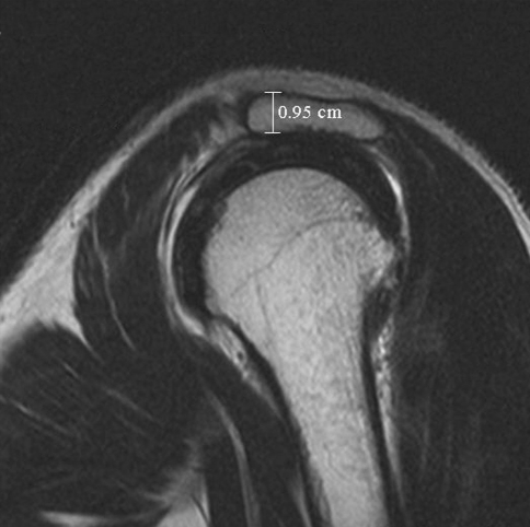
We evaluated plain radiographs, including shoulder true AP, lateral, axial, supraspinatus outlet, and 30° caudal tilt view; MRA or CTA also was performed in all patients. Acromial thickness, shape, and the distinct morphologic characteristics of acromial spurs were recorded by one musculoskeletal radiologist (JAC) using radiographic images. Acromial thickness was measured at the widest portion of the acromion on the perpendicular plane to the long axis of the acromion on the oblique sagittal image plane of MRA or CTA just lateral to the acromioclavicular joint; the measurement did not include the acromial spur (Fig. 1). Four of us (JYK, JAC, HKL, SKL) classified acromial shape as flat, curved, or hooked as described by Bigliani et al. [2]. Interobserver and intraobserver reliability of the morphologic classification of the acromial spur were calculated by kappa coefficient. For the interobserver reliability, acromial spur types were redetermined independently by two orthopaedic surgeons (HKL, SKL) after the classification scheme of acromial spurs was explained to them. The kappa coefficients for interobserver and intraobserver reliability in the coronal plane were 0.766/0.734 and 0.963/0.824 in the sagittal plane, respectively.
Fig. 1.
Acromial thickness was measured at the widest portion of the acromion on the perpendicular plane to the long axis of the acromion on the oblique sagittal image plane just lateral to the acromioclavicular joint.
We used Student’s t-test to compare continuous variables (age, acromial thickness, and tear size) between the two groups. The chi square test was used to evaluate discrete variables (gender, acromial shape, acromial spur, and types) between groups. The two-way ANOVA test was used to verify interactions between variables. Analyses were performed using the SPSS software package (Version 12.0; SPSS Inc, Chicago, IL).
Results
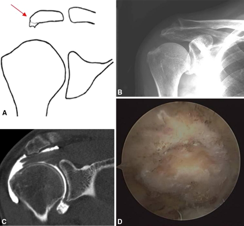
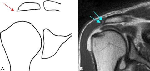
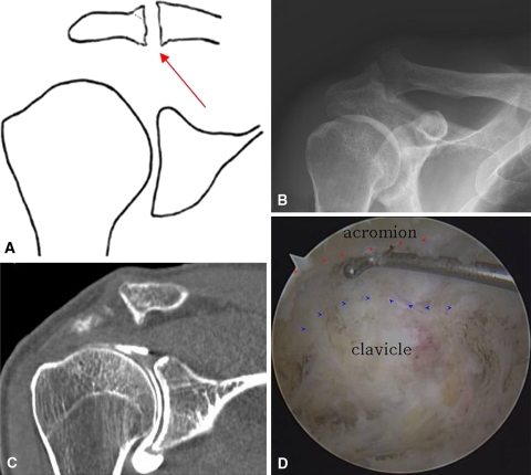
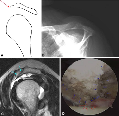
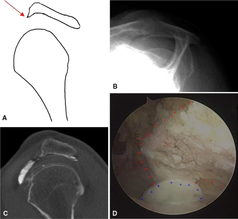
We observed distinct morphologic types of acromial spurs and categorized them into heel, lateral/anterior traction, lateral/anterior bird beak, and medial types. The quadrangular spur protruded inferiorly from the undersurface of the anterolateral acromion like the heel of a shoe in the heel-type spur (Fig. 2). Laterally protruding spurs were seen with the lateral traction and lateral bird beak-type spurs in the coronal plane. The lateral traction-type spur was congruent with the acromial undersurface or parallel to the rotator cuff tendons (Fig. 3). The lateral bird beak-type spur was incongruent with the acromial undersurface and protruded inferiorly with an acute angle at the lateral end (Fig. 4). The medial-type spur usually was associated with acromioclavicular joint arthritis, and these spurs originated from the medial end of the acromion and distal clavicle (Fig. 5). The anterior traction (Fig. 6) and anterior bird beak-type (Fig. 7) spurs were seen in the sagittal plane; they were anteriorly protruding acromial spurs similar to the lateral traction and lateral bird beak types in the coronal plane.
Fig. 2A–D.
(A) The inferior bony projection that looks like the heel of a shoe is shown in this diagram. This kind of spur was categorized as a heel-type spur. The heel-type spur can be seen on the (B) true AP radiograph of the shoulder, and with (C) CT arthrography and (D) arthroscopy.
Fig. 3A–B.
(A) The acromial spur is located at the lateral end of the acromion and is congruent with the acromial undersurface or parallel to the direction of the rotator cuff. This kind of spur was categorized as a traction-type spur. (B) Traction spurs can be seen with MR arthrography.
Fig. 4A–C.
(A) The acromial spur is located at the lateral end of the acromion. It is not congruent with the acromial undersurface or the rotator cuff. This spur was categorized as a bird beak spur and can be seen with (B) CT arthrography and (C) arthroscopy.
Fig. 5A–D.
(A) This diagram illustrates the spur located at the medial end of the acromion or distal end of the clavicle. It was categorized as a medial-type spur and can be seen on a (B) simple radiograph, or with (C) CT arthrography and (D) arthroscopy.
Fig. 6A–D.
(A) The diagram illustrates the anterior bony projection along with the coracoacromial ligament and the spur, which is congruent with the acromial undersurface or parallel to the rotator cuff. This kind of spur was categorized as a traction-type spur in the sagittal plane. Traction spurs in the sagittal plane spur were identified on (B) supraspinatus outlet views and with (C) MR arthrography and (D) arthroscopy.
Fig. 7A–D.
(A) The anteroinferior bony projection has the appearance of a bird beak. It is neither congruent with the acromial undersurface nor parallel to the rotator cuff. This type of spur was categorized as a bird beak-type spur in the sagittal plane. Bird beak-type spurs in the sagittal plane can be observed on (B) the supraspinatus outlet view and with (C) CT arthrography and (D) arthroscopy.
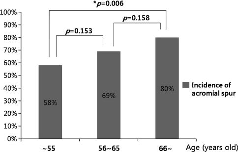
We detected acromial spurs in 142 of the 208 patients (68%), and their incidence increased with age. The acromial spurs were detected more frequently (p = 0.006) in patients older than 65 years (80%) than in patients younger than 55 years (58%) (Fig. 8). The overall spur incidence was similar for males and females and for each type of spur for males and females. The acromial spurs were detected more commonly in patients with full-thickness tears (83 of the 106 patients [78%] versus 59 of the 102 control subjects [58%]) (Table 2). Spurs were more frequent (p = 0.002) in patients with full-thickness cuff tears than in the control group. However, they were not related (p = 0.700, 0.248, respectively) with the size or retraction of the cuff tears (Table 3). The heel-type spur overall was the most common and was detected in 95 of the 208 subjects (46%). These heel-type spurs were observed more frequently (p = 0.004) in the full-thickness tear group (56%) than in the control group (35%). However, other types of acromial spurs occurred with similar frequency between the two groups (Table 2).
Fig. 8.
The incidence of acromial spurs increased with advancing age. Patients younger than 55 years had a lower incidence of spurs than older patients. There was no major difference between the 55- to 65-year-old group and the older than 65-year group.
Table 2.
Relationship between the type of acromial spur and rotator cuff tear
| Classification of acromial spurs | Rotator cuff tear group (n = 106) | Control group (n = 102) | Total (n = 208) |
|---|---|---|---|
| Acromial spur | 83* (78%) | 59 (58%) | 142 (68%) |
| Coronal spur | |||
| Heel | 59* (56%) | 36 (35%) | 95 (46%) |
| Traction | 4 (4%) | 2 (2%) | 6 (3%) |
| Bird beak | 17 (16%) | 8 (8%) | 25 (12%) |
| Medial | 7 (7%) | 5 (5%) | 12 (6%) |
| Sagittal spur | |||
| Bird beak | 29 (27%) | 21 (21%) | 50 (24%) |
| Traction | 27 (26%) | 16 (16%) | 43 (21%) |
* Significant difference between the two groups (p < 0.05).
Table 3.
Size of full-thickness rotator cuff tears according to the acromial spur
| Index group | Anteroposterior dimension (cm) | Retraction (cm) |
|---|---|---|
| Acromial spur (+) (n = 83) | 2.24 | 2.27 |
| Acromial spur (−) (n = 23) | 2.2 | 1.96 |
p > 0.05.
One hundred twenty-five patients (60%) had a flat-type acromion, 60 (28%) had the curved type, and 23 (11%) had the hooked type. There were no differences (p = 0.124) in the distribution of each acromial shape between the groups (Table 4). The mean acromion thickness was 8.0 mm (range, 5.0–11.3 mm). It was 8.3 mm (range, 5.0–11.3 mm) in the full-thickness rotator cuff tear group and 7.8 mm (range, 5.5–10.6 mm) in the control group. The acromions in the full-thickness rotator cuff tear group were thicker (p = 0.014) than those in the control group (Table 4). Acromions were thicker in men than in women although the two-way ANOVA showed gender was not a confounding factor because there was no interaction between groups and genders.
Table 4.
Relationship among acromial thickness, shape, and full-thickness rotator cuff tear
| Morphologic characteristics of acromion | Rotator cuff tear group (n = 106) | Control group (n = 102) | Total (n = 208) |
|---|---|---|---|
| Acromial shape | |||
| Flat | 70 (66%) | 55 (54%) | 125 (60%) |
| Curved | 24 (23%) | 36 (35%) | 60 (28%) |
| Hooked | 12 (11%) | 11 (11%) | 23 (11%) |
| Acromial thickness (mm) | 8.3* (5.0–11.3 ± 1.46) | 7.8 (5.5–10.6 ± 1.37) | 8.0 (5.0–11.3 ± 1.44) |
* Significant difference between the two groups (p < 0.05).
Discussion
Acromial spurs apparently form by traction of the coracoacromial ligament and reportedly are related to rotator cuff tears [5, 9, 20, 21, 26], although it is debatable whether it is the cause of a rotator cuff tear. We noticed similar morphological characteristics despite a wide variety of shapes. This suggests there could be several mechanisms of spur formation, and some kinds of spurs could affect the occurrence of rotator cuff tears more compared with other kinds. Therefore, we designed the current study to classify the morphologic characteristics of acromial spurs and to evaluate the correlations between acromial spurs and rotator cuff tears. This could provide useful guidance regarding the diagnosis and treatment of rotator cuff tears.
There are certain limitations to our study. First, spur classification by morphology is somewhat subjective, although interobserver and intraobserver reliability were 0.766/0.734 in the coronal plane and 0.963/0.824 in the sagittal plane, respectively. Not all spurs might fit the specific criteria we adopted. There were times when the traction and bird beak-type spurs could not be readily distinguished. However, based on our experience, most spurs were distinctive morphologically on MR or CT imaging. Furthermore, the heel-type spur can be detected easily on the true AP radiograph of the shoulder and provides an important clue to a suspected full-thickness rotator cuff tear. Second, our control group was comprised of patients who had shoulder pain without full-thickness rotator cuff tears confirmed by MRA or CTA. These may not reflect a healthy population, although we could not obtain MRA or CTA from healthy subjects without shoulder symptoms to confirm the presence or absence of a rotator cuff tear. We believe our study suggests one particular shape of spur (heel-type acromial spur) is seen more frequently in patients with full-thickness cuff tears than without tears. However, additional study with a more substantial number of patients, including a healthy population, would be needed to completely confirm the relationship between acromial spurs and rotator cuff tears. Finally, there were 59 patients with acromial spurs (58%) in the control group, and the heel-type spur was the most commonly found (35%). More than half of patients without full-thickness rotator cuff tears had an acromial spur, and approximately one-third of them had a heel-type spur. In other words, acromial spurs could occur without rotator cuff tears. This suggests acromial spurs should not be considered just the consequence of superior instability from rotator cuff tears. Nevertheless, we could not conclude that an acromial spur is the direct cause of the rotator cuff tear. However, careful followup of the control group can reveal the natural history of each kind of spur in the patients without rotator cuff tears. Unfortunately, we did not gather followup data of the control group, because patients in the control group were selected retrospectively. This is an observational study, and we believe more time is needed to follow up the patients in the control group.
Ogawa et al. [21] classified acromial spurs according to size and emphasized only large spurs measuring 5 mm or greater are of diagnostic value because of the high rate of association with the bursal-side and complete rotator cuff tears. We tried to classify acromial spurs according to their morphologic characteristics, because we noticed some acromial spurs share their typical shape. The mechanisms of each type of spur formation are unclear, but we presumed traction spurs were formed by the combination of traction of the coracoacromial ligament or deltoid muscle and interaction with the humeral head in humeroscapular motion interface beneath the coracoacromial arch. We supposed the formation of heel-type spurs contributed to direct abutment of the humeral head to the acromion by superior-directed microinstability. The mechanism of bird beak spur formation is believed to be the combination of traction spurs and heel-type spurs. Medial spurs supposedly result from osteophytes caused by acromial joint arthritis. Considering the above potential mechanisms, acromial spurs could be produced without rotator cuff tears; however, heel-type spurs seemed to be related more to rotator cuff tears. We found more heel-type spurs in patients with full-thickness rotator cuff tears. We could not determine whether the heel-type spur increases the possibility of full-thickness tears developing or whether the incidence of the heel-type spur is increased by adaptive change to superior instability of the shoulder as a result of full-thickness cuff tears. However, we observed an association of full-thickness rotator cuff tears and the heel-type acromial spur. Patients with partial rotator cuff tears and heel-type spurs seen on MRI or with heel-type spurs seen on true AP radiographs (Fig. 3) of the shoulder should be followed as they could progress to or might already have full-thickness rotator cuff tears.
There are numerous theories regarding the causative factors of rotator cuff tears, and none fully explain the etiology. Acromial spurs are a potential causative factor of rotator cuff tears. However, there is ongoing debate regarding whether acromial spurs are the cause or effect of rotator cuff tears. Panni et al. [24] and Bonsell et al. [4] suggest a spur is a degenerative change that increases with age, and is not associated with rotator cuff tears. In a case report regarding spur recurrence after acromioplasty, Anderson and Bowen [1] suggested morphologic features of the acromion may be a reactive change attributable to a primary cuff lesion. However, Ozaki et al. [23] and Liotard et al. [14] concluded the acromial spur was a degenerative change that could erode the rotator cuff. Jim et al. [13] and Ogawa et al. [21] reported large spurs measuring greater than 5 mm were associated with rotator cuff tears. Our data suggest the incidence of acromial spurs increases with age, and acromial spurs, especially the heel type, are more frequent in patients with full-thickness rotator cuff tears.
Since Neer [15, 16] reported the impingement theory and rotator cuff tears, numerous studies [2, 3, 8, 12, 17, 21, 23, 25, 26] have elucidated the details of the association between the acromial shape and rotator cuff disease. These authors emphasized the morphologic features of the acromion are closely associated with rotator cuff tears. However, Hirano et al. [12] reported rotator cuff injuries and the hook-type acromion were not associated; others suggested acromial length or angle is more important than acromial shape in rotator cuff tears [7, 19]. We believe it is difficult to obtain consistent images with the supraspinatus outlet view, because the projection angle can easily vary with the patient’s posture and the way the technician places the patient and cassette, and this could lead to getting different or distorted images. MR and CT are more common today for diagnosis of shoulder disorders, they provide more consistent and accurate information, and they can provide three-dimensional images of the acromion, spur shapes, and cuff and labral disorders. This might tend to focus more on the flat shape, because the acromial shape was determined by coronal, axial and sagittal cut images, although no specific acromial shape was more frequent in the full-thickness cuff tears. Also, Nicholson et al. [17] suggested acromial thickness is a primary anatomic structural feature and does not substantially change with age. We found patients with full-thickness rotator cuff tears had thicker acromions than patients without tears. A narrowed subacromial space might increase subacromial pressure and decrease vascularity of the cuff tendon. Also, a thicker acromion may be highly related to the attrition of the cuff at its undersurface. Therefore, we believe acromial thickness should be considered a factor in rotator cuff tear.
Morphologic features of acromial spurs can be classified as heel, lateral/anterior traction, lateral/anterior bird beak, or medial type on the coronal or sagittal plane. The incidence of acromial spurs increased with age, and, especially heel-type spurs, were detected more frequently in patients with full-thickness rotator cuff tears. These data provide simple and highly useful information for diagnosis of full-thickness rotator cuff tears, and a heel-type spur might be a risk factor for a full-thickness rotator cuff tear.
Acknowledgments
We thank Ki Hyun Jo, MD, Suk Jae Lee, MD, Hye Ran Kim, and Sang Mi Shim for support with data collection and patient recruitment. We also thank Pacific Edit for reviewing English in the manuscript before submission. Statistical analyses were performed by Hyun Kang, PhD, from the statistics department of our institution.
Footnotes
Each author certifies that he or she has no commercial associations (eg, consultancies, stock ownership, equity interest, patent/licensing arrangements, etc) that might pose a conflict of interest in connection with the submitted article.
Each author certifies that his or her institution has approved the human protocol for this investigation and that all investigations were conducted in conformity with ethical principles of research.
This work was performed at Seoul National University College of Medicine, Seoul, Korea.
References
- 1.Anderson K, Bowen MK. Spur reformation after arthroscopic acromioplasty. Arthroscopy. 1999;15:788–791. doi: 10.1016/S0749-8063(99)70018-6. [DOI] [PubMed] [Google Scholar]
- 2.Bigliani LU, Morrison DS, April EW. The morphology of the acromion and its relationship to rotator cuff tears. Orthop Trans. 1986;10:228. [Google Scholar]
- 3.Bigliani LU, Ticker JB, Flatow EL, Soslowsky LJ, Mow VC. [Relationship of acromial architecture and diseases of the rotator cuff] [in German] Orthopade. 1991;20:302–309. [PubMed] [Google Scholar]
- 4.Bonsell S, Pearsall AW, IV, Heitman RJ, Helms CA, Major NM, Speer KP. The relationship of age, gender, and degenerative changes observed on radiographs of the shoulder in asymptomatic individuals. J. Bone Joint Surg. Br. 2000;82:1135–1139. doi: 10.1302/0301-620X.82B8.10631. [DOI] [PubMed] [Google Scholar]
- 5.Chambler AF, Pitsillides AA, Emery RJ. Acromial spur formation in patients with rotator cuff tears. J. Shoulder Elbow Surg. 2003;12:314–321. doi: 10.1016/S1058-2746(03)00030-2. [DOI] [PubMed] [Google Scholar]
- 6.Chansky HA, Iannotti JP. The vascularity of the rotator cuff. Clin. Sports Med. 1991;10:807–822. [PubMed] [Google Scholar]
- 7.Edelson JG, Taitz C. Anatomy of the coraco-acromial arch: relation to degeneration of the acromion. J. Bone Joint Surg. Br. 1992;74:589–594. doi: 10.1302/0301-620X.74B4.1624522. [DOI] [PubMed] [Google Scholar]
- 8.Epstein RE, Schweitzer ME, Frieman BG, Fenlin JM, Mitchell DG. Hooked acromion: prevalence on MR images of painful shoulders. Radiology. 1993;187:479–481. doi: 10.1148/radiology.187.2.8475294. [DOI] [PubMed] [Google Scholar]
- 9.Fealy S, April EW, Khazzam M, Armengol-Barallat J, Bigliani LU. The coracoacromial ligament: morphology and study of acromial enthesopathy. J. Shoulder Elbow Surg. 2005;14:542–548. doi: 10.1016/j.jse.2005.02.006. [DOI] [PubMed] [Google Scholar]
- 10.Harryman DT, 2nd, Sidles JA, Clark JM, McQuade KJ, Gibb TD, Matsen FA., III Translation of the humeral head on the glenoid with passive glenohumeral motion. J. Bone Joint Surg. Am. 1990;72:1334–1343. [PubMed] [Google Scholar]
- 11.Hayes PR, Flatow EL. Attrition sign in impingement syndrome. Arthroscopy. 2002;18:E44. doi: 10.1053/jars.2002.36462. [DOI] [PubMed] [Google Scholar]
- 12.Hirano M, Ide J, Takagi K. Acromial shapes and extension of rotator cuff tears: magnetic resonance imaging evaluation. J. Shoulder Elbow Surg. 2002;11:576–578. doi: 10.1067/mse.2002.127097. [DOI] [PubMed] [Google Scholar]
- 13.Jim YF, Chang CY, Wu JJ, Chang T. Shoulder impingement syndrome: impingement view and arthrography study based on 100 cases. Skeletal Radiol. 1992;21:449–451. doi: 10.1007/BF00190989. [DOI] [PubMed] [Google Scholar]
- 14.Liotard JP, Cochard P, Walch G. Critical analysis of the supraspinatus outlet view: rationale for a standard scapular Y-view. J. Shoulder Elbow Surg. 1998;7:134–139. doi: 10.1016/S1058-2746(98)90223-3. [DOI] [PubMed] [Google Scholar]
- 15.Neer CS., II Anterior acromioplasty for the chronic impingement syndrome in the shoulder: a preliminary report. J. Bone Joint Surg. Am. 1972;54:41–50. [PubMed] [Google Scholar]
- 16.Neer CS., II Impingement lesions. Clin. Orthop. Relat. Res. 1983;173:70–77. [PubMed] [Google Scholar]
- 17.Nicholson GP, Goodman DA, Flatow EL, Bigliani LU. The acromion: morphologic condition and age-related changes. A study of 420 scapulas. J. Shoulder Elbow Surg. 1996;5:1–11. doi: 10.1016/S1058-2746(96)80024-3. [DOI] [PubMed] [Google Scholar]
- 18.Nixon JE, DiStefano V. Ruptures of the rotator cuff. Orthop. Clin. North Am. 1975;6:423–447. [PubMed] [Google Scholar]
- 19.Nyffeler RW, Werner CM, Sukthankar A, Schmid MR, Gerber C. Association of a large lateral extension of the acromion with rotator cuff tears. J. Bone Joint Surg. Am. 2006;88:800–805. doi: 10.2106/JBJS.D.03042. [DOI] [PubMed] [Google Scholar]
- 20.Ogata S, Uhthoff HK. Acromial enthesopathy and rotator cuff tear: a radiologic and histologic postmortem investigation of the coracoacromial arch. Clin. Orthop. Relat. Res. 1990;254:39–48. [PubMed] [Google Scholar]
- 21.Ogawa K, Yoshida A, Inokuchi W, Naniwa T. Acromial spur: relationship to aging and morphologic changes in the rotator cuff. J. Shoulder Elbow Surg. 2005;14:591–598. doi: 10.1016/j.jse.2005.03.007. [DOI] [PubMed] [Google Scholar]
- 22.Ouellette H, Labis J, Bredella M, Palmer WE, Sheah K, Torriani M. Spectrum of shoulder injuries in the baseball pitcher. Skeletal Radiol. 2008;37:491–498. doi: 10.1007/s00256-007-0389-0. [DOI] [PubMed] [Google Scholar]
- 23.Ozaki J, Fujimoto S, Nakagawa Y, Masuhara K, Tamai S. Tears of the rotator cuff of the shoulder associated with pathological changes in the acromion: a study in cadavera. J. Bone Joint Surg. Am. 1988;70:1224–1230. [PubMed] [Google Scholar]
- 24.Panni AS, Milano G, Lucania L, Fabbriciani C, Logroscino CA. Histological analysis of the coracoacromial arch: correlation between age-related changes and rotator cuff tears. Arthroscopy. 1996;12:531–540. doi: 10.1016/S0749-8063(96)90190-5. [DOI] [PubMed] [Google Scholar]
- 25.Toivonen DA, Tuite MJ, Orwin JF. Acromial structure and tears of the rotator cuff. J. Shoulder Elbow Surg. 1995;4:376–383. doi: 10.1016/S1058-2746(95)80022-0. [DOI] [PubMed] [Google Scholar]
- 26.Tucker TJ, Snyder SJ. The keeled acromion: an aggressive acromial variant—a series of 20 patients with associated rotator cuff tears. Arthroscopy. 2004;20:744–753. doi: 10.1016/j.arthro.2004.06.018. [DOI] [PubMed] [Google Scholar]


