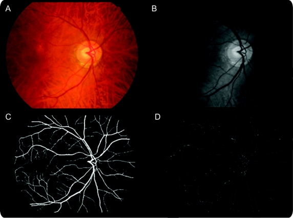
Figure 1 Retinal images showing the processing, segmentation, and skeletonization steps
Top left image (A) is the original color image which is processed to grayscale (top right B) which is then segmented to produce the bottom left image (C). Finally, this image is skeletonized to produce the bottom right image (D) upon which fractal analyses are based.

