Abstract
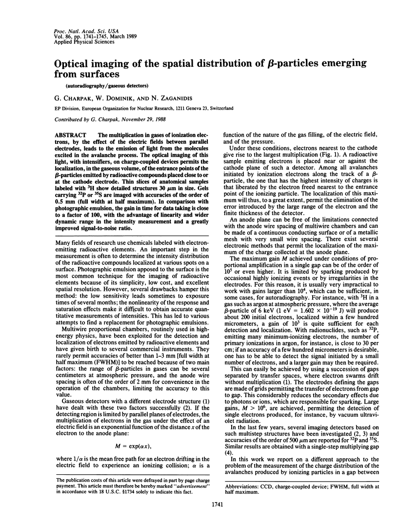
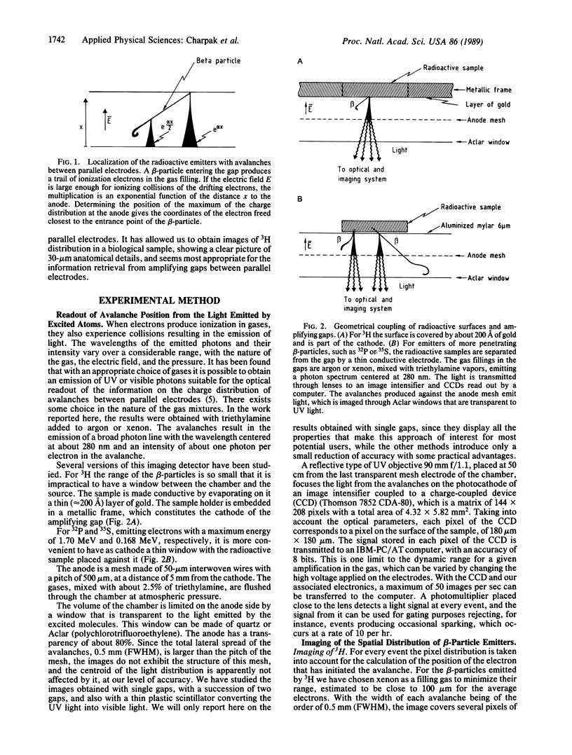
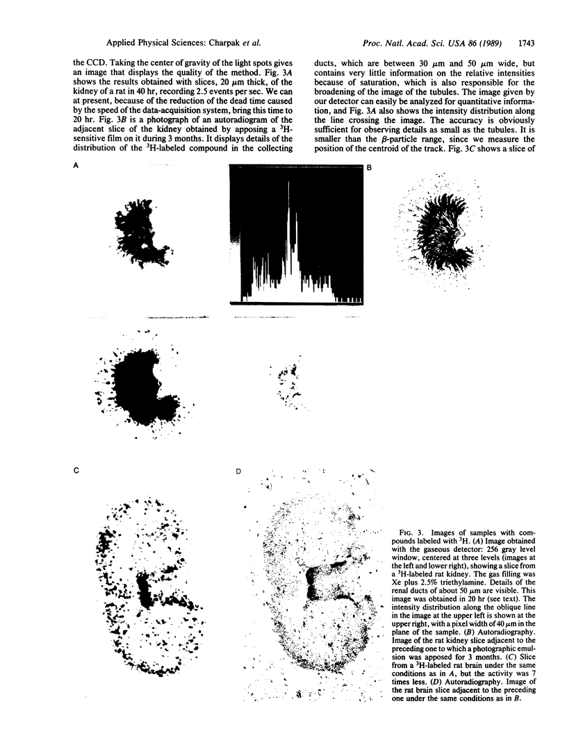
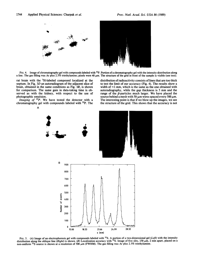
The multiplication in gases of ionization electrons, by the effect of the electric fields between parallel electrodes, leads to the emission of light from the molecules excited in the avalanche process. The optical imaging of this light, with intensifiers, on charge-coupled devices permits the localization, in the gaseous volume, of the entrance points of the beta-particles emitted by radioactive compounds placed close to or at the cathode electrode. Thin slices of anatomical samples labeled with 3H show detailed structures 30 microns in size. Gels carrying 32P or 35S are imaged with accuracies of the order of 0.5 mm (full width at half maximum). In comparison with photographic emulsion, the gain in time for data taking is close to a factor of 100, with the advantage of linearity and wider dynamic range in the intensity measurement and a greatly improved signal-to-noise ratio.
Full text
PDF