Abstract
Serial ultrasonographic examinations were performed daily on 9 Miniature Schnauzer bitches from the 15th day of gestation until parturition to determine the time the gestational structures were first detected. The gestational age was timed from the day of ovulation (day 0), which was estimated to occur when the plasma progesterone concentration was >4.0 ng/ml. The gestational length in 9 Miniature Schnauzer bitches was found to be 63.0 ± 1.7 (range 61-65) days. The initial detection of the fetal and extra-fetal structures were as follows: gestational sac at day 18.0 ± 0.9 (17-19); zonary placenta in the uterine wall at day 24.9 ± 1.1 (23-26); yolk sac membrane at day 25.0 ± 0.9 (24-26); amnionic membrane at day 27.7 ± 1.0 (26-29); embryo initial detection at day 22.6 ± 0.5 (22-23); heartbeat at day 23.4 ± 0.5 (23-24); fetal movement at day 32.5 ± 0.8 (32-34); stomach at day 31.2 ± 1.6 (29-33); urinary bladder at day 32.6 ± 1.8 (31-35); skeleton at day 34.9 ± 1.6 (34-38) and kidney at day 42.2 ± 0.7 (41-43).
Keywords: gestational structures, Miniature Schnauzer bitches, ultrasonography
Introduction
The early determination of pregnancy and the gestational age are important for reproductive management in small animal practices. In addition, predicting the parturition date can help in managing parturition or planning a Cesarean section in pregnant bitches with multiple mating or an unknown mating time [12].
Ultrasonography is a useful imaging modality for determining pregnancy [4,7,13], estimating the litter size [5,13], fetal development [14, 18], uterine examination after parturition [16], a reproductive examination and the direct detection of ovulation [1].
In veterinary medicine, estimation of the gestational age based on the anatomic appearance and predicting the parturition date by an ultrasonographic examination has been reported [12,18]. Therefore, the range for detecting the features of pregnancy (≤5 days) might also be reliable for estimating the gestational age [11,20].
However, there are no reports of gestational structures using ultrasonography in Miniature Schnauzer bitches. The aim of this study was to make an early determination of pregnancy, to establish the time for the initial detection of the extra-fetal and fetal structures using ultrasonography, and to provide the basic data for estimating the gestational age in Miniature Schnauzer bitches.
Materials and Methods
Experimental animals
Nine healthy Miniature Schnauzer bitches, aged 1 to 3 years old, weighing 5.0 to 6.9 kg were housed in individual in indoor-outdoor runs. The dogs were fed a standard commercial dog food twice daily, with water available ad libitum. Each dog was examined twice daily for any swelling of the vulva and the presence of a vaginal discharge, which signified the onset of proestrus.
The bitches were mated if these signs were present. The bitches became pregnant and whelped 2-6 pups each. Serial ultrasonographic examinations were performed on a total of 34 litters to make an early diagnosis of pregnancy and determine the time of the initial detection of the extra-fetal and fetal structures.
The estimation of optimal mating time and ovulation time
Vaginal cytology
To estimate the optimal mating time, serial vaginal smears were performed every other day from the onset of proestrus to the onset of diestrus. Vaginal cytological evaluations were performed as described by Schutte [17] and mating was performed when the cornification index was ≥90%, as described by Kim et al. [10].
Plasma progesterone concentration measurement
Ovulation (day 0) was estimated by measuring the plasma progesterone concentration daily from the onset of proestrus to the onset of diestrus using a 125I radioimmunoassay (RIA) that had been previously validated for a fertility breeding management [15].
Blood samples were collected through a cephalic venipuncture, placed immediately in chilled EDTA-coated tubes, and centrifuged for 10 min at 3,000 × g. The plasma was stored at -25℃ until analysis.
The plasma progesterone concentrations were determined in duplicate using a commercial progesterone kit (Progesterone-Coat-A-Count; Diagnostic Products, USA) with Gamma counter (EG & G Wallce, Finland), as described by Kim et al. [10].
The time of ovulation was estimated based on the when the plasma progesterone level was first >4.0 ng/ml, as described by Kim et al. [10] and Wallace et al. [19]. Ovulation was designated the first day of gestation (day 0).
Ultrasonographic examination
Serial ultrasonographic examinations were performed daily from day 15 until parturition. All the dogs were examined using real-time B-mode ultrasonography in dorsal recumbency. Ultrasonographic examinations were performed using LOGIQ 7 (GE Medical System, USA) with a 3.5C, 7L, 10L MHz transducer on 34 litters from 9 Miniature Schnauzer bitches.
The gestational age at the time of the initial detection and the appearance of the following features of pregnancy were recorded: gestational sac, zonary placenta, fetal membrane, embryo, heartbeat, limb buds, skeleton, fetal movement, and abdominal viscera.
Results
The average gestational duration of the 9 Miniature Schnauzer bitches was 63.0 ± 1.7 (range: 61-65) days, and the average litter size was 3.8 litters (total 34 litters).
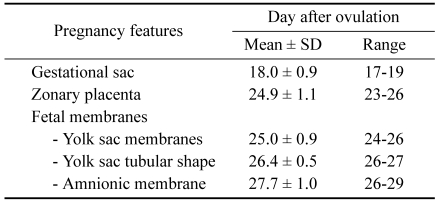
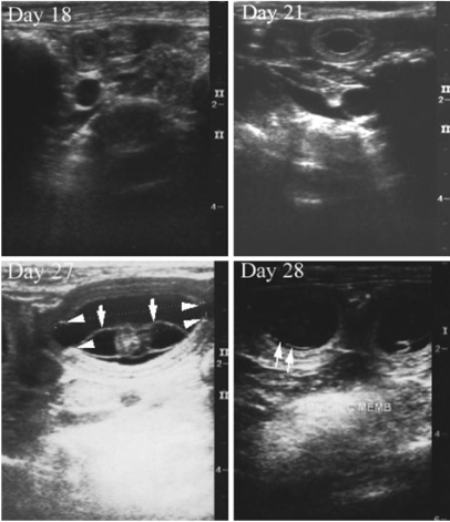
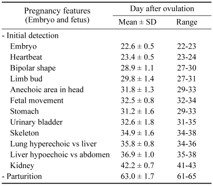
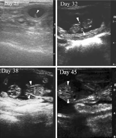
Table 1 and Fig. 1 show the time of the initial detection and presence of extra-fetal structures, and Table 2 and Fig. 2 show the time of the initial detection and presence of fetal structures.
Table 1.
Mean and range of the gestational age at the first ultrasonographic detection of the extra-fetal structures in 9 Miniature Schnauzer bitches
Fig. 1.
Ultrasonograms of the extra-fetal structures in pregnant Miniature Schnauzer bitches. Day 18: Transverse image of the first detection of an anechoic gestational sac. Day 21: Longitudinal image of the gestational sac. An echogenic inner placental layer was detected in the uterine wall. Day 27: Longitudinal image of the gestational sac contained an embryo and the tubular shape of the yolk sac membrane (white arrows). The zonary placenta (white arrowheads) was cylindrical in shape and appeared folded inward at the edges. Day 28: The amnionic membrane (white arrows) appeared faint.
Table 2.
Mean and range of the gestational age at the first ultrasonographic detection of the fetal structures in 9 Miniature Schnauzer bitches
Fig. 2.
Ultrasonograms of the fetal structures in pregnant Miniature Schnauzer bitches. Day 23: Transverse image of the gestational sac contained an oblong-shaped embryo (white arrowhead) apposed to the uterine wall. Day 32: Longitudinal image of a fetus with an anechoic area in the head and forelimb buds (white arrowheads). The fetal crown-rump length was 20 mm. Day 38: Longitudinal image of a fetus with hyperechoic skeletal structures in the head and thoracic wall (white arrowheads). Day 45: Longitudinal image of a fetal vertebral column and kidney (white arrowheads).
The time of initial detection of extra-fetal structures
As shown in Table 1, the anechogenic gestational sac was first detected on day 18.0 ± 0.9 (range: 17-19), compared with the hyperechoic uterus (Fig. 1, Day 18). The echogenic inner layers (Fig. 1, Day 21) surrounding the gestational sac was developed into a zonary placenta (Fig. 1, Day 27), which was cylindrical in shape and appeared folded inward at the edges in the longitudinal plane on day 24.9 ± 1.1 (23-26).
On day 25.0 ± 0.9 (24-26), the yolk sac membrane was first detected as an echogenic U-shape fetal membrane. In the longitudinal plane, the yolk sac membrane was changed from its initial U-shape to a tubular structure (Fig. 1, Day 27) extending from pole to pole of the chorionic cavity on day 26.4 ± 0.5 (26-27). A second, less echogenic fetal membrane, the amnionic membrane, was detected on day 27.7 ± 1.0 (26-29), which encompassed the embryo (Fig. 1, Day 28).
The time of initial detection of fetal structures
As shown in Table 2, on day 22.6 ± 0.5 (22-23), the embryo (Fig. 2, Day 23) was first detected as an oblong structure apposed to the uterine wall in the anechogenic chorionic cavity. The heartbeat, which is one of fetal vital signs, was detected as a flickering motion on day 23.4 ± 0.5 (23-24). The features of the embryo changed from an oblong to bipolar shape (Fig. 2, Day 32) on day 28.9 ± 1.1 (27-30). The limb buds were first detected on day 29.8 ± 1.4 (27-31), and fetal movement was detected on day 32.5 ± 0.8 (32-34). An anechogenic area in the head was detected on day 31.8 ± 1.3 (29-33), and the first abdominal viscera detected were the stomach and urinary bladder on day 31.2 ± 1.6 (29-33) and 32.6 ± 1.8 (31-35), respectively. The skeleton (Fig. 2, Day 38) was detected as a hyperechoic structure on day 34.9 ± 1.6 (29-33).
On day 35.8 ± 0.8 (34-36), the lung became hyperechoic, compared with the liver. At this time, the abdomen and thorax were distinct. The liver was observed to be hypoechoic, compared with the rest of the abdomen on day 36.9 ± 1.0 (35-38). The kidneys (Fig. 2, Day 45) were first detected on day 42.2 ± 0.7 (41-43).
Discussion
It is difficult to specify the length of pregnancy precisely for the following reasons: (1) the bitch can be mated over a period of several days and mating can even occur before ovulation; (2) ovulation can also occur over a period of time; (3) recently ovulated eggs cannot be fertilized but require some time, 2-5 days, for meiotic cleavage and maturation before fertilization is possible; (4) and the spermatozoa can survive several days in the reproductive tract of the bitch [2].
However, the length of canine gestation can be applied based on the time of mating, determination of the luteinizing hormone surge and the initial increase in the progesterone level [3,12,19].
The length of canine gestation appears highly variable based on the when mating occurs (57 and 72 days). A range of 15 days is a wide interval. Hence, the length of gestation can vary greatly when determined from the day of mating [12].
The surge in the luteinizing hormone (LH) level causes ovulation, which occurs approximately 2 days after this peak. Therefore, the canine gestation time is 65 ± 1 days when timed from the preovulatory LH surge in the peripheral blood [3]. The day of ovulation can be detected on the initial increase in the progesterone level. Ovulation was estimated to occur when the plasma progesterone concentration was first >4.0 ng/ml, as described by Kim et al. [10] and Wallace et al. [19].
Determining the time of ovulation based on the luteinizing hormone surge and the initial increase in progesterone provides an accurate estimation of the gestational age but this is somewhat impractical.
Therefore, ultrasonographic observations throughout pregnancy can be used to estimate the gestational age, level of fetal growth and the parturition date.
In this study, ovulation was considered the first day of gestation (day 0), when the initial progesterone concentration was ≥4.0 ng/ml. The length of gestation in Miniature Schnauzer bitches was found to be 63.0 ± 1.7 days (range: 61-65 days), which is similar to that the average gestation length in Beagle bitches, 63 ± 1 days [8].
The earliest time in which an early diagnosis of pregnancy by ultrasonographic examination in bitches can be made usually ranges from days 18-25 when an anechoic gestational sac surrounded by uterine wall is detected [4,7,9,11,20]. In this study, the gestational sac was first detected on day 17-19. The differences in the time for the early detection of pregnancy might be caused by the transducer frequency, differences in timing gestation and the dog breed. These conditions might affect the time that the pregnancy features are identified [20]. In particular, the early detection of pregnancy in this study (as early as day 17 of gestation) was achieved using a higher frequency transducer (10 MHz) with a new generation of ultrasound equipment.
In the uterine wall surrounding the gestational sac, an apparently hyperechoic inner layer was differentiated to the zonary placenta on day 24.9 ± 1.1 (23-26), which is similar to day 24-28 after ovulation [11] and day 27-30 after the preovulatory LH surge [20].
In the fetal structures, the embryo and flickering motion of the heartbeat was first detected on day 22.6 ± 0.5 (22-23) and day 23.4 ± 0.5 (23-24), respectively. The ultrasonographic detection of the embryo and heartbeat was the same for all dogs examined. Days 21 to 26 and days 21 to 27 were characterized by the finding of an embryo and heartbeat, respectively [8,9,11,20].
Yeager et al. [20] reported little anatomic differentiation within the embryo before day 30. Before day 30, the only distinguishable features of the bipolar embryo were the flickering motion of the heartbeat and the anechoic area in the head. In this study, the bipolar shape of the embryo, heartbeat and limb bud were first detected before day 30. An anechoic area in the head was detected on days 29 to 33.
Most fetal anatomic structures were detected between days 30 to 50 [20]. At this time, there was a similar time when the fetal structures were first detected (fetal movement, skeleton, stomach, urinary bladder, kidney) in the present and other studies [8,9,11,20]. For these reasons, the time the extra-fetal and fetal structures are first detected is useful for estimating the gestational age.
There are little variations (≤3 days) in the range of gestational age at the time when most pregnancy features are detected, whereas the range was 5 days in other studies [8,11,20]. This suggests that the detection of pregnant feature might be more reliable for estimating the gestational age. Differences in this result may partly explain the differences between studies. Under certain conditions, the transducer frequency might also affect the gestational age at which the pregnancy features are first detected [20]. In this study, because a higher frequency transducer was used, the time pregnancy was first detected in Miniature Schnauzer bitches was day 18 ± 0.9 (range: 17-19 day), and most pregnancy features were detected at the same gestational age or 2 to 3 days earlier than in other studies [9,11,20].
These results, as well as those related to the subsequent development of the early conceptuses and embryos, show that normal conceptuses follow a well-defined developmental course; particular characteristics develop at specific times during gestation. It is clear that an evaluation of these characteristics might be useful for predicting the time of parturition and may also be applicable to other breeds of dogs [6].
References
- 1.Boyd JS, Renton JP, Harvey MJ, Nickson DA, Eckersall PD, Ferguson JM. Problems associated with ultrasonography of the canine ovary around the time of ovulation. J Reprod Fertil Suppl. 1993;47:101–105. [PubMed] [Google Scholar]
- 2.Christiansen IJ. Reproduction in the Dog and Cat. 1st ed. London: Baillière Tindall; 1984. pp. 118–119. [Google Scholar]
- 3.Concannon P, Rendano V. Radiographic diagnosis of canine pregnancy: onset of fetal skeletal radiopacity in relation to times of bleeding, preovulatory luteinizing hormone release, and parturition. Am J Vet Res. 1983;44:1506–1511. [PubMed] [Google Scholar]
- 4.England GCW, Allen EW. Studies on canine pregnancy using B-mode ultrasound: Diagnosis of early pregnancy and the number of conceptuses. J Small Anim Pract. 1990;31:321–323. [Google Scholar]
- 5.England GCW, Porter DJ. Studies on canine pregnancy using B-mode ultrasound: development of the conceptus and determination of gestational age. J Small Anim Pract. 1990;31:324–329. [Google Scholar]
- 6.England GCW, Russo M. Ultrasonographic characteristics of early pregnancy failure in bitches. Theriogenology. 2006;66:1694–1698. doi: 10.1016/j.theriogenology.2006.01.028. [DOI] [PubMed] [Google Scholar]
- 7.England GCW, Yeager AE. Ultrasonographic appearance of the ovary and uterus of the bitch during oestrus, ovulation and early pregnancy. J Reprod Fertil Suppl. 1993;47:107–117. [PubMed] [Google Scholar]
- 8.Holst PA, Phemister RD. Onset of diestrus in the Beagle bitch: definition and significance. Am J Vet Res. 1974;35:401–406. [PubMed] [Google Scholar]
- 9.Kang BK, Choi HS, Son CH, Shin CR, Seo DH, Park IC. Ultrasonographic apperance of the gestational structures throughout pregnancy in pet dogs. I. Time of initial detection of the fetal and extra-fetal structures. Korean J Vet Clin Med. 1997;14:279–286. [Google Scholar]
- 10.Kim JH, Jeong KA, Kang HG, Oh KS, Park IC, Park SG, Han HJ, Son CH. Relationship between vaginal cytology and reproductive hormone during the estrous cycle in Korea Jin-do bitches. Korean J Vet Clin Med. 2000;17:225–233. [Google Scholar]
- 11.Ko JS, Kim BS, Lee SA, Cho YT, Kim JP, Oh KS, Kim SH, Kim JT, Park IC, Kim YH, Son CH. Ultrasonographic apperance of the gestational structures throughout pregnancy in Shih-tzu bitches. I. Time of initial detection of the fetal and extra-fetal structures. Korean J Vet Clin. 2004;21:29–34. [Google Scholar]
- 12.Luvoni GC, Beccaglia M. The prediction of parturition date in canine pregnancy. Reprod Domest Anim. 2006;41:27–32. doi: 10.1111/j.1439-0531.2006.00641.x. [DOI] [PubMed] [Google Scholar]
- 13.Luvoni GC, Grioni A. Determination of gestational age in medium and small size bitches using ultrasonographic fetal measurements. J Small Anim Pract. 2000;41:292–294. doi: 10.1111/j.1748-5827.2000.tb03204.x. [DOI] [PubMed] [Google Scholar]
- 14.Moriyoshi M, Waki Y, Nakao T, Kawata K. Observation of the growth process of a beagle embryo and fetus by ultrasonography. J Vet Med Sci. 1996;58:443–445. doi: 10.1292/jvms.58.443. [DOI] [PubMed] [Google Scholar]
- 15.Okkens AC, Teunissen JM, Van Osch W, Van Den Brom WE, Dieleman SJ, Kooistra HS. Influence of litter size and breed on the duration of gestation in dogs. J Reprod Fertil Suppl. 2001;57:193–197. [PubMed] [Google Scholar]
- 16.Pharr JW, Post K. Ultrasonography and radiography of the canine postpartum uterus. Vet Radiol Ultrasound. 1992;33:35–40. [Google Scholar]
- 17.Schutte AP. Canine vaginal cytology. I. Technique and cytological morphology. J Small Anim Pract. 1967;8:301–306. doi: 10.1111/j.1748-5827.1967.tb04554.x. [DOI] [PubMed] [Google Scholar]
- 18.Son CH, Jeong KA, Kim JH, Park IC, Kim SH, Lee CS. Establishment of the prediction table of parturition day with ultrasonography in small pet dogs. J Vet Med Sci. 2001;63:715–721. doi: 10.1292/jvms.63.715. [DOI] [PubMed] [Google Scholar]
- 19.Wallace SS, Mahaffey MB, Miller DM, Thompson FN, Chakraborty PK. Ultrasonographic appearance of the ovaries of dogs during the follicular and luteal phases of the estrous cycle. Am J Vet Res. 1992;53:209–215. [PubMed] [Google Scholar]
- 20.Yeager AE, Mohammed HO, Meyers-Wallen V, Vannerson L, Concannon PW. Ultrasonographic appearance of the uterus, placenta, fetus, and fetal membranes throughout accurately timed pregnancy in beagels. Am J Vet Res. 1992;53:342–351. [PubMed] [Google Scholar]


