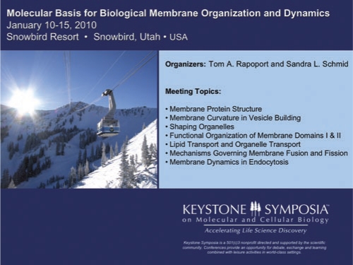
The Keystone Symposium on the Molecular Basis for Biological Membrane Organization and Dynamics held in January this year offered new insights into the molecular machines at work within cells. Topics included the machinery responsible for the dynamic shape of organelles, the budding and fusion of vesicular carriers, and the intricate sorting systems that ensure the correct delivery of cellular components.
Abstract
The Keystone Symposium on the Molecular Basis for Biological Membrane Organization and Dynamics held in January this year offered new insights into the molecular machines at work in cells. Topics included the machinery responsible for the dynamic shape of organelles, the budding and fusion of vesicular carriers, and the intricate sorting systems that ensure the correct delivery of cellular components.
 |
The acquisition of intracellular membranes allowed eukaryotic cells to delegate former plasma membrane activities to other organelles. The thin, flexible membrane of the endoplasmic reticulum (ER), for example, proved ideal for protein insertion and translocation, and for inserting lipids into bilayer leaflets. However, with membrane assembly assigned to the ER, eukaryotic cells had to solve the problem of providing the plasma membrane with its specific lipid and protein components.
The solution was a vesicular transport pathway between the ER and the plasma membrane. Modern eukaryotic cells also contain several additional membranes including the Golgi complex and the endocytic pathway. All of these membranes are interconnected by bidirectional vesicular traffic, yet each maintains a unique lipid and protein complement. New insights into these topics and the molecular machinery driving these systems were discussed at the recent Keystone meeting on the Molecular Basis for Biological Membrane Organization and Dynamics, held this January. This stimulating 5-day symposium was organized by Tom Rapoport (Harvard Medical School) and Sandra Schmid (The Scripps Research Institute) and gave the participants ample time to deliberate these complex cellular processes, both in the lecture room and on the sunny slopes of Snowbird, Utah.
Curvature and budding
The dynamic shape of organelles is determined largely by mechanisms that confer curvature and tension. Yoko Shibata (Harvard Medical School) reported that ER tubules are stabilized by spiral scaffolds of wedges, formed by the double hairpin structures of the reticulons and DP1/Yop1. These membrane proteins interact with membrane-bound, dynamin-like proteins, which include the atlastins in mammals and Sey1 in yeast, which are additionally required for the formation of the dynamic ER network. Similar mechanisms might shape ER sheets, the nuclear envelope and mitochondria. The high curvature in the nuclear pore is stabilized by the nuclear pore complex, which is related structurally to the COPII vesicle coat (Thomas Schwartz, MIT).
Emanuele Cocucci (Immune Disease Institute, Harvard Medical School) showed that in clathrin-mediated endocytosis, two AP2 adaptors and one clathrin triskelion are sufficient to initiate coat assembly at the plasma membrane. This event can result in a clathrin-coated bud and vesicle, or a flat clathrin-coated plaque, which cannot interconvert. The alternative endocytic coat protein caveolin binds to cellular membranes by its hydrophobic loop and triple palmitoyl residues. However, a cytosolic multiprotein complex consisting of various cavins is needed to stabilize caveolin-induced membrane deformation to produce caveolae in vivo. Rob Parton (U. Queensland, Australia) showed that the cavin complex is not recruited to the Golgi pool of caveolin. The loss of caveolin 1 in a number of cancer cells seems to be accompanied by a loss of cavin 1. Debbie Brown (Stony Brook U., New York) reported that re-expression of caveolin 1 by itself gave rise to long microtubule-dependent and short actin-dependent tubules, which might reflect interactions that are normally modulated by cavin 1.
Flippases are members of the P4 family of P-type ATPases situated in the various sphingolipid- and cholesterol-rich membranes. They translocate the aminophospholipids phosphatidylserine and phosphatidylethanolamine from the exoplasmic to the cytosolic membrane leaflet, and there are various experimental and theoretical lines of evidence that they provide (part of) the driving force for budding in the late secretory and endocytic pathways. Poul Nissen (U. Aarhus, Denmark) described the massive conformational changes that occur when cation-transporting P-type ATPases undergo their catalytic cycle, although how such proteins translocate lipids across the bilayer remains unclear. Lipid transbilayer translocation alone is unable to drive fission. By contrast, Michael Kozlov (Tel-Aviv U.) reported that N-BAR-containing proteins, such as endophilin and amphiphysin, can induce fission by themselves. These proteins bend the membrane by the shallow insertion of amphipathic helices and membrane scaffolding through their concave surface. Moreover, many fission events depend on specialized proteins such as dynamin. Interestingly, Schmid reported that shallow insertion of a hydrophobic loop of the PH domain of dynamin is crucial for high membrane curvature generation and subsequent membrane fission.
The mitochondrial division machinery requires dynamin-related proteins (DRPs). Jodi Nunnari (U. California, Davis) described that co-assembly with specific DRP-associated proteins nucleates and promotes the self-assembly of DRPs into helical structures that drive membrane scission. Some proteins that dip into the bilayer might curve the membrane at high concentrations, but may sense curvature when present at low concentrations. Bruno Antonny (Institute of Molecular and Cellular Pharmacology, Valbonne, France) reported how the ALPS domain of ARF-GAP, an amphipathic helix with small, uncharged residues on its polar side, only inserts into membranes with a radius ≤35 nm, suggesting a way to programme coat disassembly by curvature (Ambroggio et al, 2010).
…massive conformational changes […] occur when cation-transporting P-type ATPases undergo their catalytic cycle…
Tubulation of endosomes is driven by the sorting nexins of the retromer complex—SNX1, -2, -5 and -6—which drive membrane tubulation through their carboxy-terminal BAR domains, as reported by Matthew Seaman (U. Cambridge, UK). Budding and fission also occur in the opposite direction, away from the cytosol. When internalized receptors and other cargo are destined for lysosomal degradation, they are ubiquitinated and sorted by the ESCRT complexes 0, I, II and III into multivesicular bodies. Thomas Wollert (NIH) described reconstitution of the complete process in giant unilamellar vesicles. The results explain how the ESCRTs direct membrane budding and scission without being consumed in the reaction (Wollert & Hurley, 2010).
Sorting
The sorting of membrane proteins and lipids that are destined for transport by a vesicular pathway necessarily involves the inclusion of the molecules to be transported into the budding membrane vesicle. Proteins are generally enriched in the bud by interacting directly or indirectly with the cytoplasmic protein coat responsible for the budding of the transport vesicle. Randy Schekman (U. California, Berkeley) reported the in vitro reconstitution of ER stress-induced transport of the transcription factor ATF6 in COPII vesicles, whereby a reducing agent and ATP induced ATF6 release from the chaperone BiP and access of ATF6 to the COPII budding machinery. Interestingly, Beverly Wendland (Johns Hopkins U., Baltimore) described how the specialized endocytic adaptors, muniscins, contain domains involved in cargo selection and membrane tubulation. They have both a C-terminal domain homologous to cargo-binding μ-homology domains and an amino-terminal domain homologous to the crescent-shaped membrane-tubulating EFC/F-BAR domains (Reider et al, 2009).
Because the plasma membrane is enriched in sphingolipids and cholesterol, lipids must be sorted between the ER and the plasma membrane. The regular membrane sphingolipids, such as sphingomyelin and complex glycolipids, are transported on the lumenal side of membrane vesicles, and thus they must be enriched in the anterograde pathway. Their concentration is reduced in retrograde COPI vesicles (Brügger et al, 2000), and Kai Simons (MPI Molecular Cell Biology and Genetics, Dresden) reported that sphingolipids and sterols are enriched in TGN-derived vesicles destined for the plasma membrane (Klemm et al, 2009). In the absence of evidence that plasma membrane proteins can bind to more than one sphingolipid molecule, the basis for the sphingolipid enrichment must lie in their ability to segregate into more ordered domains in the Golgi membrane, the inclusion of such domains into transport vesicles, and the incorporation of the cognate SNARE and Rab proteins that provide the proper targeting information (Ohya et al, 2009). Patricia Bassereau (Institut Curie, Paris) showed that drawing tubules of high curvature from a giant liposome can induce a phase segregation when the mixture is close to a demixing point, with the more fluid phase in the tubule. However, the presence in the liposome of the glycolipid-binding cholera and Shiga toxins, which prefer a negative curvature, resulted in accumulation of the toxin bound to its glycolipid ligand in the tubule. Most interestingly, accumulation of Gb3 by Shiga toxin led to simultaneous accumulation in the tubule of the bulk sphingolipid sphingomyelin: interaction-based sorting can overcome curvature-based sorting and ordered lipid rafts can be enriched in highly curved membranes.
…the ALPS domain of ARF-GAP […] only inserts into membranes with a radius ≤35 nm, suggesting a way to programme coat disassembly by curvature
Because membrane proteins do not generally partition into ordered lipid phases in model membranes, the question arises whether proteins partition into lipid rafts. Simons discussed the isolation of giant vesicles derived from plasma membranes by formaldehyde-induced membrane blebbing (giant plasma membrane vesicles; GPMVs) and plasma membrane spheres generated after osmotic swelling. Only the latter supported the selective inclusion of raft transmembrane proteins with the ganglioside GM1 into one phase, which probably indicates a shortcoming of the most commonly used raft model systems. He then provided evidence that ‘lubrication' of membrane proteins with palmitoyl chains, a GPI-lipid anchor, or by the specific binding of a raft lipid can make them raftophilic.
Fusion and contact sites
Fusion is also stimulated by the destabilization of the surface by membrane bending. Harvey McMahon (U. Cambridge, UK) described how the Ca2+ sensors synaptotagmin 1 and Doc2b deform synaptic (plasma) membranes by shallow insertion into the cytosolic leaflet during synaptic vesicle exocytosis. Fusion is then triggered by the combined actions of SNAREs and these two proteins, which bind to membranes in a Ca2+-dependent manner through their multiple C2 domains (McMahon et al, 2010). Synaptic vesicle fusion—and especially the role of Munc18-1 and Munc13-1 in forming the core of the fusion machinery with the SNAREs—was discussed by Josep Rizo (U. Texas Southwestern Medical Center), while Bill Wickner (Dartmouth College, Hanover, USA) presented an update on how four SNAREs, two SNARE chaperone systems and two phosphoinositides are required for homotypic yeast vacuole fusion, in addition to a complex lipid requirement. For comparison: a fusion machinery of 17 proteins was functionally reconstituted as a model of mammalian endosome fusion (Ohya et al, 2009). An interesting twist to membrane fusion was presented by Yoshinori Ohsumi (Tokyo Institute of Technology), who explained the enzymology behind the lipidation of Atg8, a ubiquitin-like protein, with phosphatidylethanolamine. Lipidated Atg8 induces membrane tethering and hemifusion in vitro, and probably also in vivo during autophagy (Nakatogawa et al, 2009).
Contact between highly curved membranes does not necessarily result in fusion, as exemplified by the close contact of the ER with several other organelles through specialized proteins. Instead, such sites might function only to transport molecules. The best-characterized contact sites are those between the ER and the mitochondria. Dennis Voelker (U. Colorado Health Science Center, Denver) discussed evidence from his group that transport of the phospholipid phosphatidylserine from the ER to the mitochondrial inner membrane depends on ubiquitination of ER and mitochondrial proteins, and on polyphosphoinositides. Phosphatidylethanolamine derived from decarboxylation of this phosphatidylserine by the mitochondrial inner membrane decarboxylase PSD cannot be replaced by phosphatidylethanolamine synthesized in the ER, which indicates specificity in the contact site-mediated lipid transport. Gerrit van Meer (Utrecht U., The Netherlands) suggested that the transport of ceramide from the ER to the trans-Golgi, and of glucosylceramide from the trans-Golgi back to the ER, are both mediated by dedicated transfer proteins at contact sites between the ER and the trans-Golgi. Will Prinz (National Institute of Diabetes and Digestive and Kidney Diseases, Bethesda, USA) discussed transport at contact sites of sterols by oxysterol-binding protein-related proteins from the plasma membrane to the ER. Oxysterol-binding protein-related proteins can bind simultaneously to two membranes. Whether they really function in transport or sensing is not clear (Schulz et al, 2009). Finally, Tobias Meyer (Stanford U.) described how the ER membrane-spanning STIM1 and STIM2 proteins sense the lumenal Ca2+ concentration and form tetramers at ER plasma membrane contact sites on Ca2+ release from the ER. Their oligomerization is sufficient to recruit and activate plasma membrane ORAI Ca2+channels, which causes a Ca2+ influx that acts as a feedback module to keep basal cytosolic and ER Ca2+ concentrations within tight limits.
…transport of the phospholipid phosphatidylserine […] depends on ubiquitination of ER and mitochondrial proteins, and on polyphosphoinositides
Technical development
Several scientists reported the reconstitution of their membrane protein of interest into nanodiscs in order to study structure and function. The idea behind the technique is to use a small piece of bilayer surrounded by a fence of amphipathic helices based on the structure of high-density lipoprotein (Bayburt & Sligar, 2009). Stephen Sligar (U. Illinois at Urbana-Champaign) emphasized the reproducibility and versatility of the technique. On the basis of ApoA1, his group has designed ‘membrane scaffold proteins' that, depending on their length, self-assemble with phospholipids into soluble discoidal bilayers of defined size (8–16 nm in diameter), known as nanodiscs. Kyle Brewer (U. Texas Southwestern Medical Center) used nanodiscs to study the orientation of the C2 domains of synaptotagmin by nuclear magnetic resonance, and Roland Beckmann (U. Munich) reported the reconstitution of a functional SecYEG complex monomer with ribosome-nascent chain complexes in ApoA1-based nanodiscs.
Given its diverse mix of presentations, this Keystone Symposium demonstrated clearly that membrane research is an interdisciplinary field. The combined contributions of cell biologists, biochemists, biophysicists and theoreticians are all essential to reach a true understanding of membranes at the molecular level in all their complexity: a concept we might call membrane emotion.
References
- Ambroggio E et al. (2010) EMBO J 29: 292–303 [DOI] [PMC free article] [PubMed] [Google Scholar]
- Bayburt TH, Sligar SG (2009) FEBS Lett doi:10.1016/j.febslet.2009.10.024 [Google Scholar]
- Brügger B et al. (2000) J Cell Biol 151: 507–517 [DOI] [PMC free article] [PubMed] [Google Scholar]
- Klemm RW et al. (2009) J Cell Biol 185: 601–612 [DOI] [PMC free article] [PubMed] [Google Scholar]
- McMahon HT, Kozlov MM, Martens S (2010) Cell 140: 601–605 [DOI] [PubMed] [Google Scholar]
- Nakatogawa H et al. (2009) Nat Rev Mol Cell Biol 10: 458–467 [DOI] [PubMed] [Google Scholar]
- Ohya T et al. (2009) Nature 459: 1091–1097 [DOI] [PubMed] [Google Scholar]
- Reider A et al. (2009) EMBO J 28: 3103–3116 [DOI] [PMC free article] [PubMed] [Google Scholar]
- Schulz TA et al. (2009) J Cell Biol 187: 889–903 [DOI] [PMC free article] [PubMed] [Google Scholar]
- Wollert T, Hurley JH (2010) Nature 464: 864–869 [DOI] [PMC free article] [PubMed] [Google Scholar]


