

Abstract
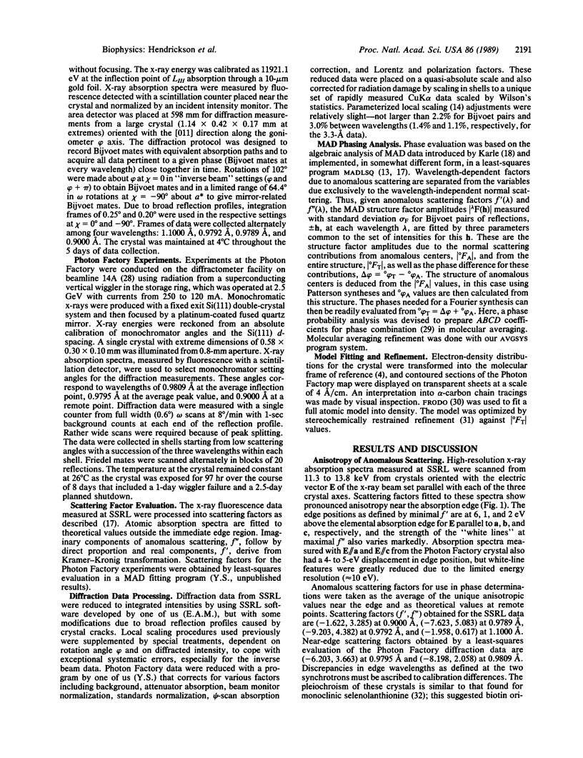
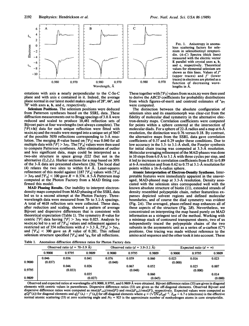
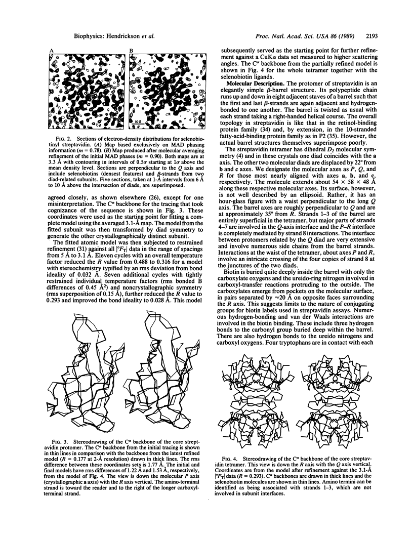
A three-dimensional crystal structure of the biotin-binding core of streptavidin has been determined at 3.1-A resolution. The structure was analyzed from diffraction data measured at three wavelengths from a single crystal of the selenobiotinyl complex with streptavidin. Streptavidin is a tetramer with subunits arrayed in D2 symmetry. Each protomer is an 8-stranded beta-barrel with simple up-down topology. Biotin molecules are bound at one end of each barrel. This study demonstrates the effectiveness of multiwavelength anomalous diffraction (MAD) procedures for macromolecular crystallography and provides a basis for detailed study of biotin-avidin interactions.
Full text
PDF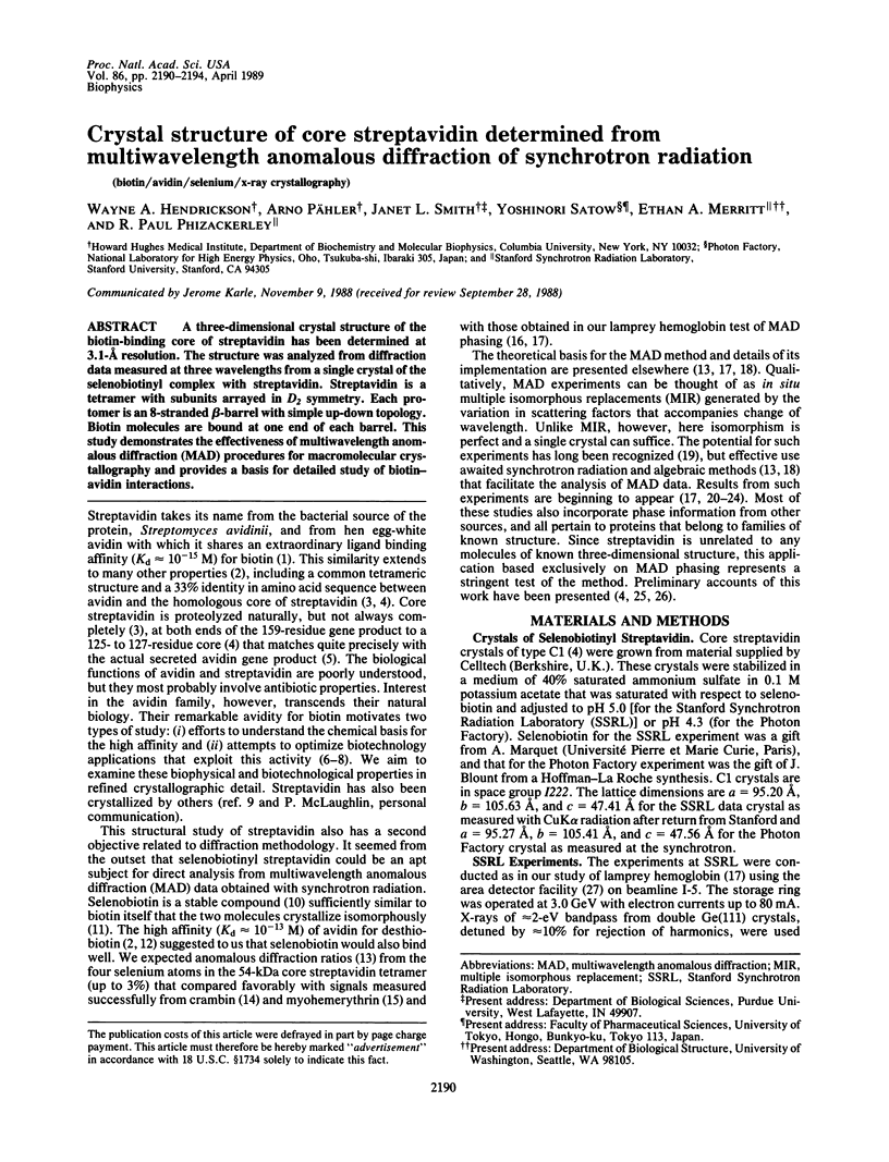
Selected References
These references are in PubMed. This may not be the complete list of references from this article.
- Bayer E. A., Wilchek M. The use of the avidin-biotin complex as a tool in molecular biology. Methods Biochem Anal. 1980;26:1–45. doi: 10.1002/9780470110461.ch1. [DOI] [PubMed] [Google Scholar]
- CHAIET L., WOLF F. J. THE PROPERTIES OF STREPTAVIDIN, A BIOTIN-BINDING PROTEIN PRODUCED BY STREPTOMYCETES. Arch Biochem Biophys. 1964 Jul 20;106:1–5. doi: 10.1016/0003-9861(64)90150-x. [DOI] [PubMed] [Google Scholar]
- DeTitta G. T., Parthasarathy R., Blessing R. H., Stallings W. Carboxybiotin translocation mechanisms suggested by diffraction studies of biotin and its vitamers. Proc Natl Acad Sci U S A. 1980 Jan;77(1):333–337. doi: 10.1073/pnas.77.1.333. [DOI] [PMC free article] [PubMed] [Google Scholar]
- Gope M. L., Keinänen R. A., Kristo P. A., Conneely O. M., Beattie W. G., Zarucki-Schulz T., O'Malley B. W., Kulomaa M. S. Molecular cloning of the chicken avidin cDNA. Nucleic Acids Res. 1987 Apr 24;15(8):3595–3606. doi: 10.1093/nar/15.8.3595. [DOI] [PMC free article] [PubMed] [Google Scholar]
- Green N. M. Avidin. Adv Protein Chem. 1975;29:85–133. doi: 10.1016/s0065-3233(08)60411-8. [DOI] [PubMed] [Google Scholar]
- Guss J. M., Merritt E. A., Phizackerley R. P., Hedman B., Murata M., Hodgson K. O., Freeman H. C. Phase determination by multiple-wavelength x-ray diffraction: crystal structure of a basic "blue" copper protein from cucumbers. Science. 1988 Aug 12;241(4867):806–811. doi: 10.1126/science.3406739. [DOI] [PubMed] [Google Scholar]
- Hendrickson W. A., Smith J. L., Phizackerley R. P., Merritt E. A. Crystallographic structure analysis of lamprey hemoglobin from anomalous dispersion of synchrotron radiation. Proteins. 1988;4(2):77–88. doi: 10.1002/prot.340040202. [DOI] [PubMed] [Google Scholar]
- Hendrickson W. A., Smith J. L., Sheriff S. Direct phase determination based on anomalous scattering. Methods Enzymol. 1985;115:41–55. doi: 10.1016/0076-6879(85)15006-8. [DOI] [PubMed] [Google Scholar]
- Hofmann K., Titus G., Montibeller J. A., Finn F. M. Avidin binding of carboxyl-substituted biotin and analogues. Biochemistry. 1982 Mar 2;21(5):978–984. doi: 10.1021/bi00534a024. [DOI] [PubMed] [Google Scholar]
- Jones T. A., Bergfors T., Sedzik J., Unge T. The three-dimensional structure of P2 myelin protein. EMBO J. 1988 Jun;7(6):1597–1604. doi: 10.1002/j.1460-2075.1988.tb02985.x. [DOI] [PMC free article] [PubMed] [Google Scholar]
- Kahn R., Fourme R., Bosshard R., Chiadmi M., Risler J. L., Dideberg O., Wery J. P. Crystal structure study of Opsanus tau parvalbumin by multiwavelength anomalous diffraction. FEBS Lett. 1985 Jan 1;179(1):133–137. doi: 10.1016/0014-5793(85)80207-6. [DOI] [PubMed] [Google Scholar]
- Korszun Z. R. The tertiary structure of azurin from Pseudomonas denitrificans as determined by Cu resonant diffraction using synchrotron radiation. J Mol Biol. 1987 Jul 20;196(2):413–419. doi: 10.1016/0022-2836(87)90701-7. [DOI] [PubMed] [Google Scholar]
- Murthy H. M., Hendrickson W. A., Orme-Johnson W. H., Merritt E. A., Phizackerley R. P. Crystal structure of Clostridium acidi-urici ferredoxin at 5-A resolution based on measurements of anomalous X-ray scattering at multiple wavelengths. J Biol Chem. 1988 Dec 5;263(34):18430–18436. [PubMed] [Google Scholar]
- Newcomer M. E., Jones T. A., Aqvist J., Sundelin J., Eriksson U., Rask L., Peterson P. A. The three-dimensional structure of retinol-binding protein. EMBO J. 1984 Jul;3(7):1451–1454. doi: 10.1002/j.1460-2075.1984.tb01995.x. [DOI] [PMC free article] [PubMed] [Google Scholar]
- Pähler A., Hendrickson W. A., Kolks M. A., Argaraña C. E., Cantor C. R. Characterization and crystallization of core streptavidin. J Biol Chem. 1987 Oct 15;262(29):13933–13937. [PubMed] [Google Scholar]
- Templeton L. K., Templeton D. H. Biaxial tensors for anomalous scattering of X-rays in selenolanthionine. Acta Crystallogr A. 1988 Nov 1;44(Pt 6):1045–1051. doi: 10.1107/s0108767388007810. [DOI] [PubMed] [Google Scholar]
- Weber P. C., Cox M. J., Salemme F. R., Ohlendorf D. H. Crystallographic data for Streptomyces avidinii streptavidin. J Biol Chem. 1987 Sep 15;262(26):12728–12729. [PubMed] [Google Scholar]