Abstract
In terrestrial subsurface environments where nitrate is a critical groundwater contaminant, few cultivated representatives are available to verify the metabolism of organisms that catalyze denitrification. In this study, five species of denitrifying bacteria from three phyla were isolated from subsurface sediments exposed to metal radionuclide and nitrate contamination as part of the U.S. Department of Energy's Oak Ridge Integrated Field Research Challenge (OR-IFRC). Isolates belonged to the genera Afipia and Hyphomicrobium (Alphaproteobacteria), Rhodanobacter (Gammaproteobacteria), Intrasporangium (Actinobacteria), and Bacillus (Firmicutes). Isolates from the phylum Proteobacteria were complete denitrifiers, whereas the Gram-positive isolates reduced nitrate to nitrous oxide. rRNA gene analyses coupled with physiological and genomic analyses suggest that bacteria from the genus Rhodanobacter are a diverse population of denitrifiers that are circumneutral to moderately acidophilic, with a high relative abundance in areas of the acidic source zone at the OR-IFRC site. Based on genome analysis, Rhodanobacter species contain two nitrite reductase genes and have not been detected in functional-gene surveys of denitrifying bacteria at the OR-IFRC site. Nitrite and nitrous oxide reductase gene sequences were recovered from the isolates and from the terrestrial subsurface by designing primer sets mined from genomic and metagenomic data and from draft genomes of two of the isolates. We demonstrate that a combination of cultivation and genomic and metagenomic data is essential to the in situ characterization of denitrifiers and that current PCR-based approaches are not suitable for deep coverage of denitrifiers. Our results indicate that the diversity of denitrifiers is significantly underestimated in the terrestrial subsurface.
Nitrate is among the most prevalent of groundwater contaminants that threaten drinking water resources on a global scale (48). Nitrate and uranium are priority cocontaminants at U.S. Department of Energy (DOE)-managed nuclear legacy waste sites where nitric acid was used for the extraction and processing of radioactive metals (47). Current remediation practices favor reductive immobilization of U(VI) catalyzed by indigenous microbial communities, along with natural attenuation and monitoring, and previous studies have indicated that U(VI) reduction does not proceed until nitrate is depleted (1, 18, 29, 47, 63). Conversely, uranium can be remobilized into groundwaters through oxidation of U(IV) precipitates by intermediate compounds produced during microbial denitrification (53), and some subsurface microorganisms can catalyze the oxidation of U(IV) coupled to nitrate reduction (5). Thus, when nitrate is present as a cocontaminant, the fate and transport of uranium in the contaminated terrestrial subsurface is intimately linked to the presence and metabolic activity of denitrifying bacteria.
Denitrifying microbial populations have been extensively profiled in soils, wastewater treatment systems, and marine environments (16, 26, 37, 46, 58). However, microorganisms that mediate denitrification and the mechanisms or most important controls of the process remain understudied in terrestrial aquifers (20, 48). Relatively few denitrifying bacteria from the terrestrial subsurface have been isolated and physiologically characterized in detail (17, 55). Furthermore, attempts to correlate microbial community structure and abundance, as assayed by molecular analyses of functional denitrification genes, to biogeochemical measurements in the subsurface have not been successful (44, 65).
Due to the paraphyletic nature and broad taxonomic distribution of denitrifying organisms, rRNA gene sequences are not suitable for demonstration of a denitrifying genotype (45). Instead, functional genes coding for proteins that catalyze denitrification are common targets for characterizing denitrifying populations by molecular analyses. Despite some successes (55), the application of functional-gene analyses for characterizing denitrifying communities has been limited by small genetic databases and the disproportionate representation of environmental sequences without reference sequences from isolated organisms. Likewise, the amplification of functional genes from confirmed, isolated denitrifying bacteria with commonly used primer sets has met with mixed success (21, 24-26, 57). Thus, the development of more-general primer sets capable of amplifying such gene targets from a wider range of organisms mediating denitrification remains a challenge. As a result of the limits of both cultivation-based and molecular approaches, the diversity, abundance, and distribution of denitrifying bacteria in the environment are likely underestimated.
This study investigated denitrifying bacteria and denitrification genes in contaminated, terrestrial subsurface sediments with the following objectives: (i) enrichment, cultivation, and purification of novel denitrifying bacteria from the Oak Ridge Integrated Field Research Challenge (OR-IFRC) subsurface; (ii) cultivation-directed amplification and phylogenetic analysis of gene targets for denitrifiers in subsurface sediments and groundwater; and (iii) development of improved primer sets for the detection of denitrifiers in environmental samples. We sought to improve access to the cultivatable diversity of denitrifiers in the subsurface by using simple carbon substrates and a wide range of nitrate concentrations and by minimizing the salt and major nutrient concentrations in a minimal synthetic groundwater medium. In addition, we sought to expand the known phylogenetic target range of functional-gene primers through a combination of genomic- and metagenomic-sequence data mining coupled with analyses of these genes from the recovered isolates. We demonstrate that a polyphasic approach incorporating cultivation and genomic and metagenomic data is essential to the characterization of denitrifiers and that current broad-spectrum PCR-based approaches are not suitable for deep coverage of denitrifying microorganisms in the terrestrial subsurface.
MATERIALS AND METHODS
Sediment sampling.
Sampling was conducted as part of the Department of Energy's Oak Ridge Integrated Field Research Challenge (OR-IFRC) in Oak Ridge, TN, where the subsurface has been widely contaminated with a diverse array of mixed contaminants. The OR-IFRC site, managed by the Oak Ridge National Laboratory (ORNL), is located adjacent to the Y-12 industrial complex in Oak Ridge, TN. The contaminated area lies adjacent to a parking lot that caps three former waste ponds (S-3 ponds) which contained uranium and nitric acid wastes generated during nuclear weapon production (http://www.esd.ornl.gov/orifrc/). Sediment cores were recovered from the saturated zone using a Geoprobe drill equipped with polyurethane sleeves lining the corer. A core from the most highly contaminated area (area 3, adjacent to the S-3 ponds) was recovered on 7 February 2008 (well FWB124; lat 35.97734171, long 84.27339281; 1.23 to 15.08 m below the surface), and a core from a less contaminated area was recovered on 12 September 2007 (borehole FB107; lat 35.97552296, long 84.27459978; 5.56 to 6.53 m below the surface). Cores were aseptically sectioned under strictly anoxic conditions in a Coy anaerobic chamber (Coy Laboratory Products, Grass Lake, MI) and stored anaerobically in gas-tight containers at 4°C prior to overnight shipment to Florida State University. Groundwater was sampled from well FW106 (lat 35.97729757, long 84.2734838) within area 3 during part of a larger sampling operation on 13 May 2009. The subsurface of area 3 is generally acidic (i.e., with a pH below 4.0) and highly and variably contaminated with nitrate (may exceed 100 mM) and uranium (may exceed 200 μM), along with many other contaminants (for a list of identified contaminants, see reference 63). The area 2 experimental plot within the OR-IFRC site is located west and down-gradient of the S-3 pond site and has a circumneutral pH, though levels of nitrate (up to 26 mM) and uranium (0.5 to 4.5 μM) remain elevated relative to those in background areas (60). Methods and results from the geochemical characterization of the subsurface sediments used as inocula for our cultures are presented in the text and Table S1 in the supplemental material.
Enrichment and isolation of denitrifying bacteria.
A minimal synthetic groundwater medium (SGWM) with trace element and vitamin mixes was prepared and dispensed according to Widdel and Bak (62), with the modifications of reducing the overall salt concentration (see below) and omitting resazurin, selenite, and tungstate. Unless otherwise indicated, this medium was used for all cultivation experiments. The medium contained the following components per liter: NaCl (100 mg), NH4Cl (100 mg), KH2PO4 (50 mg), KCl (10 mg), MgCl2·6H2O (40 mg), CaCl2 (40 mg), NaHCO3 (2.5 g), trace element solution (TES; 1 ml), vitamin B12 (1 ml), vitamin mix (1 ml), and thiamine (1.0 ml). The medium was cooled after being autoclaved under strictly anoxic conditions and was then saturated with N2-CO2 (80:20), resulting in a final pH of 6.9.
Four different concentrations of nitrate (0.5, 1.0, 10, and 25 mM) were used with four carbon sources (10 mM acetate; 10 mM lactate; a mixture of acetate, propionate, and butyrate at 10 mM each; and 20 mM ethanol), resulting in 16 enrichment conditions in SGWM. Each enrichment was performed in 60-ml-capacity serum vials and contained 30 ml of completely anoxic SGWM, as described above. Each serum vial was inoculated with contaminated sediment to a final concentration of 10% (wt/vol) and incubated at 30°C in the dark. Growth was monitored by measuring the optical density of the medium at a wavelength of 600 nm (OD600) using the NanoDrop ND-1000 spectrophotometer (Thermo Scientific, Wilmington, DE) and by colorimetric measurement of nitrate (10). Transfers to fresh medium were performed every 10 days using a 10% inoculum (vol/vol). After four successive transfers, isolation of distinct denitrifying bacteria was conducted by plating on agar medium composed of SGWM and 10 mM HEPES (Fisher Scientific, Fairlawn, NJ).
Spread and streak plate methods were used for isolation. Plates were incubated in an anaerobic chamber with 3% H2 in N2 at room temperature. Pure cultures of the strains were acquired by repeatedly transferring morphologically distinct colonies to fresh plates. To test for aerobic growth, strains were inoculated in the SGWM medium with the appropriate electron donor and incubated aerobically at 30°C. The purity of each culture was tested using multiple restreakings, as well as by colony PCR amplification with bacterial small-subunit (SSU) rRNA gene primers and subsequent direct sequencing without cloning. Isolates were subsequently preserved at −80°C in 20% anaerobic glycerol.
Growth rates and nitrogen gas production.
Strains were screened to assess growth and nitrate utilization rates. Isolates were transferred to SGWM containing 10 mM nitrate and supplemented with the electron donor that produced the maximum growth. Lactate (10 mM) was used as the electron donor for Bacillus, Afipia, and Intrasporangium isolates, whereas acetate (10 mM) and ethanol (20 mM) were used for Rhodanobacter and Hyphomicrobium isolates, respectively. Nitrate was omitted from control cultures to test for fermentative growth. For gas measurement, mid-log-phase cultures were inoculated into SGWM at 1% (vol/vol) for Bacillus, Afipia, and Intrasporangium and at 5% (vol/vol) for Rhodanobacter and Intrasporangium, with nitrate (10 mM) and the appropriate electron donor. Cell densities and nitrate concentrations were measured as described above.
Concurrently, production of dinitrogen gas (N2) and nitrous oxide (N2O) was studied under denitrifying conditions (anoxic, nitrate amended) by growing representative isolates in SGWM amended with 15N-enriched NO3− (98 atom%; Cambridge Isotope Laboratories, Inc., Andover, MA) and a low concentration of ammonium (0.25 mM). Cultures and uninoculated controls were prepared in 10-ml Hungate tubes. At the initial time point (immediately after inoculation) and after 10 to 15 days of growth, based on visual inspection of cell density, growth was terminated in duplicate cultures by the addition of 1% (wt/vol) HgCl2. Gas headspace samples for assessing the production of N2O were extracted through the rubber septa using a 100-μl syringe with a conical, side-port needle. Samples were immediately analyzed by gas chromatography using a Shimadzu GC-8A gas chromatograph with a Porapak Q column and electron capture detection. The reduction of nitrate to N2 was indicated by the accumulation of excess 15N-N2 in cultures amended with 15N-NO3−. Dissolved 15N-N2 concentrations were determined using a membrane inlet mass spectrometer configured and calibrated according to An et al. (4).
DNA extraction, PCR amplification, primer design, and gene sequencing.
Genomic DNA (gDNA) was extracted from bacterial isolates, sediment, and groundwater samples using Mo Bio DNA isolation kits (Mo Bio Laboratories, Carlsbad, CA) according to the manufacturer's instructions. Extracted genomic DNA (approximately 10 ng per reaction) was used as a template for small-subunit (SSU) rRNA gene amplification using the 27F and 1492R general bacterial primers (Table 1). PCRs were conducted using DreamTaq green DNA polymerase (Fermentas, Inc., Glen Burnie, MD), with a working concentration of 2 mM magnesium, 200 μM deoxynucleoside triphosphates, and 0.5 μM each primer. Annealing temperatures and primer sequences for each primer set (functional-gene primer sets are numbered 1 to 7) are provided in Table 1. For rRNA gene amplification, 30 reaction cycles were employed. PCR yields were column purified using the UltraClean PCR clean-up kit (Mo Bio). For each representative isolate, multiple sequencing reactions were performed, the sequence data were aligned, and a composite sequence was determined using the software package Sequencher (Gene Codes, Ann Arbor, MI). The basic local alignment search tool (BLAST; 2) was used to identify closely related sequences and generate sequence similarity estimates.
TABLE 1.
PCR primers used in this study
| Primer set | Individual primer | Gene | Sequence (5′→3′) | Primer position | Annealing temp (°C) | Intended target | Source |
|---|---|---|---|---|---|---|---|
| 27F | rrs | AGA GTT TGA TCM TGG CTC AG | 8-27a | 55 | Bacteria | Lane (34) | |
| 1492R | GGT TAC CTT GTT ACG ACT T | 1510-1492a | |||||
| 1 | nirK1F | nirK | GGM ATG GTK CCS TGG CA | 526-542b | 56 | Universal | Braker et al. (7) |
| nirK5R | GCC TCG ATC AGR TTR TGG | 1040-1023b | |||||
| 2 | F1aCu | nirK | ATC ATG GTS CTG CCG CG | 568-584b | 50 | Universal | Hallin and Lindgren (21) |
| R3Cu | GCC TCG ATC AGR TTG TGG TT | 1040-1021b | |||||
| 3 | MG763-F1aCu | nirK | ATC CTG GTC GAG CCG CC | 568-584b | 73 | Rhodanobacter | This study |
| MG763-R3Cu | GCG CGG AAG ATC GAG TGG TC | 1040-1021b | |||||
| 4 | HD-nirkF1 | nirK | CCA GCT CAA CCT TCT CGT TC | 439-458c | 55 | Hyphomicrobium denitrificans | This study |
| HD-nirKR1 | TTG CAT GAC GTA GAA CTC GC | 1040-1021c | |||||
| 5 | nirS1F | nirS | CCT AYT GGC CGC CRC ART | 763-780d | 61 | Universal | Braker et al. (7) |
| nirS6R | CGT TGA ACT TRC CGG T | 1653-1638d | |||||
| 6 | nosZ2F | nosZ | CGC RAC GGC AAS AAG GTS MSS GT | 1603-1625e | 65 | Universal | Henry et al. (23) |
| nosZ2R | CAK RTG CAK SGC RTG GCA GAA | 1869-1849e | |||||
| 7 | Rh-nosZ-F | nosZ | CGG CTG GGG TGT CAC CAA | 303-320e | 63 | Rhodanobacter | This study |
| Rh-nosZ-R | CAT GTG CAT CGC ATG GCA GAA | 1869-1849e |
According to the rrs gene of Escherichia coli (E05133).
According to the nirK gene of Alcaligenes faecalis (D13155).
According to the nirK gene of Hyphomicrobium denitrificans (AB076606).
According to the nirS gene of Pseudomonas stutzeri (X56813).
According to the nosZ gene of Pseudomonas fluorescens (AF197468).
DNA from each of the isolates was subjected to PCR amplification (35 cycles) using primers targeting nitrite reductase (nirK and nirS) and nitrous oxide reductase (nosZ) genes. PCR amplification was conducted as described above, with various annealing temperatures (Table 1). PCR amplicons were cleaned as described above and sequenced directly. Primers specific to the nirK gene of Hyphomicrobium denitrificans were designed using a previously published sequence of H. denitrificans strain A3151 (primer set 4 [64]). A primer set for this gene was generated using the online software Primer-BLAST at the NCBI website (51). Primers for nirK genes from bacteria of the genus Rhodanobacter were designed initially from metagenomic sequence data retrieved from groundwater recovered in well FW106 in area 3 (22). These primer sequences were subsequently confirmed with draft genome sequence data from Rhodanobacter sp. strain 2APBS1. Primers for nosZ genes from bacteria of the genus Rhodanobacter were designed from the draft genome of Rhodanobacter sp. 2APBS1 (primer set 7). The conserved locations were identified by performing gene sequence alignments with the software package ClustalW2 (35). Genomic DNA extracted from environmental samples (well FW106 groundwater and FWB124 sediment) was used as a template for PCR amplification with the Rhodanobacter nirK primers developed in this study (primer set 3). These PCR products were cloned using the Lucigen pSMARTGC cloning kit (Lucigen Corporation, Middleton, WI) according to the manufacturer's instructions. Gene sequences were recovered from each clone via PCR amplification with plasmid-bound primers.
Phylogenetic analyses.
Recovered SSU rRNA gene sequences and close relatives identified by BLAST searches were aligned using the GreenGenes SSU rRNA gene alignment tool (15) and imported into the ARB software package (39). Selected sequences were exported from ARB using a bacterial 50% conservation filter (38) and imported into the MEGA 4.0 software package (56). Bootstrapped neighbor-joining phylogenetic trees were constructed within MEGA using the maximum-composite-likelihood substitution model with complete deletion of gapped positions (946 informational positions). Additionally, Bayesian analyses (MrBayes version 3.1 [50]) were performed on the filtered sequence data as described previously (19).
Phylogenetic trees of genes coding for copper-containing nitrite reductase (nirK) and nitrous oxide reductase (nosZ) were generated based on inferred amino acid sequences obtained from the Integrated Microbial Genomes website (http://img.jgi.doe.gov/cgi-bin/pub/main.cgi), from GenBank (National Center for Biotechnology Information; http://www.ncbi.nlm.nih.gov/), and from sequencing analyses performed for this study. Inferred amino acid sequences were aligned using ClustalW2, and a phylogenetic tree was generated in MEGA by using pair-wise deletion of sequence data. The aligned sequence data were also analyzed using Bayesian analysis to provide additional statistical support for nodes.
Genome sequencing.
Total genomic DNA from Rhodanobacter sp. 2APBS1 and Intrasporangium sp. strain 4LS1 was isolated using the PowerMax soil DNA isolation kit (Mo Bio Laboratories). DNA libraries were prepared for each genomic DNA sample by following the 454 GS FLX long paired-end library preparation protocol and then subjected to pyrosequencing on the GS FLX system (454 Life Sciences, Branford, CT). Nucleotide sequence reads, contigs, and quality scores were reported according to standard 454 operating procedures. The Newbler de novo assembly application (part of the 454 Genome Sequencer FLX software package) was used to assemble the sequence reads into DNA contigs. Open reading frames were then predicted and annotated as described previously (33).
Nucleotide sequence accession numbers.
SSU rRNA gene sequences were submitted to GenBank under the accession numbers FJ851425 to FJ851445. Representative nirK and nosZ gene sequences were submitted to GenBank under the accession numbers GQ404513 to GQ404524 and GU814013, and gene sequences recovered from draft genomes of Rhodanobacter sp. 2APBS1 and Intrasporangium sp. 4LS1 were submitted to GenBank under the accession numbers GU233006 to GU233009 (see Table 2).
TABLE 2.
Phenotypic and genotypic characterization of denitrifying isolates
| Genus (class or phylum) and isolatea | Isolation conditions |
Denitrificationc |
Aerobic growthd | SSU rRNA (rrs) | Primer set (GenBank accession no.)e |
GenBank accession no. for draft genome sequence |
||||
|---|---|---|---|---|---|---|---|---|---|---|
| Electron donorb | Nitrate concn (mM) | N2O production | N2 production | nirK | nosZ | nirK | nosZ | |||
| Afipia (Alphaproteobacteria) | ||||||||||
| 1NLS2* | Lactate | 0.5 | — | — | — | FJ851425 | 1 | 6 | ||
| 2LS1 | Lactate | 1 | — | — | — | FJ851426 | 2 | 6 | ||
| 3AS3* | Acetate | 10 | Neg | Pos | Pos | FJ851427 | 1 (GQ404513) | 6 (GQ404519) | ||
| 4AS1 | Acetate | 25 | Neg | Pos | — | FJ851428 | 1 (GQ404514) | 6 | ||
| 4LS2 | Lactate | 25 | Neg | Pos | — | FJ851429 | 1 | 6 | ||
| 4LS4 | Lactate | 25 | — | — | — | FJ851430 | 2 | 6 (GQ404520) | ||
| 4LS5 | Lactate | 25 | Neg | Pos | — | FJ851431 | 1 | 6 | ||
| Hyphomicrobium (Alphaproteobacteria) | ||||||||||
| 1NES1* | Ethanol | 0.5 | Neg | Pos | VW | FJ851438 | 4 (GU814013) | 6 (GQ404521) | ||
| 2NES1* | Ethanol | 1 | Neg | Pos | — | FJ851439 | 4 | 6 (GQ404522) | ||
| Rhodanobacter (Gammaproteobacteria) | ||||||||||
| 2AS1* | Acetate | 1 | Pos | Pos | — | FJ851443 | 3 (GQ404515) | 7 (GQ404523) | ||
| 2APBS1* | APB | 1 | — | — | Pos | FJ851444 | 3 (GQ404516) | 7 (GQ404524) | GU233006, GU233007 | GU233008 |
| 3LS1 | Lactate | 10 | Pos | Pos | — | FJ851445 | 3 | 7 | ||
| Intrasporangium (Actinobacteria) | ||||||||||
| 3APBS1* | APB | 10 | Pos | Neg | Pos | FJ851441 | Neg | Neg | ||
| 3AS1 | Acetate | 10 | Pos | Neg | — | FJ851440 | Neg | Neg | ||
| 4LS1* | Lactate | 25 | Pos | Neg | — | FJ851442 | Neg | Neg | GU233009 | |
| Bacillus (Firmicutes) | ||||||||||
| 1NLA3E* | Lactate | 0.5 | Pos | Neg | Pos | FJ851432 | Neg | Neg | ||
| 2NLA3E* | Lactate | 1 | — | — | — | FJ851433 | Neg | Neg | ||
| AS31 | Lactate | 0.5 | — | — | — | FJ851434 | Neg | Neg | ||
| A3S2 | Lactate | 0.5 | — | — | — | FJ851435 | Neg | Neg | ||
| A3S3 | Lactate | 0.5 | — | — | — | FJ851436 | Neg | Neg | ||
| AS34 | Lactate | 0.5 | — | — | — | FJ851437 | Neg | Neg | ||
An asterisk indicates that the isolate is presented in the phylogenetic tree (Fig. 1).
Electron donor concentrations: acetate, 10 mM; lactate, 10 mM; ethanol, 20 mM; acetate, propionate, butyrate (APB), 10 mM.
Production of nitrous oxide or nitrogen gas was tested by gas chromatography, as described in Materials and Methods. Neg, negative; Pos, positive; —, not tested.
Pos, positive growth on agar medium under aerobic conditions; VW, very weak growth; —, not tested.
RESULTS
Isolation of novel denitrifying bacteria.
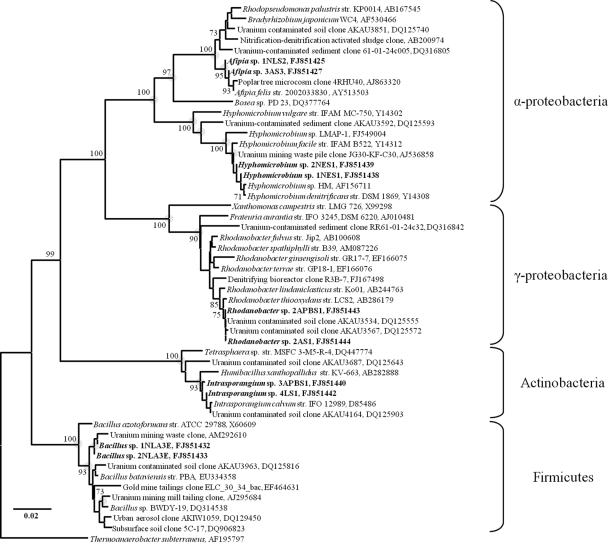
Denitrifying bacteria capable of reducing nitrate to nitrous oxide or nitrogen gas were isolated from sediment obtained from the OR-IFRC site using a systematic enrichment strategy (Table 2; Fig. 1). From each enrichment, more than 100 colonies were recovered, and based on colony morphology and growth pattern, 21 colonies were selected for further analyses. Analysis of bacterial SSU rRNA gene sequences revealed that the characterized isolates belong to five genera within three phyla. SSU rRNA gene sequences from all of the recovered isolates from area 3 samples were nearly identical and were most similar (>98.1%) to the SSU rRNA gene sequence of Bacillus bataviensis, a phenol-degrading, aerobic, denitrifying firmicute (GenBank accession no. EU334358) (Fig. 1). The environmental sequence most similar to the SSU rRNA gene of Bacillus isolates was recovered from a uranium-contaminated waste pile (>99.2% similarity to GenBank accession no. AM292610) (Fig. 1).
FIG. 1.
Bootstrapped (1,000 iterations) neighbor-joining tree of denitrifying isolate SSU rRNA gene sequences. Isolate sequences recovered in this study are in bold, with additional genetic and phenotypic properties provided in Table 2. Nodes supported by bootstrap values greater than 70% are indicated by numeric values, and nodes supported by Bayesian analysis, with posterior probability values greater than 95%, are indicated with gray circles. The scale bar represents 0.02 substitutions per nucleotide position.
The isolates recovered from area 2 subsurface sediment enrichments belonged to the genera Afipia, Hyphomicrobium, Rhodanobacter, and Intrasporangium. The Hyphomicrobium isolates showed high SSU rRNA gene sequence similarity (>99.3%) to a known denitrifier, H. denitrificans strain DSM1869 (GenBank accession no. Y14308), and to an isolate capable of degrading organophosphate pesticides (99.7% similar to Hyphomicrobium sp. strain LMAP-1, GenBank accession no. FJ549004). Sequences from Intrasporangium isolates were nearly identical (>99.4% similarity) to the SSU rRNA gene sequence of Intrasporangium calvum, an aerobic actinomycete that has not been well characterized (32, 40), and to an environmental sequence recovered from a clone library generated from OR-IFRC area 2 sediment (>99.4% similarity to GenBank accession no. DQ125903 [8]). The capacity for denitrification has not been shown previously in the genus Intrasporangium or the family Intrasporangiaceae (54). Some members of the family Intrasporangiaceae are capable of nitrate reduction to nitrite but not denitrification (31). An analysis of the draft genome of Intrasporangium sp. 4LS1 revealed the presence of a partial denitrification pathway including nitrate, nitrite, and nitric oxide reductase genes. No nitrous oxide reductase gene was found, consistent with the lack of N2 production by these isolates (Table 2), but a final determination awaits complete genome sequence data.
Isolates from the genus Afipia belong to the Bradyrhizobium, Agromonas, Nitrobacter, and Afipia (BANA) cluster of the class Alphaproteobacteria (52) and have SSU rRNA gene sequences that were nearly identical (>99.7% sequence similarity) to those of Afipia felis (GenBank accession no. AY513503), a cat pathogen (6), and to an uncultured rhizosphere organism (GenBank accession no. AJ863320). The BANA cluster includes methylotrophs (42) and organisms capable of heterotrophic nitrate assimilation (41). Some representatives of the genus Afipia are denitrifiers, including a soil isolate with 98% rRNA gene sequence similarity to the sequences of the isolates recovered in this study (GenBank accession no. AB162068).
Isolates from the genus Rhodanobacter were identical by SSU rRNA gene sequencing to a clone recovered from the area 2 subsurface at the OR-IFRC site (GenBank accession no. DQ125555 [8]) and highly similar (>98.9%) to the SSU rRNA gene sequence of Rhodanobacter thiooxydans, an organism capable of thiosulfate oxidation and nitrate, but not nitrite, reduction (36). No validly described species of Rhodanobacter has been shown to denitrify (3, 14, 28, 36, 43, 61); however, Cheneby et al. (11) previously isolated several denitrifying bacteria from agricultural soil (GenBank accession no. AY122296, AY122297, and AY122309), and based on their SSU rRNA gene sequences, these organisms belong within the genus Rhodanobacter. An analysis of the draft genome of Rhodanobacter sp. 2APBS1 revealed the presence of a complete denitrification pathway including nitrate, nitrite, nitric oxide, and nitrous oxide reductase genes.
Growth rates and production of gaseous nitrogen by bacterial isolates.
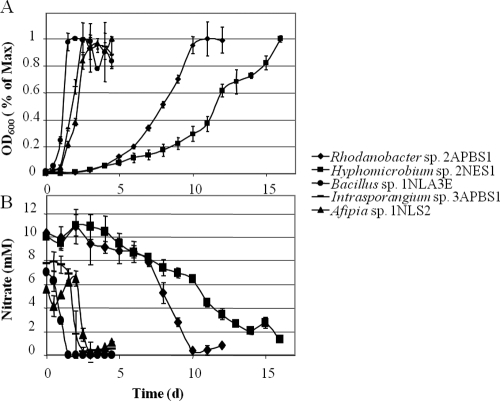
Representatives of the five novel taxa were tested for nitrate consumption and growth under denitrifying (i.e., anoxic, nitrate-amended) conditions. Denitrification capability was demonstrated by measurement of nitrous oxide and nitrogen gas production. All isolates were capable of converting nitrate to gaseous nitrogen species and grew optimally when ethanol (Hyphomicrobium), lactate (Afipia, Bacillus, and Intrasporangium), or acetate (Rhodanobacter) was used as the sole carbon substrate and electron donor. Little or no growth was observed in the absence of nitrate. Growth rates of the isolates diverged significantly, and nitrate removal rates mirrored cell growth (Fig. 2). Afipia, Intrasporangium, and Bacillus strains grew rapidly (i.e., doubling times of 6 h or less), whereas Rhodanobacter and Hyphomicrobium strains grew more slowly even under optimal conditions (i.e., doubling times of 1 day or greater) (Fig. 2). Proteobacterial isolates were capable of complete denitrification and production of N2, whereas Gram-positive isolates produced N2O but not N2. Rhodanobacter isolates accumulated N2O during denitrification under the growth conditions in this study.
FIG. 2.
Growth under denitrifying conditions of representatives of the five denitrifying organisms recovered in this study. (A) Growth curves, as measured by determining the optical density at 600 nm (OD600), for each isolate. Lactate (10 mM) was used as the electron donor for Bacillus, Afipia, and Intrasporangium isolates. Acetate (10 mM) and ethanol (20 mM) were used for Rhodanobacter and Hyphomicrobium isolates, respectively. (B) Nitrate removal during growth of the five denitrifiers. Production of gaseous end products as a result of denitrification was demonstrated (Table 2).
Functional-gene analysis of isolates, environmental clones, and groundwater metagenomes.
Functional genes involved in denitrification were PCR amplified from genomic DNA extracted from the isolates using primers targeting conserved regions of the nitrite reductase and nitrous oxide reductase genes. The results, reported as positive or negative for PCR amplification, are presented in Table 2. The isolates were tested initially for genes of two different forms of nitrite reductases (i.e., the cytochrome cd1 gene, nirS, and the copper-containing nitrite reductase gene nirK) using several previously published primer sets (Table 1). Only genomic DNA from the Afipia isolates was amplified when either of the previously published nirK primer sets was employed (i.e., primer sets 1 and 2 in Table 2). Nitrite reductase gene amplification using published nirS primers was not successful for any of the isolates (primer set 5 in Table 2). Based on a known sequence of nirK in H. denitrificans (64), a primer set for this gene was designed and successfully used to amplify the nirK gene from genomic DNA extracted from the Hyphomicrobium isolates (primer set 4 in Table 2). The gene sequence from Hyphomicrobium sp. strain 1NES1 (GenBank accession no. GU814013) was 88% identical (with 95% positive matches) in its inferred amino acid sequence to the nirK product sequence of H. denitrificans (GenBank accession no. AB076606). Nitrous oxide reductase genes were also amplified from both alphaproteobacterial taxa, using previously published primers (primer set 6 in Table 2). However, nosZ gene amplifications using these primers were not successful for the Gram-positive or Rhodanobacter isolates.
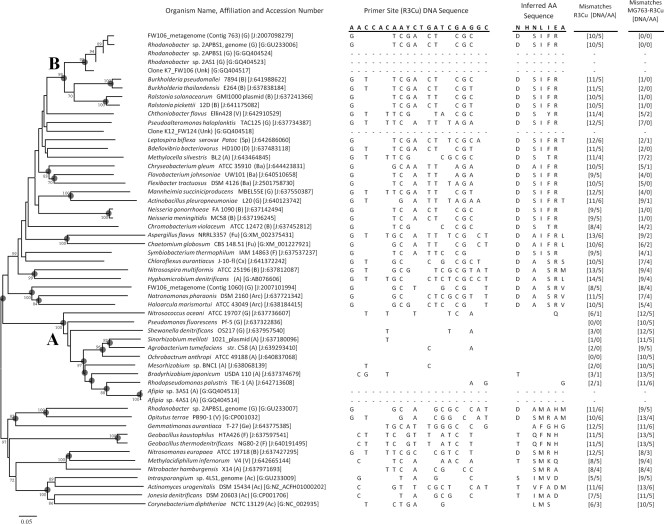
We were initially unable to amplify either nitrite or nitrous oxide reductase genes from the Rhodanobacter and Gram-positive isolates. Complete gene sequences, when present, were recovered from draft genome sequences of Rhodanobacter sp. 2APBS1 and Intrasporangium sp. 4LS1. We attempted to design primers specific to Rhodanobacter denitrification genes by examining sequence data recovered from the draft Rhodanobacter genome and from metagenome analyses of acidic and circumneutral groundwater recovered from OR-IFRC site wells (FW106 and FW301, respectively [22]). No nitrite reductase genes were detected in the larger background well metagenome (JGI taxon object identifier [ID] 2007427000; ∼106 Mb), while two putative nirK genes and no nirS genes were identified in the acidic groundwater metagenome from well FW106 (JGI taxon object ID 2006543007; ∼9.5 Mb). The nirK genes from the groundwater metagenome were found on a small contig with no other genes (contig 763; gene object ID 2007098279 [Fig. 3]) and on a large contig (∼568,000 bp) containing a complete denitrification pathway (contig 1060; gene object ID 2007101994 [Fig. 3]). Phylogenetic analyses showed that the unaffiliated nirK gene found on the small contig was highly similar to one of the two nirK genes recovered from the draft genome of Rhodanobacter sp. 2APBS1 (GenBank accession no. GU233006 [Fig. 3, cluster B]). The metagenome nirK gene located on the large contig in close proximity to other genes in the denitrification pathway was phylogenetically distant from the small contig gene and was most similar to nirK genes from Archaea (Fig. 3). Within the Rhodanobacter sp. 2APBS1 draft genome, a second nirK gene was identified (GenBank accession no. GU233007), but phylogenetic analyses revealed that this gene was highly divergent from any other nirK gene associated with Rhodanobacter and was most similar to the nirK gene detected within the genome of a Verrucomicrobia isolate, Opitutus terrae PB90-1 (Fig. 3). Using primers targeting cluster B Rhodanobacter nirK genes (primer set 3 in Table 1), we were able to amplify nirK genes from genomic DNA extracted from groundwater recovered from well FW106 and from genomic DNA extracted from sediment recovered from an area 3 contaminated sediment (borehole FWB124; clones K7 and K12 [Fig. 3]).
FIG. 3.
Unrooted phylogeny of partial and complete nirK sequences. The trees were generated based on aligned amino acid (AA) sequences inferred from DNA sequences. The numerical values at the nodes represent bootstrap values (1,000 iterations), and nodes with gray circles are supported by Bayesian posterior probability values greater than 95%. The scale bar represents 5% distance after Poisson correction. Organism affiliations are in parentheses (Ac, Actinobacteria; Ba, Bacteroidetes; Cx, Chloroflexi; F, Firmicutes; Ge, Gemmatimonadetes; A, Alphaproteobacteria; B, Betaproteobacteria; D, Deltaproteobacteria; G, Gammaproteobacteria; Sp, Spirochaetes; V, Verrucomicrobia; Arc, Archaea; Fu, fungi). Accession numbers are in brackets, and the database is indicated (G, GenBank; J, JGI gene object ID). Adjacent to each organism is the DNA and inferred amino acid sequence for the class I nirK primer R3Cu (primer set 2 in Table 1), with the primer sequence shown for the sense strand. DNA and amino acid mismatches relative to the R3Cu primer sequence and inferred AA sequence are indicated. In addition, DNA and AA mismatches relative to the MG763-R3Cu primer sequence are shown (primer set 3 in Table 1). Differences from the primer DNA sequence and inferred AA sequence are indicated for each sequence by showing the different base or residue. No sequence data at the primer sites are included for sequences recovered in this study, as these primer sequences were used for amplification of the gene fragment.
The reason for the lack of nirK gene amplification from some isolates is clearly revealed in an examination of sequence mismatches between isolate gene sequences and representative primer sequences (primer R3Cu [Fig. 3] and primer F1aCu [see Table S2 in the supplemental material]). These and other published nirK primers target only class I nirK genes (Fig. 3, cluster A). Such primers will likely miss some of the class I genes as well due to the presence of synonymous mutations in the primer sites (e.g., Shewanella denitrificans with 8 combined nucleotide mismatches with primer set 2 but no amino acid mismatches [Fig. 3 and see Table S2 in the supplemental material]). In the primer R3Cu (Table 1), the only inferred conserved amino acid is the histidine residue which serves as a type II copper atom ligand (59). Within the class II nirK genes, a high level of sequence diversity is noted, and the class II nirK primers developed herein (primer set 3) appear to be highly specific to certain nirK genes from the genera Rhodanobacter, Burkholderia, and Ralstonia (Fig. 3, cluster B). The clustering of these genes is strongly supported by bootstrap and Bayesian analyses. No amplification of the nirK gene was achieved for the Bacillus isolates, and we note that no such genes for the genus are currently available. Nitrite reductase genes from the Firmicutes do not all cluster together; for example, the nirK genes from Symbiobacterium thermophilum and Geobacillus spp. are highly divergent (Fig. 3). The nirK gene recovered from Intrasporangium sp. 4LS1 clustered with nirK genes from other Actinobacteria (Fig. 3).
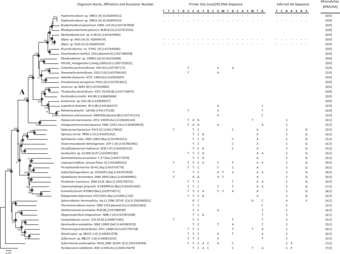
We generated a phylogenetic tree based on nosZ gene sequences recovered in this study and from complete or nearly complete gene sequences recovered from the JGI and GenBank databases (Fig. 4). Nitrous oxide reductase genes were recovered from all proteobacterial isolates but neither of the Gram-positive isolates. Partial nosZ gene sequences were amplified and sequenced from two Rhodanobacter isolates, and sequences were identical to the full gene recovered from the draft genome of Rhodanobacter sp. 2APBS1. Phylogenetic analyses revealed that the nosZ gene sequence from Rhodanobacter sp. 2APBS1 was most similar to the nosZ sequence recovered from the OR-IFRC acidic-groundwater metagenome. Together, these sequences clustered with sequences of other representative members of the gammaproteobacteria (Fig. 4). The target range of a selected primer set (primer set 6) was examined by in silico comparison of DNA and inferred amino acid sequences from each organism to the primer sequences (Fig. 4; see also Table S3 in the supplemental material). The inferred target range of this primer and other commonly used nosZ gene primers are genes from alpha-, beta-, and gammaproteobacteria (Fig. 4, cluster A). Not all nosZ genes from proteobacteria are amplified by this primer set, as demonstrated, for example, by the lack of amplification from the Rhodanobacter isolates. Comparison of the Rhodanobacter sp. 2APBS1 nosZ gene sequence to primer set 6 revealed five mismatches with the forward primer (see Table S3 in the supplemental material) and none with the reverse primer (Fig. 4). Genes from other taxa, including the Archaea, have four to eight mismatches with the highly degenerate primer nosZ2R (a combination of 32 variants), and similarly high sequence divergence is observed at other priming sites (Fig. 4; Table S3).
FIG. 4.
Unrooted phylogeny of partial and complete nosZ sequences. The trees were generated based on aligned amino acid (AA) sequences inferred from DNA sequences. Numerical values at the nodes represent bootstrap values (1,000 iterations), and nodes with gray circles are supported by Bayesian posterior probability values greater than 95%. The scale bar represents 5% distance after Poisson correction. Organism affiliations are in parentheses (Ac, Actinobacteria; Aq, Aquificae; Ba, Bacteroidetes; Cx, Chloroflexi; Def, Deferribacteres; F, Firmicutes; Ge, Gemmatimonadetes; A, Alphaproteobacteria; B, Betaproteobacteria; G, Gammaproteobacteria; D, Deltaproteobacteria; E, Epsilonproteobacteria; S, Spirochaetes; V, Verrucomicrobia; Arc, Archaea; Fu, fungi). Accession numbers are in brackets, and the database is indicated (G, GenBank; J, JGI gene object ID). Adjacent to each organism is the DNA and inferred amino acid sequence for the primer nosZ2R (primer set 6 in Table 1), with the primer sequence shown for the sense strand. DNA and amino acid mismatches relative to the nosZ2R primer sequence and inferred AA sequence are indicated. Differences from the primer DNA sequence and inferred AA sequence are indicated for each sequence by showing the different base or residue. No sequence data at the primer sites are included for alphaproteobacterial sequences recovered in this study, as these primer sequences were used for amplification of the gene fragment. Amino acid positions indicated by an “X” represent multiple possible amino acid residues encoded by degeneracies in the DNA primer sequence. At the two “X” positions, the primer codon represents either an M or L.
DISCUSSION
The scope of denitrifying bacterial diversity and activity remains uncertain, and terrestrial subsurface environments are understudied in comparison to other ecosystems, such as soils or wastewater treatment systems. An improved understanding of denitrifying microbial communities is required in order to predict and control denitrification mechanisms for the remediation of contaminated groundwaters. A multifaceted approach, in which cultivation-based and molecular analyses were combined with genomic and metagenomic data mining, was taken to examine the native denitrifying community at the OR-IFRC site. A total of 21 bacterial isolates, representing five genera (Afipia, Bacillus, Hyphomicrobium, Intrasporangium, and Rhodanobacter) within three phyla, were isolated and subjected to genetic and phenotypic analyses. All of the isolates reduce nitrate to gaseous end products, either N2 or N2O. The isolates are all facultative anaerobes, consistent with the highly variable redox conditions in the subsurface at the OR-IFRC site. This is the first report demonstrating denitrification activity of bacteria from the genus Intrasporangium. Furthermore, our analyses of structural and functional-gene sequences have revealed that members of the genus Rhodanobacter comprise a diverse community of bacteria that are circumneutral to moderately acidophilic and can have a very high relative abundance in some areas of the acidic source zone at the OR-IFRC site (9, 22). Although a single study reported that some isolates within the genus Rhodanobacter are capable of denitrification (11), members of the genus have not been routinely associated with denitrification activity. The data presented in this study and metagenomic data from the OR-IFRC site further indicate the denitrification potential within the genus Rhodanobacter (22), though these organisms have not been previously identified in functional denitrification gene surveys of the OR-IFRC site (55, 65). Due to the large number of mismatches between established denitrification gene primers and the gene sequences from Rhodanobacter spp., we were unable to amplify nitrite and nitrous oxide reductase genes from the Rhodanobacter isolates using these commonly used primer sets.
The lack of amplification of functional denitrification genes from many denitrifying bacteria isolated from agricultural soils and wastewater treatment systems has been reported (21, 24, 26, 57). Our initial, unsuccessful attempts to amplify denitrification genes from the Rhodanobacter strains isolated in this study highlight the fact that key organisms with denitrifying capabilities are not targeted with commonly used primer sets. Primers developed from genomic and metagenomic data enabled us to amplify nirK genes from Rhodanobacter isolates and directly from environmental genomic DNA. Comparison of these gene sequences to those from full-genome studies revealed that the amplification difficulties arise because the commonly used nirK gene primers target only the class I form of copper nitrite reductases (CuNiR), and this form has not been found in bacteria from the genus Rhodanobacter. Two clusters of CuNiR were previously established by Philippot (45), including cluster or class I genes generally assumed to encode soluble periplasmic proteins and class or cluster II genes which encode an outer membrane-bound form in some organisms (e.g., Neisseria gonorrhoeae [27]). Additionally, Philippot (45) demonstrated the presence of more-divergent sequences from Gram-positive bacteria and from an ammonia-oxidizing organism (Nitrosomonas europaea). Commonly used nirK gene primers have been designed to target class I CuNiR genes, and the current data set of class I sequences is composed largely of alphaproteobacterial sequences (7, 30). Due to large sequence differences present even in conserved primer sites (often, but not always, located in copper-binding domains), it is not possible to amplify class II nirK genes with class I primer sets (Fig. 3) (30). The class I primer sets are also unsuitable for targeting nirK genes from a third cluster of nirK genes that are divergent from both class I and II genes (Fig. 3) (45). This cluster includes highly divergent genes from Actinobacteria, from the firmicutes genus Geobacillus, from Verrucomicrobia and a member of the class Gemmatimonadetes, and from ammonia- or nitrite-oxidizing proteobacteria (Nitrosomonas and Nitrobacter). Curiously, one of the two putative nitrite reductase genes identified in the draft genome of Rhodanobacter sp. 2APBS1 belongs to this “third cluster” of nitrite reductase genes and is most similar to genes from Opitutus terrae PB90-1 (Verrucomicrobia) and to Gemmatimonas aurantiaca (Gemmatimonadetes). Neither the O. terrae nor the G. aurantiaca isolates have been shown to denitrify (12, 66), and this leaves uncertainty about the function of this putative nirK gene within Rhodanobacter sp. 2APBS1. We note that some third-cluster nitrite reductase genes are likely functional, as indicated by denitrification activity in the Intrasporangium spp. recovered in this study (Table 2; Fig. 2) and in other Actinobacteria (e.g., Jonesia denitrificans [49]).
For environmental systems such as the OR-IFRC site, where the predominant organisms may contain class II nirK genes, community composition analyses based on functional-gene primers can produce inaccurate quantitation and characterization of the subsurface denitrifying community. This problem is not unique to the contaminated subsurface. Of great concern is the DNA sequence divergence among the recovered nirK genes, which places considerable restraints on the design of suitable, broad-spectrum primers. Part of this sequence divergence is common to functional genes as a result of synonymous mutations. For nirK genes, copper-binding residues appear to hold the greatest hope for locating conserved sequences, and we identified a potential priming location where two copper-binding residues are adjacent (see Table S4 in the supplemental material). Although this region is much more highly conserved across a wide range of class I and class II nirK genes than among commonly used primers, we observed up to six mismatches with known nirK genes, such as those from Actinobacteria, including Intrasporangium sp. 4LS1, recovered in this study. As a result of the remarkable sequence divergence in nirK genes, we believe that direct PCR amplification or quantitative PCR amplification using currently employed primer sets cannot be reasonably employed to explore the true diversity and abundance of denitrifying bacteria in environmental systems.
For the contaminated subsurface, the limited metagenomic data from the site provide a PCR-independent indication that nirK genes are the dominant form in the highly contaminated source zone (two nirK and no nirS genes were detected in the acidic area 3 groundwater, and no nir genes were detected in a background well metagenome [22]). In addition, our results suggest that denitrifier abundance and diversity data based on nir gene amplification should be reevaluated. Likewise, the conventional wisdom that cytochrome cd1 nitrite reductases (nirS) are environmentally more abundant than copper-containing reductases (13) should be reevaluated in the context of these findings. In parallel, we observed similar impediments to functional-gene detection with commonly employed nosZ gene primers, which exclusively target genes from alpha-, beta-, and gammaproteobacteria, suggesting that previous denitrifier community studies using nosZ analyses most likely do not elucidate the true diversity and abundance of denitrifying bacteria in the environment.
Supplementary Material
Acknowledgments
This research was supported by the Office of Science (BER), U.S. Department of Energy, grant DE-FG02-07ER64373, and by the Integrated Field-Scale Subsurface Research Challenge at Oak Ridge, operated by the Environmental Sciences Division, Oak Ridge National Laboratory (ORNL), under U.S. Department of Energy contract DE-AC05-00OR22725.
We kindly acknowledge assistance from Tonia Mehlhorn and Kenneth Lowe for sampling and uranium measurements and from Chris Hemme and Jizhong Zhou for access to metagenome sequence data.
Footnotes
Published ahead of print on 19 March 2010.
Supplemental material for this article may be found at http://aem.asm.org/.
REFERENCES
- 1.Akob, D. M., H. J. Mills, T. M. Gihring, L. Kerkhof, J. W. Stucki, A. S. Anastácio, K.-J. Chin, K. Küsel, A. V. Palumbo, D. B. Watson, and J. E. Kostka. 2008. Functional diversity and electron donor dependence of microbial populations capable of U(VI) reduction in radionuclide-contaminated subsurface sediments. Appl. Environ. Microbiol. 74:3159-3170. [DOI] [PMC free article] [PubMed] [Google Scholar]
- 2.Altschul, S. F., T. L. Madden, A. A. Schaffer, J. H. Zhang, Z. Zhang, W. Miller, and D. J. Lipman. 1997. Gapped BLAST and PSI-BLAST: a new generation of protein database search programs. Nucleic Acids Res. 25:3389-3402. [DOI] [PMC free article] [PubMed] [Google Scholar]
- 3.An, D. S., H. G. Lee, S. T. Lee, and W. T. Im. 2009. Rhodanobacter ginsenosidimutans sp. nov., isolated from soil of a ginseng field in South Korea. Int. J. Syst. Evol. Microbiol. 59:691-694. [DOI] [PubMed] [Google Scholar]
- 4.An, S. M., W. S. Gardner, and T. Kana. 2001. Simultaneous measurement of denitrification and nitrogen fixation using isotope pairing with membrane inlet mass spectrometry analysis. Appl. Environ. Microbiol. 67:1171-1178. [DOI] [PMC free article] [PubMed] [Google Scholar]
- 5.Beller, H. R. 2005. Anaerobic, nitrate-dependent oxidation of U(IV) oxide minerals by the chemolithoautotrophic bacterium Thiobacillus denitrificans. Appl. Environ. Microbiol. 71:2170-2174. [DOI] [PMC free article] [PubMed] [Google Scholar]
- 6.Birkness, K. A., V. G. George, E. H. White, D. S. Stephens, and F. D. Quinn. 1992. Intracellular growth of Afipia felis, a putative etiologic agent of cat scratch disease. Infect. Immun. 60:2281-2287. [DOI] [PMC free article] [PubMed] [Google Scholar]
- 7.Braker, G., A. Fesefeldt, and K.-P. Witzel. 1998. Development of PCR primer systems for amplification of nitrite reductase genes (nirK and nirS) to detect denitrifying bacteria in environmental samples. Appl. Environ. Microbiol. 64:3769-3775. [DOI] [PMC free article] [PubMed] [Google Scholar]
- 8.Brodie, E. L., T. Z. DeSantis, D. C. Joyner, S. M. Baek, J. T. Larsen, G. L. Andersen, T. C. Hazen, P. M. Richardson, D. J. Herman, T. K. Tokunaga, J. M. Wan, and M. K. Firestone. 2006. Application of a high-density oligonucleotide microarray approach to study bacterial population dynamics during uranium reduction and reoxidation. Appl. Environ. Microbiol. 72:6288-6298. [DOI] [PMC free article] [PubMed] [Google Scholar]
- 9.Cardenas, E., W.-M. Wu, M. B. Leigh, J. Carley, S. Carroll, T. Gentry, J. Luo, D. Watson, B. Gu, M. Ginder-Vogel, P. K. Kitanidis, P. M. Jardine, J. Zhou, C. S. Criddle, T. L. Marsh, and J. A. Tiedje. 2008. Microbial communities in contaminated sediments, associated with bioremediation of uranium to submicromolar levels. Appl. Environ. Microbiol. 74:3718-3729. [DOI] [PMC free article] [PubMed] [Google Scholar]
- 10.Cataldo, D. A., M. Haroon, L. E. Schrader, and V. L. Youngs. 1975. Rapid colorimetric determination of nitrate in plant tissue by nitration of salicylic acid. Commun. Soil Sci. Plant Anal. 6:71-80. [Google Scholar]
- 11.Cheneby, D., L. Philippot, A. Hartmann, C. Henault, and J. C. Germon. 2000. 16S rDNA analysis for characterization of denitrifying bacteria isolated from three agricultural soils. FEMS Microbiol. Ecol. 34:121-128. [DOI] [PubMed] [Google Scholar]
- 12.Chin, K. J., W. Liesack, and P. H. Janssen. 2001. Opitutus terrae gen. nov., sp. nov., to accommodate novel strains of the division ‘Verrucomicrobia’ isolated from rice paddy soil. Int. J. Syst. Evol. Microbiol. 51:1965-1968. [DOI] [PubMed] [Google Scholar]
- 13.Coyne, M. S., A. Arunakumari, B. A. Averill, and J. M. Tiedje. 1989. Immunological identification and distribution of dissimilatory heme cd1 and nonheme copper nitrite reductases in denitrifying bacteria. Appl. Environ. Microbiol. 55:2924-2931. [DOI] [PMC free article] [PubMed] [Google Scholar]
- 14.De Clercq, D., S. Van Trappen, I. Cleenwerck, A. Ceustermans, J. Swings, J. Coosemans, and J. Ryckeboer. 2006. Rhodanobacter spathiphylli sp. nov., a gammaproteobacterium isolated from the roots of Spathiphyllum plants grown in a compost-amended potting mix. Int. J. Syst. Evol. Microbiol. 56:1755-1759. [DOI] [PubMed] [Google Scholar]
- 15.DeSantis, T. Z., P. Hugenholtz, K. Keller, E. L. Brodie, N. Larsen, Y. M. Piceno, R. Phan, and G. L. Andersen. 2006. NAST: a multiple sequence alignment server for comparative analysis of 16S rRNA genes. Nucleic Acids Res. 34:W394-W399. [DOI] [PMC free article] [PubMed] [Google Scholar]
- 16.Drake, H. L., and M. A. Horn. 2006. Earthworms as a transient heaven for terrestrial denitrifying microbes: a review. Eng. Life Sci. 6:261-265. [Google Scholar]
- 17.Fields, M. W., T. F. Yan, S. K. Rhee, S. L. Carroll, P. M. Jardine, D. B. Watson, C. S. Criddle, and J. Z. Zhou. 2005. Impacts on microbial communities and cultivable isolates from groundwater contaminated with high levels of nitric acid-uranium waste. FEMS Microbiol. Ecol. 53:417-428. [DOI] [PubMed] [Google Scholar]
- 18.Finneran, K. T., R. T. Anderson, K. P. Nevin, and D. R. Lovley. 2002. Potential for bioremediation of uranium-contaminated aquifers with microbial U(VI) reduction. Soil Sediment Contam. 11:339-357. [Google Scholar]
- 19.Green, S. J., F. C. Michel, Y. Hadar, and D. Minz. 2007. Contrasting patterns of seed and root colonization by bacteria from the genus Chryseobacterium and from the family Oxalobacteraceae. ISME J. 1:291-299. [DOI] [PubMed] [Google Scholar]
- 20.Griebler, C., and T. Lueders. 2009. Microbial biodiversity in groundwater ecosystems. Freshwat. Biol. 54:649-677. [Google Scholar]
- 21.Hallin, S., and P.-E. Lindgren. 1999. PCR detection of genes encoding nitrite reductase in denitrifying bacteria. Appl. Environ. Microbiol. 65:1652-1657. [DOI] [PMC free article] [PubMed] [Google Scholar]
- 22.Hemme, C. L., Y. Deng, T. J. Gentry, M. W. Fields, L. Y. Wu, S. Barua, K. Barry, S. Green-Tringe, D. B. Watson, Z. He, T. C. Hazen, J. M. Tiedje, E. M. Rubin, and J. Zhou. 25 February 2010, posting date. Metagenomic insights into evolution of a heavy metal-contaminated groundwater microbial community. ISME J. doi: 10.1038/ismej.2009.154. [DOI] [PubMed]
- 23.Henry, S., D. Bru, B. Stres, S. Hallet, and L. Philippot. 2006. Quantitative detection of the nosZ gene, encoding nitrous oxide reductase, and comparison of the abundances of 16S rRNA, narG, nirK, and nosZ genes in soils. Appl. Environ. Microbiol. 72:5181-5189. [DOI] [PMC free article] [PubMed] [Google Scholar]
- 24.Heylen, K., D. Gevers, B. Vanparys, L. Wittebolle, J. Geets, N. Boon, and P. De Vos. 2006. The incidence of nirS and nirK and their genetic heterogeneity in cultivated denitrifiers. Environ. Microbiol. 8:2012-2021. [DOI] [PubMed] [Google Scholar]
- 25.Heylen, K., B. Vanparys, D. Gevers, L. Wittebolle, N. Boon, and P. De Vos. 2007. Nitric oxide reductase (norB) gene sequence analysis reveals discrepancies with nitrite reductase (nir) gene phylogeny in cultivated denitrifiers. Environ. Microbiol. 9:1072-1077. [DOI] [PubMed] [Google Scholar]
- 26.Heylen, K., B. Vanparys, L. Wittebolle, W. Verstraete, N. Boon, and P. De Vos. 2006. Cultivation of denitrifying bacteria: optimization of isolation conditions and diversity study. Appl. Environ. Microbiol. 72:2637-2643. [DOI] [PMC free article] [PubMed] [Google Scholar]
- 27.Hoehn, G. T., and V. L. Clark. 1992. The major anaerobically induced outer membrane protein of Neisseria gonorrhoeae, Pan 1, is a lipoprotein. Infect. Immun. 60:4704-4708. [DOI] [PMC free article] [PubMed] [Google Scholar]
- 28.Im, W. T., S. T. Lee, and A. Yokota. 2004. Rhodanobacter fulvus sp. nov., a beta-galactosidase-producing gammaproteobacterium. J. Gen. Appl. Microbiol. 50:143-147. [DOI] [PubMed] [Google Scholar]
- 29.Istok, J. D., J. M. Senko, L. R. Krumholz, D. Watson, M. A. Bogle, A. Peacock, Y. J. Chang, and D. C. White. 2004. In situ bioreduction of technetium and uranium in a nitrate-contaminated aquifer. Environ. Sci. Technol. 38:468-475. [DOI] [PubMed] [Google Scholar]
- 30.Jones, C. M., B. Stres, M. Rosenquist, and S. Hallin. 2008. Phylogenetic analysis of nitrite, nitric oxide, and nitrous oxide respiratory enzymes reveal a complex evolutionary history for denitrification. Mol. Biol. Evol. 25:1955-1966. [DOI] [PubMed] [Google Scholar]
- 31.Jung, S. Y., H. S. Kim, J. J. Song, S. G. Lee, T. K. Oh, and J. H. Yoon. 2006. Kribbia dieselivorans gen. nov., sp. nov., a novel member of the family Intrasporangiaceae. Int. J. Syst. Evol. Microbiol. 56:2427-2432. [DOI] [PubMed] [Google Scholar]
- 32.Kalakoutskii, L. V., I. P. Kirillov, and N. A. Krassilnikov. 1967. A new genus of Actinomycetales—Intrasporangium gen. nov. J. Gen. Microbiol. 48:79-85. [Google Scholar]
- 33.Kouvelis, V. N., E. Saunders, T. S. Brettin, D. Bruce, C. Detter, C. Han, M. A. Typas, and K. M. Pappas. 2009. Complete genome sequence of the ethanol producer Zymomonas mobilis NCIMB 11163. J. Bacteriol. 191:7140-7141. [DOI] [PMC free article] [PubMed] [Google Scholar]
- 34.Lane, D. J. 1991. 16S/23S rRNA sequencing, p. 115-147. In E. Stackebrandt and M. Goodfellow (ed.), Nucleic acid techniques. John Wiley & Sons, Chichester, United Kingdom.
- 35.Larkin, M. A., G. Blackshields, N. P. Brown, R. Chenna, P. A. McGettigan, H. McWilliam, F. Valentin, I. M. Wallace, A. Wilm, R. Lopez, J. D. Thompson, T. J. Gibson, and D. G. Higgins. 2007. ClustalW and ClustalX version 2.0. Bioinformatics 23:2947-2948. [DOI] [PubMed] [Google Scholar]
- 36.Lee, C. S., K. K. Kim, Z. Aslam, and S. T. Lee. 2007. Rhodanobacter thiooxydans sp. nov., isolated from a biofilm on sulfur particles used in an autotrophic denitrification process. Int. J. Syst. Evol. Microbiol. 57:1775-1779. [DOI] [PubMed] [Google Scholar]
- 37.Liu, X., S. M. Tiquia, G. Holguin, L. Wu, S. C. Nold, A. H. Devol, K. Luo, A. V. Palumbo, J. M. Tiedje, and J. Zhou. 2003. Molecular diversity of denitrifying genes in continental margin sediments within the oxygen-deficient zone off the Pacific coast of Mexico. Appl. Environ. Microbiol. 69:3549-3560. [DOI] [PMC free article] [PubMed] [Google Scholar]
- 38.Ludwig, W., and H. P. Klenk. 2001. Overview: a phylogenetic backbone and taxonomic framework for prokaryotic systematics, p. 49-65. In G. Garrity (ed.), Bergey's manual of systematic bacteriology, 2nd ed. Springer, New York, NY.
- 39.Ludwig, W., O. Strunk, R. Westram, L. Richter, H. Meier, Y. Kumar, A. Buchner, T. Lai, S. Steppi, G. Jobb, W. Forster, I. Brettske, S. Gerber, A. W. Ginhart, O. Gross, S. Grumann, S. Hermann, R. Jost, A. Konig, T. Liss, R. Lussmann, M. May, B. Nonhoff, B. Reichel, R. Strehlow, A. Stamatakis, N. Stuckmann, A. Vilbig, M. Lenke, T. Ludwig, A. Bode, and K. H. Schleifer. 2004. ARB: a software environment for sequence data. Nucleic Acids Res. 32:1363-1371. [DOI] [PMC free article] [PubMed] [Google Scholar]
- 40.Maszenan, A. M., R. J. Seviour, B. K. C. Patel, P. Schumann, J. Burghardt, Y. Tokiwa, and H. M. Stratton. 2000. Three isolates of novel polyphosphate-accumulating Gram-positive cocci, obtained from activated sludge, belong to a new genus, Tetrasphaera gen. nov., and description of two new species, Tetrasphaera japonica sp. nov. and Tetrasphaera australiensis sp. nov. Int. J. Syst. Evol. Microbiol. 50:593-603. [DOI] [PubMed] [Google Scholar]
- 41.Mergaert, J., A. Boley, M. C. Cnockaert, W. R. Muller, and J. Swings. 2001. Identity and potential functions of heterotrophic bacterial isolates from a continuous-upflow fixed-bed reactor for denitrification of drinking water with bacterial polyester as source of carbon and electron donor. Syst. Appl. Microbiol. 24:303-310. [DOI] [PubMed] [Google Scholar]
- 42.Moosvi, S. A., I. R. McDonald, D. A. Pearce, D. P. Kelly, and A. P. Wood. 2005. Molecular detection and isolation from Antarctica of methylotrophic bacteria able to grow with methylated sulfur compounds. Syst. Appl. Microbiol. 28:541-554. [DOI] [PubMed] [Google Scholar]
- 43.Nalin, R., P. Simonet, T. M. Vogel, and P. Normand. 1999. Rhodanobacter lindaniclasticus gen. nov., sp. nov., a lindane-degrading bacterium. Int. J. Syst. Bacteriol. 49:19-23. [DOI] [PubMed] [Google Scholar]
- 44.Palumbo, A. V., J. C. Schryver, M. W. Fields, C. E. Bagwell, J.-Z. Zhou, T. Yan, X. Liu, and C. C. Brandt. 2004. Coupling of functional gene diversity and geochemical data from environmental samples. Appl. Environ. Microbiol. 70:6525-6534. [DOI] [PMC free article] [PubMed] [Google Scholar]
- 45.Philippot, L. 2002. Denitrifying genes in bacterial and archaeal genomes. Biochim. Biophys. Acta 1577:355-376. [DOI] [PubMed] [Google Scholar]
- 46.Philippot, L., J. Cuhel, N. P. A. Saby, D. Cheneby, A. Chronakova, D. Bru, D. Arrouays, F. Martin-Laurent, and M. Simek. 2009. Mapping field-scale spatial patterns of size and activity of the denitrifier community. Environ. Microbiol. 11:1518-1526. [DOI] [PubMed] [Google Scholar]
- 47.Riley, R. G., and J. M. Zachara. 1992. Chemical contaminants on DOE lands and selection of contaminant mixtures for subsurface research. DOE/ER-0547T. U.S. Department of Energy, Washington, DC.
- 48.Rivett, M. O., S. R. Buss, P. Morgan, J. W. N. Smith, and C. D. Bemment. 2008. Nitrate attenuation in groundwater: a review of biogeochemical controlling processes. Water Res. 42:4215-4232. [DOI] [PubMed] [Google Scholar]
- 49.Rocourt, J., U. Wehmeyer, and E. Stackebrandt. 1987. Transfer of Listeria denitrificans to a new genus, Jonesia gen. nov., as Jonesia denitrificans comb. nov. Int. J. Syst. Bacteriol. 37:266-270. [Google Scholar]
- 50.Ronquist, F., and J. P. Huelsenbeck. 2003. MrBayes 3: Bayesian phylogenetic inference under mixed models. Bioinformatics 19:1572-1574. [DOI] [PubMed] [Google Scholar]
- 51.Rozen, S., and H. J. Skaletsky. 2000. Primer3 on the WWW for general users and for biologist programmers. Methods Mol. Biol. 132:365-386. [DOI] [PubMed] [Google Scholar]
- 52.Saito, A., H. Mitsui, R. Hattori, K. Minamisawa, and T. Hattori. 1998. Slow-growing and oligotrophic soil bacteria phylogenetically close to Bradyrhizobium japonicum. FEMS Microbiol. Ecol. 25:277-286. [Google Scholar]
- 53.Senko, J. M., J. D. Istok, J. M. Suflita, and L. R. Krumholz. 2002. In-situ evidence for uranium immobilization and remobilization. Environ. Sci. Technol. 36:1491-1496. [DOI] [PubMed] [Google Scholar]
- 54.Shapleigh, J. P. 2006. The denitrifying prokaryotes, p. 769-792. In M. Dworkin, S. Falkow, E. Rosenberg, K.-H. Schleifer, and E. Stackebrandt (ed.), The prokaryotes. Springer, New York, NY.
- 55.Spain, A. M., A. D. Peacock, J. D. Istok, M. S. Elshahed, F. Z. Najar, B. A. Roe, D. C. White, and L. R. Krumholz. 2007. Identification and isolation of a Castellaniella species important during biostimulation of an acidic nitrate- and uranium-contaminated aquifer. Appl. Environ. Microbiol. 73:4892- 4904. [DOI] [PMC free article] [PubMed] [Google Scholar]
- 56.Tamura, K., J. Dudley, M. Nei, and S. Kumar. 2007. MEGA4: Molecular Evolutionary Genetics Analysis (MEGA) software version 4.0. Mol. Biol. Evol. 24:1596-1599. [DOI] [PubMed] [Google Scholar]
- 57.Throbck, I. N., K. Enwall, A. Jarvis, and S. Hallin. 2004. Reassessing PCR primers targeting nirS, nirK and nosZ genes for community surveys of denitrifying bacteria with DGGE. FEMS Microbiol. Ecol. 49:401-417. [DOI] [PubMed] [Google Scholar]
- 58.Tiedje, J. 1988. Ecology of denitrification and dissimilatory nitrate reduction to ammonium, p. 179-244. In A. J. B. Zehnder (ed.), Biology of anaerobic microorganisms. John Wiley & Sons, New York, NY.
- 59.Velasco, L., S. Mesa, M. J. Delgado, and E. J. Bedmar. 2001. Characterization of the nirK gene encoding the respiratory, Cu-containing nitrite reductase of Bradyrhizobium japonicum. Biochim. Biophys. Acta 1521:130-134. [DOI] [PubMed] [Google Scholar]
- 60.Watson, D. B., J. E. Kostka, M. W. Fields, and P. M. Jardine. 2004. The Oak Ridge Field Research Center conceptual model. NABIR Field Research Center, Oak Ridge, TN.
- 61.Weon, H. Y., B. Y. Kim, S. B. Hong, Y. A. Jeon, S. W. Kwon, S. J. Go, and B. S. Koo. 2007. Rhodanobacter ginsengisoli sp. nov. and Rhodanobacter terrae sp. nov., isolated from soil cultivated with Korean ginseng. Int. J. Syst. Evol. Microbiol. 57:2810-2813. [DOI] [PubMed] [Google Scholar]
- 62.Widdel, F., and F. Bak. 1992. Gram-negative mesophilic sulfate-reducing bacteria, p. 3352-3378. In A. Balows, H. G. Truper, M. Dworkin, W. Harder, and K.-H. Schleifer (ed.), The prokaryotes, 2nd ed., vol. 4. Springer, Heidelberg, Germany. [Google Scholar]
- 63.Wu, W. M., J. Carley, T. Gentry, M. A. Ginder-Vogel, M. Fienen, T. Mehlhorn, H. Yan, S. Caroll, M. N. Pace, J. Nyman, J. Luo, M. E. Gentile, M. W. Fields, R. F. Hickey, B. H. Gu, D. Watson, O. A. Cirpka, J. Z. Zhou, S. Fendorf, P. K. Kitanidis, P. M. Jardine, and C. S. Criddle. 2006. Pilot-scale in situ bioremediation of uranium in a highly contaminated aquifer. 2. Reduction of U(VI) and geochemical control of U(VI) bioavailability. Environ. Sci. Technol. 40:3986-3995. [DOI] [PubMed] [Google Scholar]
- 64.Yamaguchi, K., K. Kataoka, M. Kobayashi, K. Itoh, A. Fukui, and S. Suzuki. 2004. Characterization of two type 1 Cu sites of Hyphomicrobium denitrificans nitrite reductase: a new class of copper-containing nitrite reductases. Biochemistry 43:14180-14188. [DOI] [PubMed] [Google Scholar]
- 65.Yan, T., M. W. Fields, L. Wu, Y. Zu, J. M. Tiedje, and J. Zhou. 2003. Molecular diversity and characterization of nitrite reductase gene fragments (nirK and nirS) from nitrate- and uranium-contaminated groundwater. Environ. Microbiol. 5:13-24. [DOI] [PubMed] [Google Scholar]
- 66.Zhang, H., Y. Sekiguchi, S. Hanada, P. Hugenholtz, H. Kim, Y. Kamagata, and K. Nakamura. 2003. Gemmatimonas aurantiaca gen. nov., sp. nov., a gram-negative, aerobic, polyphosphate-accumulating micro-organism, the first cultured representative of the new bacterial phylum Gemmatimonadetes phyl. nov. Int. J. Syst. Evol. Microbiol. 53:1155-1163. [DOI] [PubMed] [Google Scholar]
Associated Data
This section collects any data citations, data availability statements, or supplementary materials included in this article.


