Abstract
Dynamic knee joint stability may be affected by the onset of metabolic fatigue during sports participation that could increase the risk for knee injury. The purpose of this investigation was to determine the effects of metabolic fatigue on knee muscle activation, peak knee joint angles, and peak knee internal moments in young women during 2 jumping tasks. Fifteen women (mean age: 24.6 ± 2.6 years) participated in one nonfatigued session and one fatigued session. During both sessions, peak knee landing flexion and valgus joint angles, peak knee extension and varus/valgus internal moments, electromyographic (EMG) muscle activity of the quadriceps and hamstrings, and quadriceps/hamstring EMG cocontraction ratio were measured. The tasks consisted of a single-legged drop jump from a 40-cm box and a 20-cm, up-down, repeated hop task. The fatigued session included a Wingate anaerobic protocol followed by performance of the 2 tasks. Although participants exhibited greater knee injury–predisposing factors during the fatigued session, such as lesser knee flexion joint angles, greater knee valgus joint angles, and greater varus/valgus internal joint moments for both tasks, only knee flexion during the up-down task was statistically significant (p = 0.028). Metabolic fatigue may perhaps predispose young women to knee injuries by impairing dynamic knee joint stability. Training strength-endurance components and the ability to maintain control of body movements in either rested or fatigued situations might help reduce injuries in young women athletes.
Keywords: ACL, hop, landing, neuromuscular, drop jump
Introduction
Fatigue is one of the many factors affecting lower limb dynamic joint stability during athletic tasks (20). Several researchers have reported greater lower-extremity injury incidence because playing time and fatigue are increased in sports such as rugby (11), soccer (27), and field hockey (21), affecting athletic productivity and performance (30). It has been hypothesized that reduced muscle force (29,30), less coordination (7,27,28), delayed neuromuscular activation (5), increased knee shear forces and moments (5), and impaired joint stability (5) are main factors responsible for lower-extremity injury rates during fatigued conditions. However, the specific fatigue-induced mechanisms thought to be responsible for increased knee injury, especially anterior cruciate ligament (ACL) tears, remain controversial for both sexes (5). Fatigue involves both peripheral and central nervous system factors (18), which are difficult to recreate in a laboratory (30). Several investigators (5,10,20,29,36) have evaluated the effects of fatigue with varied fatigue protocols. Less knee flexion (1,6,30), decreased vertical jump height (29), increased electromyographic (EMG) activity of quadriceps and hamstrings (23,29), and impaired balance (12,35) have been found by some investigators after the onset of fatigue, whereas others have reported no differences in biomechanical and performance measures between nonfatigued and fatigued conditions (7,10,36). Furthermore, the detrimental effects of fatigue not only could increase the risk for injury but also might impair jumping performance and athletic execution by decreasing knee power (28), jump height (5), and muscle work (7).
The dynamic control of the knee joint primarily depends on the eccentric shock attenuation capability of the muscles that surrounds it, especially the quadriceps muscle group (7,23). If eccentric capacity is lost because of fatigue, injury might result in damage to ligaments, cartilage, and bone (5,7). Typically, single-leg landings are performed with stiffer knees, increasing the probability for ligamentous injury (7). Because many of the sporting activities are single legged in nature (7), if knee muscles lose their ability to withstand the shocks imposed upon the lower extremity during landing after a jump, the risk for knee injury might double (8). Although most protocols used to assess the effects of fatigue in athletic performance have targeted local knee muscles through eccentric loading or joint stress, metabolic fatigue has been also related to impair physical performance (12,17,26,35,37) through several mechanisms.
The purpose of this study was to evaluate the effects of metabolic fatigue on knee muscle neuromuscular activation, peak knee flexion and valgus joint angles, and peak knee extension and varus/valgus internal joint moments during 2 landing tasks performed by young women. Based on previous findings (1,6,21,24,30), it was hypothesized that women would exhibit greater normalized EMG activity of the gluteus maximus, quadriceps, and hamstring muscles and greater quadriceps and hamstring EMG cocontraction ratios, as a neuromuscular compensation to reach dynamic knee joint stability during fatigue. In addition, greater kinematic and kinetic injury–predisposing factors such as a reduction in knee flexion, greater knee valgus, and larger knee extension and varus/valgus internal moments were expected during the fatigued session compared with the nonfatigued session.
Methods
Experimental Approach to the Problem
This investigation was a single-factor (session) repeated-measures design intended to compare peak knee flexion and valgus joint angles, peak knee internal joint moments, and knee muscle EMG activity during nonfatigued and fatigued conditions in young healthy women during the landing phase in 2 functional hop tasks. Metabolic fatigue has been found to impair balance and dynamic stability during the performance of balance (12,35), velocity (17), neuromuscular activation (26), and landing (5) tasks. Local factors associated with fatigue and impaired performance are eccentric damage of thigh muscles and direct joint stress. In women's sports that demonstrate a high incidence of ACL injury and where high-intensity short bouts of activity are required, metabolic fatigue could be an important knee injury–predisposing factor. To test our hypotheses, peak knee joint angles, knee internal moments, and EMG of selected thigh muscles were selected as dependent variables because they have shown to be directly related to altered landing mechanics that predispose persons to ACL injuries (5,6). To accomplish this, each participant performed 2 jumping/landing functional tasks in separate nonfatigued and fatigued sessions less than 5 days apart. All dependent variables between the nonfatigued and fatigued sessions were compared to evaluate possible differences in landing strategies.
Subjects
Fifteen physically active young adult women engaged in recreational exercise activities such as jogging and weightlifting (mean age: 24.6 ± 2.6 years; height: 164.7 cm; body mass: 58.4 kg) with no history of back or lower-extremity surgery and no recent injury participated in this study. The subjects participating in this investigation did not have any past experience in competitive sports where high levels of metabolic fatigue are experienced. In addition, they were enrolled in graduate programs during the academic year. Therefore, we expected that the effects of metabolic fatigue would be higher in this population than in competitive athletes. Each participant signed an informed consent approved by the institutional review board before participation and was informed of all possible experimental risks and discomforts of participating in this investigation. As an inclusion criterion for participation, each participant had to have a difference of less than 3 mm of anterior tibial displacement between knees as measured by a knee arthrometer (KT-1000; MEDmetric, Corp., San Diego, CA, USA) as an indication of a stable knee. In addition, each participant was asked to perform a single hop for distance and a crossover hop for distance as clearance for participation. Participants not able to stick a landing during the single and crossover hops were excluded from the study. Three women were excluded from the study because of their inability to stick their landing during 1 of the 2 hops required.
Procedures
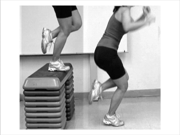
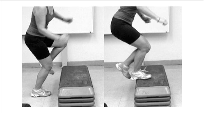
After informed consent procedures, weight, height, and distance between anterior superior iliac spines were measured. The distance between the anterior superior iliac spines was used to calculate the hip joint center. Leg dominance was operationally defined as the leg preferred to perform a single hop for distance (24,25). Each subject performed 2, separate, randomly ordered nonfatigued and fatigued sessions from 3 to 5 days apart. Each subject was asked to not exercise before the testing session, schedule study visits at the same time of the day for both sessions, and wear the same athletic shoes. The warm-up protocol for both sessions consisted of 5 minutes of pedaling at 40–60 revolutions per minute (rpm) on a cycle ergometer followed by completing 10 half squats, 5 continuous vertical jumps (countermovement jumps), and 2 practice trials of both jump tasks. The tasks considered in this investigation were a 40-cm single-legged drop jump (Figure 1) and a 20-cm up-down hop task (Figure 2). The drop jump was selected for its capability of creating high eccentric loading on the lower extremities during landing (33). The up-down hop task was selected for its high sensitivity to detect dynamic knee instability (15,16). These tasks were randomly ordered. The drop jump (Figure 1) consisted of standing initially on the 40-cm platform with both feet, and then standing on the jumping leg when the command “on your mark” was given. After the command “set,” the subject was instructed to jump when she felt ready to do so. Each subject was instructed to perform a maximal effort vertical jump as high as she could upon landing single legged on the force plate. Five trials of the drop jump were used because it has been demonstrated that the average values for 5 trials have exhibited good reliability (intraclass correlation [ICC] > 0.77) for all kinematic and kinetic variables (24). For the up-down hop task (Figure 2), each subject performed 10 repetitive jumps up to and down from a 20-cm step. One trial of 10 repeated up-down hops has demonstrated reliable results (ICC > 0.75) for all kinematic and kinetic variables when the average value of 10 hops is considered (24). The fatigued session was similar to the nonfatigued session, except for the addition of a 30-second Wingate anaerobic protocol with the purpose of inducing metabolic fatigue before task performance. The 30-second Wingate anaerobic protocol was selected as the fatigue-inducing task because of its capability of producing metabolic fatigue in a short period (14,37). The Wingate protocol was performed as described by Inbar et al. (14) after the generic warm-up session. During the Wingate protocol, each subject performed a 5-minute warm-up on a cycle ergometer (Monark 828 E; Monark Exercise AB, Vansbro, Sweden) at a speed of 40–60 rpm. Each subject performed a 30-second pedaling bout at maximal effort against a predetermined resistance immediately after the warm-up. The resistance on the cycle ergometer was determined by multiplying each subject's weight by the constant 0.090 kilopond. The resultant resistance was added in kilograms to the flywheel of the ergometer before the 30-second bout. The total amount of revolutions for each 5-second interval throughout the entire 30-second trial was displayed on the ergometer's electronic meter and recorded on a spreadsheet. Verbal encouragement was given to the subjects by the research team throughout the fatigue protocol. The subjects began the first trial of the functional tasks immediately after the maximal pedaling test (in less than 30 seconds). All remaining trials of the functional tasks were performed within 30 seconds of each other to prevent recovery. To confirm that each subject reached a desirable level of exhaustion with the Wingate protocol, a fatigue index was calculated. The fatigue index is an indication of decreased power output throughout the 30-second maximal pedaling bout (14). The fatigue index was calculated by the following formula: ([highest peak power − lowest peak power] × 100)/highest peak power. The average fatigue index for the subjects in this study was 34.94%. Based on previously reported findings in elite female track and field athletes, these levels are associated with high levels of lactate accumulation that are sufficient to impair athletic performance. Therefore, the subjects in this investigation reached the expected levels of fatigue.
Figure 1.

Forty-centimeter single-leg drop jump. Subjects were asked to drop with the predetermined dominant limb from the 40-cm platform onto the force plate. Upon landing, they were asked to perform a maximal vertical jump.
Figure 2.

Twenty-centimeter up-down hop task. Subjects were required to jump with their dominant limb 10 consecutive times up to and down from the 20-cm step. All 10 jumps were considered a trial.
Instrumentation
Twelve retroreflective markers were secured over both anterior superior iliac spines, second sacral vertebra, bilateral greater trochanters, lateral femoral epicondyles, mid-distance between greater trochanters and lateral femoral epicondyles, medial femoral epicondyles, lateral malleoli, mid-distance between lateral femoral epicondyles and lateral malleoli, medial malleoli, calcaneal tuberosities, and second metatarsophalangeal joints on each subject, for each of the 2 sessions. The same member of the research team placed the markers on each subject in both sessions to enhance consistency in marker placement over body prominences and anatomical landmarks across sessions. The motion analysis system consisted of 5 digital cameras (60 Hz sampling rate) time synchronized to the force plate (AMTI, Watertown, MA, USA) at 1 kHz sampling rate. Video and force plate data were captured with VISOL MultiDV Capture and KwonGRF 2.1 software, respectively (VISOL, Seoul, Korea). Recording space was calibrated according to manufacturer's recommendation with a 12-point 81.5-cm3 cube using an 11-parameter direct linear transformation algorithm before data collection. A static trial was captured on each day with subjects standing still with arms across the chest and feet shoulder-width apart. The static trial was used to define the neutral position (zero rotation) for each segment of the lower extremities to estimate its joint centers and calibrate its coordinates to the laboratory. After the static trial, bilateral medial femoral epicondylar and medial malleolar markers were removed to prevent interference between markers and legs during the tasks. Surface EMG was recorded from 4, bipolar, self-adhesive, Ag/AgCl preamplified surface electrodes (M-00-S; Ambu, Ølstykke, Denmark; overall gain: 2,000 mV, total electrode contact area: 4.1 × 3.4 cm, and sensor area: 1.32 cm2). Before electrode placement, the skin was cleaned with a gauze soaked in alcohol. Electrodes were placed on the skin over the gluteus maximus, quadriceps, and lateral and medial hamstrings according to the Cram et al. (8) recommendations. The reference electrode was placed over the anterior tibial crest. All electrodes were secured with hypoallergenic adhesive tape to reduce movement artifact. EMG was collected with a computerized telemetry system (Noraxon, Inc., Scottsdale, AZ, USA). Raw muscle activity was transmitted via frequency modulation (FM) signal from a transmitter that subjects wore on a belt. In the transmitter, the signal was filtered at a bandwidth of 10–500 Hz with 130 dB common rejection mode. The receiver converted the signal from analog to digital through an external universal serial bus (USB) analog-to-digital (A/D) converter, and raw muscle signals were displayed on the computer monitor.
Data Reduction
Joint angles and knee internal joint moment data were synchronized and analyzed with Kwon3D 3.1 (VISOL). Joint angles were derived from the 3-dimensional trajectory of retroreflective markers. Frequency contents were screened using residual analysis followed by a filtering process through a second-order low-pass Butterworth filter at a frequency of 6 Hz. Peak knee joint angles were defined in sagittal, frontal, and transverse planes as the first, second, and third rotations, respectively. Peak knee joint rotations were expressed relative to each subject's standing posture (neutral position). Rotational transformation matrices between thigh and shank segments were computed based on the unit vectors of the local frames of these 2 segments. Euler angles (orientation angles) were calculated from the rotational transformation matrices using the mediolateral-anteroposterior-longitudinal axis (x-y-z) rotational sequence. Internal knee joint moments were derived by inverse dynamic methods instrumented in the software normalized to body mass (N·m·kg−1).
The kinematic and kinetic data of interest for the drop jump were the peak angle and moments values of the entire ground contact phase. The ground contact phase was operationally defined from the moment of initial contact to foot from the force plate. For the up-down hop task, the first 2 and last 2 jumps of the total 10 jumps were excluded to account for acceleration and deceleration during the task. The peak knee joint angles and internal moments during the middle 6 jumps were averaged for analysis.
EMG raw data were amplified (1,000 times), and full-wave rectified using Myoresearch software (Noraxon, Inc.). EMG data were normalized by using a dynamic normalization procedure in which the average signal for each muscle group in the window of interest (ground contact phase) was divided by the maximum signal generated on the specific trial analyzed. This method has been widely used to analyze EMG activity during dynamic tasks (2,9,19,28,29) and has been shown to reduce subject variability when compared with maximal isometric voluntary contractions (2,31) and to control for the variability between trials caused by fatigue during dynamic tasks with multiple trials (9). Because the hamstring muscle group was separated into medial and lateral compartments, the normalized results were averaged to represent the hamstring group in its entirety (2,19). In addition to the normalized EMG data, a cocontraction ratio of the quadriceps and hamstrings was calculated (2,19). The first step for calculating the cocontraction ratio involved obtaining the normalized values for both the quadriceps and hamstring muscle groups during the targeted window of time (2,19). The hamstring value was used as the divisor if its value was greater than the quadriceps. However, the quadriceps value was used as divisor if its value was greater than the hamstrings (2,19). Therefore, the cocontraction ratio value was always less than or equal to 1 and represented the relative activation of the flexor and extensor muscle groups crossing the knee joint (2,19). Therefore, a cocontraction ratio closer to 1 indicated excellent cocontraction between quadriceps and hamstrings, whereas values closer to 0 represented poor cocontraction between these muscles (2,19).
Statistical Analyses
All kinematic, kinetic, and EMG data were screened for normality assumptions using the Shapiro-Wilk test and histograms. Analyses of variance (ANOVA) were used to compare differences in all dependent variables between sessions. An a priori alpha level of 0.05 was selected for all ANOVAs. Effect sizes and power (1 − β) were also calculated for all analyses.
Results
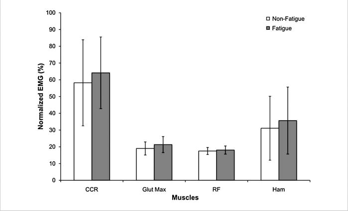
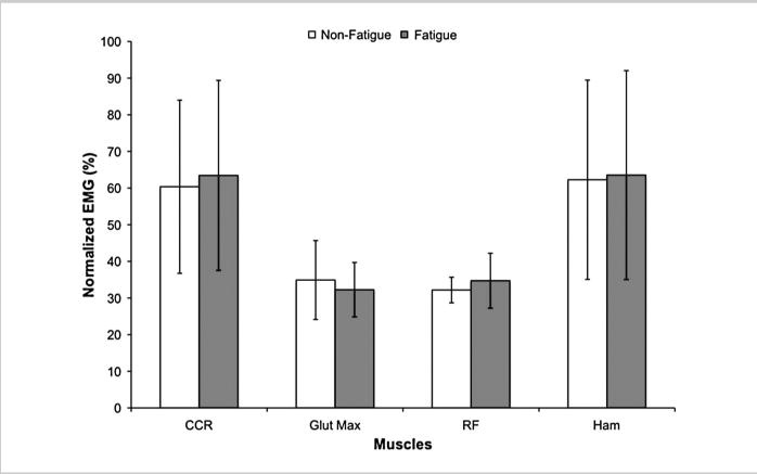
All variables met normality assumptions for both tasks. The results of the drop jump showed in its majority a trend toward greater knee injury–predisposing factors (Tables 1 and 2) and greater EMG activity (Figure 3) during the fatigued session. However, these results were not statistically significant between sessions. The results of the up-down hop task showed varied results for kinematic (Table 1), kinetic (Table 2), and EMG (Figure 4) variables with a reduction in knee flexion during the fatigued session as the only statistically significant (p = 0.028) finding.
Table 1.
Peak knee joint angles between nonfatigued and fatigued conditions (n = 15).
| Peak joint angles (°) | ||||
|---|---|---|---|---|
| 40-cm drop jump | 20-cm up-down | |||
| Nonfatigue | Fatigue | Nonfatigue | Fatigue | |
| Knee flexion | ||||
| Mean ± SD | 57.85 ± 5.68 | 56.80 ± 7.34 | 49.27 ± 5.43 | 45.46 ± 5.36* |
| ES (1 − β) | 0.059 (0.140) | 0.299 (0.624) | ||
| Knee valgus | ||||
| Mean ± SD | 9.88 ± 5.34 | 10.38 ± 6.58 | 5.80 ± 3.84 | 8.19 ± 4.57 |
| ES (1 − β) | 0.012 (0.067) | 0.164 (0.339) | ||
p = 0.029; ES = effect size; 1 − β = power.
Table 2.
Peak knee joint moments, between nonfatigued and fatigued conditions (n = 15).
| Peak joint moments (N·m·kg−1) | ||||
|---|---|---|---|---|
| 40-cm drop jump | 20-cm up-down | |||
| Nonfatigue | Fatigue | Nonfatigue | Fatigue | |
| Extension | ||||
| Mean ± SD | 3.02 ± 0.72 | 2.86 ± 0.66 | 2.80 ± 0.66 | 2.77 ± 1.02 |
| ES (1 − β) | 0.064 (0.150) | 0.066 (0.127) | ||
| Valgus | ||||
| Mean ± SD | 0.07 ± 0.05 | 0.11 ± 0.09 | 0.33 ± 0.14 | 0.33 ± 0.16 |
| ES (1 − β) | 0.198 (0.409) | 0.001 (0.051) | ||
| Varus | ||||
| Mean ± SD | 1.70 ± 0.76 | 1.83 ± 1.09 | 2.05 ± 0.88 | 2.14 ± 0.75 |
| ES (1 − β) | 0.038 (0.106) | 0.067 (0.128) | ||
N·m·kg−1 = joint moments in newton meters per kilogram of body mass; ES = effect size; 1 − β = power.
Figure 3.

Normalized EMG activity during the drop jump presented no difference between conditions. CCR = quadriceps/hamstring cocontraction ratio; Glut Max = gluteus maximus; RF = rectus femoris; HAM = hamstrings.
Figure 4.

Normalized EMG activity during the up-down hop task presented no difference between conditions. CCR = quadriceps/hamstrings cocontraction ratio; Glut Max = gluteus maximus; RF = rectus femoris; HAM = hamstrings.
Discussion
The purpose of this investigation was to evaluate the effects of fatigue on knee joint angles, internal knee moments, and neuromuscular activation during the landing phase of 2 jumping tasks performed by young women. As hypothesized, greater knee injury–predisposing factors during the fatigued condition were observed but solely during the up-down hop task. Decreased knee flexion joint angles might increase the likelihood for greater knee ligamentous stress by decreasing the ability of the hamstrings to resist anterior shear forces on the tibia (5). Smaller peak knee flexion joint angles after the onset of fatigue might be a compensation strategy to prevent collapse of the center of mass because of fatigue in the quadriceps muscle group (22,28). This compensation can be observed by minimal smaller knee joint extension moments during fatigue as a strategy to sustain performance by preventing deep knee flexion and subsequent collapse of the center of mass (23,28,29). This compensatory strategy reduces knee moments by increasing joint stiffness, further allowing the subjects to perform faster concentric contractions toward a jump (13,28,29,34). In addition, given the inherent difficulties of landing maneuvers, greater knee stiffness during landing single legged from a jump may be not only a strategy to prevent collapse of the knee joint but also an indication of strategies to stabilize anterior-posterior trunk movements keeping the trunk centered within the base of support (7). Among the possible reasons influencing the magnitude of findings in knee joint flexion between tasks could be the repetitive nature of the up-down hop test. It can be hypothesized that the up-down hop task presents a greater challenge to the motor system given the high speed of movement required to maintain control and balance. Although not statistically significant, the greater varus/valgus moment exhibited by the subjects in this investigation during the fatigue session agreed with previous findings (5,20). These greater varus/valgus moments in combination with smaller knee joint flexion angles are important factors for generating high knee loads that could reach injurious magnitudes (5,20). Therefore, it seems that fatigue places the knee in an unstable situation by decreasing its dynamic stability in the sagittal and frontal planes.
No previously published studies reported the effects of fatigue on task performance by using the cocontraction ratio. Investigators have suggested that the quadriceps/hamstrings cocontraction is used as the main strategy to stabilize the knee joint and resist external loads toward varus/valgus and flexion/extension during high-level athletic tasks (2,19). This strategy has been hypothesized to help protect knee ligaments against stresses that can induce ligament strain and failure by enhancing neuromuscular activation of stabilizing muscles (2,19). Therefore, although the tasks in this investigation have been shown to provide high lower-extremity external loads (10,13,16) in flexion/extension and varus/valgus, it would be reasonable to observe a similar or increased cocontraction ratio between quadriceps and hamstrings during the fatigued condition to prevent ligamentous injury. A different explanation for the observed results of the cocontraction ratios pertains to the operational definition used for the landing phase. Knee ligament injuries in women athletes tend to occur immediately before push off into a cutting or jumping maneuver (10), whereas others have reported the beginning stages of the landing cycle as specifically responsible for ACL injuries (3,4,6). In this investigation, the landing phase was defined as the entire time the subjects were in contact with the force plate, from the moment of initial contact to push off, without differentiating among preactivation (9), weight acceptance (eccentric) (2–4), and push off (concentric) subphases during contact time (2–4,6). The preactivation phase is defined as a motor control phase in which muscle activation is enhanced (9) with the purpose of reducing external impact forces upon landing (13). The eccentric (negative) phase is reflexive in nature and the one in which a rapid response from all stabilizing muscles is created to provide stability during potentially injurious situations (9) and resist the downward collapse of the center of mass (13). The takeoff (push off) phase is the component in which the subjects exert a concentric contraction to recover from the jump and prepare to perform an upward vertical jump (29). Therefore, further work assessing neuromuscular cocontraction between quadriceps and hamstrings should consider different phases (concentric/eccentric) of the landing cycle. Additionally, a successful trial was defined as one where the subject was able to perform the vertical jump after landing. All subjects performed more than 5 trials during the fatigued session as the first or second or both trials were repeated because the subjects fell or were not able to recover from the landing. The exclusion of unsuccessful trials from the analyses might have concealed the real impact of fatigue in landing mechanics.
Apparently, the fatigue created by the Wingate anaerobic test did not affect neuromuscular activation and dynamic knee joint stability in these young women to the level expected. Although it appears that women reached a high level of fatigue according to the fatigue index estimation, the rapid recovery from the Wingate protocol might have played a factor preventing differences between nonfatigued and fatigued sessions. There are additional factors that may have prevented significant biomechanical knee injury–predisposing factors from being observed during the fatigued session. Greater muscle control during the fatigued condition, greater eccentric control of the knee joint (10), and compensatory neuromuscular strategies (28,29) are all possible compensations to prevent dynamic instability of the lower limbs. Neuromuscular factors that might help explain this behavior are the recruitment of additional motor units (29), increased firing synchronization (29), and the “common drive” (movement pattern generator) theory (28,29). The recruitment of additional motor neurons and increased synchronization of gluteal and thigh muscles might be an attempt of the nervous system to activate, more intensively, the muscles to maintain the ability to jump after a landing (28), generating greater eccentric control (23). The common drive theory states that force-generating properties of agonist and antagonist muscles are controlled by single reflexive actions indicating that agonists and antagonists muscle groups are activated simultaneously as a functional unit, regardless of fatigue level of specific muscle groups (2,28,29). Given that the jumping tasks used in this investigation required a jump after landing, and repetitive continuous jumps, these explanations seem viable.
The small sample size (n = 15; observed power < 51%) was another factor affecting this investigation. Therefore, we recommend the use of a larger sample to explore further the effects of fatigue on dynamic knee joint stability. The lack of subject familiarization with the Wingate protocol might have also affected the results. Barfield et al. (1) recommended the use of a familiarization session with the Wingate protocol before baseline measures to minimize a learning effect. These investigators found an increase in peak and mean power from the first to the second session when the Wingate protocol was administered 7 days apart. Therefore, it is possible that the subjects in this investigation did not perform maximally during the fatigue protocol because of lack of experience and motivation, even though verbal encouragement was given during the fatigue protocol (32). There is a possibility that the fatigue protocol may have not induced sufficient fatigue and that subjects may have recovered between trials, even though the fatigue index indicated levels where lactate concentration levels are normally high.
Although the purpose of EMG and kinematics normalization procedures is to standardize all data, performance of 2 sessions in different days may have introduced error because of the nature of placing skin retroreflective markers and EMG electrodes differently on both occasions. Voluntary adjustments in landing mechanics during preplanned activities might have affected neuromuscular recruitment strategies because the subjects knew in advance the maneuver to be performed (10). Both tasks used in this study were planned maneuvers in which each subject was aware of the movement patterns needed to perform each trial. Besier et al. (2,3) reported differences in movement patterns between planned and unplanned maneuvers. The performance of unplanned tasks placed more stress on the knee joint, increasing the possibility of injury, if improper neuromuscular strategies are used (2,3). These findings might indicate that even though the tasks used in this study are capable of creating high loads to the knee joint (16), the difficulty level may not have been high enough to be altered by metabolic fatigue. Albeit the 60 Hz frequency sampling rate used may have introduced variability into the measurement for both tasks, frequency contents were screened with a residual analysis and filtered through a low-pass Butterworth filter (6 Hz).
Practical Applications
Metabolic fatigue has been identified as one of the possible contributing factors to impair dynamic control and lower-extremity injuries. Strength and conditioning specialists need to be aware of how athletic performance can be affected by fatigue and associated mechanisms leading to increased risk for injuries. It is apparent that the human body might be able to protect the lower extremities against unstable conditions through neuromuscular compensations, such as an increase in muscular activation during the onset of fatigue. However, this is not always the case because certain evidence exists in which fatigue may exacerbate predisposing factors for lower-extremity injuries. It is for this reason that strength and conditioning specialists should not underestimate training endurance and strength-endurance components in all athletes as means to reduce injuries and maximize performance. In addition, it might be possible that the use of repetitive functional exercises help the athletes develop neuromuscular strategies that could be used in situations where fatigue might pose a risk for injury.
Acknowledgments
This study was supported in part by an institutional grant from Texas Woman's University (Research Enhancement Program), and the Research Center for Minority Institutions-Clinical Research Infrastructure Initiative (RCRCII) award, 1P20RR11126 and, G12RR03051 and R25RR017589, from the National Center for Research Resources, National Institutes of Health, and The National Strength and Conditioning Association Foundation.
Footnotes
This study was conducted in the Musculoskeletal Laboratory at the Physical Therapy Program from Texas Woman's University-Houston Campus.
References
- 1.Barfield JP, Sells PD, Rowe DA, Hannigan-Downs K. Practice effect of the Wingate anaerobic test. J Strength Cond Res. 2002;16:472–473. [PubMed] [Google Scholar]
- 2.Besier TF, Lloyd DG, Ackland TR. Muscle activation strategies at the knee during running and cutting maneuvers. Med Sci Sports Exerc. 2003;35:119–127. doi: 10.1097/00005768-200301000-00019. [DOI] [PubMed] [Google Scholar]
- 3.Besier TF, Lloyd DG, Ackland TR, Cochrane JL. Anticipatory effects on knee joint loading during running and cutting maneuvers. Med Sci Sports Exerc. 2001;33:1176–1181. doi: 10.1097/00005768-200107000-00015. [DOI] [PubMed] [Google Scholar]
- 4.Besier TF, Lloyd DG, Cochrane JL, Ackland TR. External loading of the knee joint during running and cutting maneuvers. Med Sci Sports Exerc. 2001;33:1168–1175. doi: 10.1097/00005768-200107000-00014. [DOI] [PubMed] [Google Scholar]
- 5.Chappell JD, Herman DC, Knight BS, Kirkendall DT, Garrett WE, Yu B. Effect of fatigue on knee kinetics and kinematics in stop-jump tasks. Am J Sports Med. 2005;33:1022–1029. doi: 10.1177/0363546504273047. [DOI] [PubMed] [Google Scholar]
- 6.Chappell JD, Yu B, Kirkendall DT, Garrett WE. A comparison of knee kinetics between male and female recreational athletes in stop-jump tasks. Am J Sports Med. 2002;30:261–267. doi: 10.1177/03635465020300021901. [DOI] [PubMed] [Google Scholar]
- 7.Coventry E, O'Connor KM, Hart BA, Earl JE, Ebersole KT. The effect of lower extremity fatigue on shock attenuation during single-leg landing. Clin Biomech (Bristol, Avon) 2006;21:1090–1097. doi: 10.1016/j.clinbiomech.2006.07.004. [DOI] [PubMed] [Google Scholar]
- 8.Cram JR, Kasman GS, Holtz J. Introduction to Surface Electromyography. Gaithersburg, MD: Aspen Publishers; 1998. [Google Scholar]
- 9.Croce RV, Russell PJ, Decoster LC. Knee muscular response strategies differ by developmental level but not gender during jump landing. Electromyogr Clin Neurophysiol. 2004;44:339–348. [PubMed] [Google Scholar]
- 10.Fagenbaum R, Darling WG. Jump landing strategies in male and female college athletes and the implications of such strategies for anterior cruciate ligament injury. Am J Sports Med. 2003;31:233–240. doi: 10.1177/03635465030310021301. [DOI] [PubMed] [Google Scholar]
- 11.Gabbett TJ. Incidence of injury in amateur rugby league sevens. Br J Sports Med. 2002;36:23–26. doi: 10.1136/bjsm.36.1.23. [DOI] [PMC free article] [PubMed] [Google Scholar]
- 12.Greig M, Wlker-Johnson C. The influence of soccer-specific fatigue on functional stability. Physical Therapy in Sport. 2007;8:185–190. [Google Scholar]
- 13.Hass CJ, Schick EA, Tillman MD, Chow JW, Brunt D, Cauraugh JH. Knee biomechanics during landings: Comparison of pre- and postpubescent females. Med Sci Sports Exerc. 2005;37:100–107. doi: 10.1249/01.mss.0000150085.07169.73. [DOI] [PubMed] [Google Scholar]
- 14.Inbar O, Bar-Or O, Skinner JS. The Wingate Anaerobic Test. Champaign, IL: Human Kinetics; 1996. [Google Scholar]
- 15.Itoh H, Ichihashi N, Maruyama T, Kurosaka M, Hirohata K. Weakness of thigh muscles in individuals sustaining anterior cruciate ligament injury. Kobe J Med Sci. 1992;38:93–107. [PubMed] [Google Scholar]
- 16.Itoh H, Kurosaka M, Yoshiya S, Ichihashi N, Mizuno K. Evaluation of functional deficits determined by four different hop tests in patients with anterior cruciate ligament deficiency. Knee Surg Sports Traumatol Arthrosc. 1998;6:241–245. doi: 10.1007/s001670050106. [DOI] [PubMed] [Google Scholar]
- 17.Kellis E, Katis A, Vrabas IS. Effects of an intermittent exercise fatigue protocol on biomechanics of soccer kick performance. Scand J Med Sci Sports. 2006;16:334–344. doi: 10.1111/j.1600-0838.2005.00496.x. [DOI] [PubMed] [Google Scholar]
- 18.Kent-Braun JA. Central and peripheral contributions to muscle fatigue in humans during sustained maximal effort. Eur J Appl Physiol Occup Physiol. 1999;80:57–63. doi: 10.1007/s004210050558. [DOI] [PubMed] [Google Scholar]
- 19.Lloyd DG, Buchanan TS. Strategies of muscular support of varus and valgus isometric loads at the human knee. J Biomech. 2001;34:1257–1267. doi: 10.1016/s0021-9290(01)00095-1. [DOI] [PubMed] [Google Scholar]
- 20.McLean SG, Felin RE, Suedekum N, Calabrese G, Passerallo A, Joy S. Impact of fatigue on gender-based high-risk landing strategies. Med Sci Sports Exerc. 2007;39:502–514. doi: 10.1249/mss.0b013e3180d47f0. [DOI] [PubMed] [Google Scholar]
- 21.Murtaugh K. Injury patterns among female field hockey players. Med Sci Sports Exerc. 2001;33:201–207. doi: 10.1097/00005768-200102000-00005. [DOI] [PubMed] [Google Scholar]
- 22.Nyland JA, Shapiro R, Caborn DN, Nitz AJ, Malone TR. The effect of quadriceps femoris, hamstring, and placebo eccentric fatigue on knee and ankle dynamics during crossover cutting. J Orthop Sports Phys Ther. 1997;25:171–184. doi: 10.2519/jospt.1997.25.3.171. [DOI] [PubMed] [Google Scholar]
- 23.Orishimo KF, Kremenic IJ. Effect of fatigue on single-leg hop landing biomechanics. J Appl Biomech. 2006;22:245–254. doi: 10.1123/jab.22.4.245. [DOI] [PubMed] [Google Scholar]
- 24.Ortiz A, Olson SL, Libby CL, Kwon YH, Trudelle-Jackson E. Kinematic and kinetic reliability of two jumping and landing physical performance tasks in young adult women. N Am J Sports Phys Ther. 2007;2:104–112. [PMC free article] [PubMed] [Google Scholar]
- 25.Ortiz A, Olson SL, Libby CL, Trudelle-Jackson E, Kwon YH, Etnyre B, Bartlett WP. Landing mechanics between non-injured women and women with ACL reconstruction during two jump tasks. Am J Sports Med. 2008;36:149–157. doi: 10.1177/0363546507307758. [DOI] [PMC free article] [PubMed] [Google Scholar]
- 26.Rahnama N, Lees A, Reilly T. Electromyography of selected lower-limb muscles fatigued by exercise at the intensity of soccer match-play. J Electromyogr Kinesiol. 2006;16:257–263. doi: 10.1016/j.jelekin.2005.07.011. [DOI] [PubMed] [Google Scholar]
- 27.Rahnama N, Reilly T, Lees A. Injury risk associated with playing actions during competitive soccer. Br J Sports Med. 2002;36:354–359. doi: 10.1136/bjsm.36.5.354. [DOI] [PMC free article] [PubMed] [Google Scholar]
- 28.Rodacki AL, Fowler NE, Bennett SJ. Multi-segment coordination: Fatigue effects. Med Sci Sports Exerc. 2001;33:1157–1167. doi: 10.1097/00005768-200107000-00013. [DOI] [PubMed] [Google Scholar]
- 29.Rodacki AL, Fowler NE, Bennett SJ. Vertical jump coordination: Fatigue effects. Med Sci Sports Exerc. 2002;34:105–116. doi: 10.1097/00005768-200201000-00017. [DOI] [PubMed] [Google Scholar]
- 30.Ronglan L, Raastad T, Borgesen A. Neuromuscular fatigue and recovery in elite female handball players. Scand J Med Sci Sports. 2005;16:267–273. doi: 10.1111/j.1600-0838.2005.00474.x. [DOI] [PubMed] [Google Scholar]
- 31.Soderberg GL, Knutson LM. A guide for use and interpretation of kinesiologic electromyographic data. Phys Ther. 2000;80:485–498. [PubMed] [Google Scholar]
- 32.Clair Gibson A, Baden DA, Lambert MI, Lambert EV, Harley YX, Hampson D, Russell VA, Noakes TD. The conscious perception of the sensation of fatigue. Sports Med. 2003;33:167–176. doi: 10.2165/00007256-200333030-00001. [DOI] [PubMed] [Google Scholar]
- 33.Walsh M, Arampatzis A, Schade F, Bruggemann GP. The effect of drop jump starting height and contact time on power, work performed, and moment of force. J Strength Cond Res. 2004;18:561–566. doi: 10.1519/1533-4287(2004)18<561:TEODJS>2.0.CO;2. [DOI] [PubMed] [Google Scholar]
- 34.Wikstrom EA, Powers ME, Tillman MD. Dynamic stabilization time after isokinetic and functional fatigue. J Athl Train. 2004;39:247–253. [PMC free article] [PubMed] [Google Scholar]
- 35.Wilkins JC, Valovich McLeod TC, Perrin DH, Gansneder BM. Performance on the balance error scoring system decreases after fatigue. J Athl Train. 2004;39:156–161. [PMC free article] [PubMed] [Google Scholar]
- 36.Wojtys EM, Wylie BB, Huston LJ. The effects of muscle fatigue on neuromuscular function and anterior tibial translation in healthy knees. Am J Sports Med. 1996;24:615–621. doi: 10.1177/036354659602400509. [DOI] [PubMed] [Google Scholar]
- 37.Zajac A, Jarzabek R, Waskiewicz Z. The diagnostic value of the 10- and 30-second Wingate test for competitive athletes. J Strength Cond Res. 1999;13:16–19. [Google Scholar]


