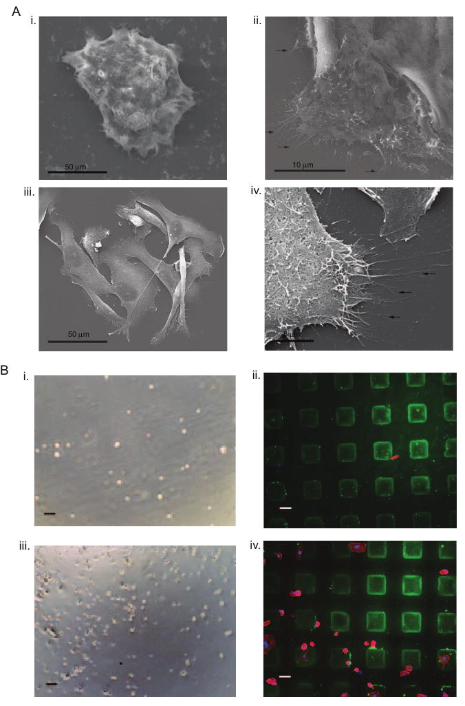
Figure 4. Cancer cell interaction with HA.

A. SEM analysis revealed adhesive protrusions of colon cancer cells cultured on HA surfaces shown at (i) low and (ii) high magnifications and of breast cancer cells cultured on HA surfaces shown at (iii) low and (iv) high magnifications (arrows indicate cell protrusions) B. Blocking of CD44 prevented (i) colon cancer cell adhesion after 24 hours of culture while (ii) HA surfaces are present (green; actin-stained colon cancer cells in red; nuclei in blue) and (iii) breast cancer cell adhesion after 24 hours of culture while (iv) HA surfaces are present (green; actin-stained breast cancer cells in red; nuclei in blue). Scale bars are 100 μm in Bi and Biii, and 50 μm in Bii and Biv.
