Abstract
Background/Aims
The inadequacy of information on the treatment of gastric candidiasis with antifungal agents promoted us to evaluate patients with fungal infections who had gastric ulcers and assess the need for proton-pump inhibitors or antifungal agents.
Methods
Sixteen patients were included in the study. The criterion for the diagnosis of candidiasis was finding yeast and hyphae in the tissue or an ulcer on histological sections of biopsy samples. Surface fungi were not considered infections.
Results
In all cases with benign ulcers, follow-up endoscopy performed 6 weeks after proton-pump-inhibitor treatment revealed that the ulcer had improved without antifungal medication. However, in patients with malignant ulcers, surgical resection was necessary for a definitive cure. Two patients with lymphoma received combined chemotherapy and a proton-pump inhibitor, which improved their condition.
Conclusions
The results of this study suggest that benign ulcers with candidiasis can be effectively treated by a proton-pump inhibitor without antifungal medication. However, surgical resection should be considered for malignant ulcers with candidiasis.
Keywords: Candidiasis, Antifungal agents, Stomach ulcer, Proton pump inhibitors
INTRODUCTION
Most studies on the infectious nature of gastric ulcer disease have focused on the role of Helicobacter pylori on pathogenesis. The effect of fungal infections on the course of gastric ulcer disease has not been clarified to date.1 A study reported by Neeman et al.2 found that fungal infection of gastric ulcers was associated with delayed healing; antimycotic therapy was advocated in such cases. By contrast, Minoli et al.3 and Gotlieb-Jensen et al.4 considered it an epiphenomenon without any significance. However, because the issue remains unresolved we retrospectively reviewed fungal infections in patients with gastric ulcers to determine the need for proton pump inhibitor and/or antifungal agents.
MATERIALS AND METHODS
We reviewed the pathology specimens and medical records of patients with gastroduodenal ulcers diagnosed by upper gastrointestinal endoscopy at Kyungpook National University Hospital from September 1998 to August 2007. Sixteen patients were included in the study. The criterion for the diagnosis of candidiasis was the finding of yeast and hyphae in the tissue or ulcer on histological sections of biopsies. Surface fungi were not considered infections. The demonstration of Candida in smears and cultures was not considered reliable evidence for candidiasis, as this organism is a common commensal organism and its presence does not imply a pathogenic role.5 Consequently, culture of biopsies for Candida was not done routinely.
Two groups of gastric candidiasis: the thrush type was characterized by whitish exudates with inflammation and the ulcerated type manifested as an ulcer with well defined margins and whitish exudates.6 A clinical response was defined as the relief of symptoms and a decrease in the ulcer size.
RESULTS
1. Basic characteri
We reviewed 16 cases of gastric candidiasis that had endoscopic biopsies. There were 9 cases of gastric candidiasis with benign ulcers and 7 cases with malignant ulcers including gastric adenocarcinoma and gastric lymphomas. The mean age of the patients with benign ulcers was 64 (42-77) and for the malignant ulcers it was 59.7 (43-76). Other conditions present were diabetes, liver cirrhosis, lung cancer, and pulmonary tuberculosis (Table 1).
Table 1.
Baseline Characteristics of Gastric Candidiasis
2. Endoscopic findings
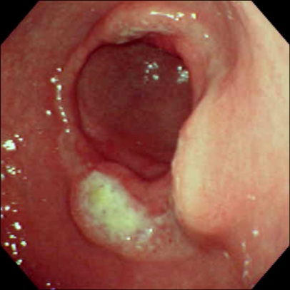
The gastric candidiasis, in both benign and malignant cases, was distributed throughout the stomach (Table 2). Most cases were the ulcerated type; there was only one case of thrush. The mean size of the benign ulcers was 43 mm. The malignant ulcers were larger, with a mean size of 66 mm. In benign ulcer, a round ulcer with thick whitish exudates was seen (Fig. 1), endoscopically we couldn't know whether infected with candia or not. Malignant ulcer with candidiasis had more larger ulcer with thick and dirty whitish exudates (Fig. 2, 3).
Table 2.
Endoscopy Findings of Gastric Candidiasis
A, antrum; AW, anterior wall; CLO, rapid urease test; DB, distal body; F, fundus; GC, greater curvature; LC, lesser curvature; M, multiple; MB, midbody; N/A, not available; PB, proximal body; PW, posterior wall; S, single.
Fig. 1.
A round ulcer with white exudates was evident at the antrum near the pylorus.
Fig. 2.

An ulcerofungating mass with thick white exudates was evident at the posterior side of the distal body.
Fig. 3.
A deep round ulcer with thick white exudates was evident at the greater curvature side of the antrum.
3. Treatment response
For all of the benign ulcers, follow-up endoscopy done 6 weeks after proton pump inhibitor treatment revealed that the ulcers showed response without antifungal medication. Just one case was treated with fluconazole 100 mg for seven days because of concomitant esophageal candidiasis (Table 3).
Table 3.
Treatment Response of Gastric Candidiasis
However, for the malignant ulcers, if the lesion was resectable, curative surgery was performed. In two cases with lymphoma, combined chemotherapy with proton pump inhibitor treatment was prescribed, and the patients improved. Though all cases of malignant ulcer were not treated with antifungal medication before surgery or chemotherapy, they did not develop the systemic fungal infection or did not need to change the treatment plan. Even though the three adenocarcinoma cases could not undergo resection and were not treated with antifungal agents, they died without progression to invasive systemic candidiasis.
DISCUSSION
The prevalence of fungal infection of gastric ulcers ranges from 4% in an autopsy series to 36%.1,4,7,8 The discrepancy between these data would appear to be due to the criteria used to diagnose fungal colonization, the age of the patients studied, concurrent disease, and the pharmacotherapy applied. Assessment of the factors that might enhance fungal infection, in patients with gastric ulcer disease, revealed that fungal infection was associated with increased age. It is assumed that in the elderly, because of weakening of the host defense mechanisms, the fungus more easily invades the ulcer.2,8 Other factors, such as gender, smoking, tea or coffee intake, underlying disease, antibiotic usage, and immunosuppressive therapy, have not been shown to influence the rate of fungal infections of gastric ulcers.9-11 In this study, the mean age of the patients with gastric candidiasis was 62. Of note, is that the cases with benign ulcers were older than were those with malignant ulcers. However in this study, most of benign ulcer have underlying disease such as diabetes mellitus, liver cirrhosis, pulmonary disease. It also could be explained the weakening of the host defense mechanisms and the increased opportunistic infection of oropharyngeal or esophageal candidiasis.
Previous studies have reported that fungal infection delays gastric ulcer healing,2 whereas others debate this issue.1,3,4,9 On microscopic examination, fungal material was found mainly in the exudative substance at the ulcer base, and fungal infiltration could be identified in adjacent normal mucosa or submucosa in only a few patients.12 Thus, when present in gastric ulcers, the fungus may have a very low capability for tissue-invasion and act as an "opportunistic microbe."13 The presence of the fungus does not seem to have a negative effect on the outcome of medical therapy. In the present study, the gastric ulcers improved with proton pump therapy without specific treatment for the candidiasis.
The role of acid suppression in promoting gastric Candida infection is also controversial. An earlier study suggested a predisposition to Candida overgrowth with H2-receptor antagonist therapy. However, a recent prospective study, did not report higher Candida culture rates in patients receiving H2-blockers or proton pump inhibitor treatment compared to no treatment with acid suppressants.14 In the present study, the cases with benign ulcer responded to proton pump inhibitor treatment alone.
Minoli et al.3 reported that the gastric ulcers with fungal infection appeared malignant and appeared larger on endoscopic inspection in a significantly higher percentage of cases than "normal" ulcers. In this study, malignant ulcer was larger and had thick whitish exudates. But we couldn't find out the exact prevalence of gastric candidiasis and possible interaction between candida and malignant ulcers.
In conclusion, the results of this study suggest that benign gastric ulcer disease with candidiasis was effectively treated with proton pump inhibitor therapy without anti-fungal medication. The malignant ulcers with candidiasis should be considered for surgical resection. Therefore, the candidiasis may act as an opportunistic microbe and does not require additional treatment with antifungal medication.
References
- 1.Di Febo G, Miglioli M, Calo G, et al. Candida albicans infection of gastric ulcer frequency and correlation with medical treatment. Results of a multicenter study. Dig Dis Sci. 1985;30:178–181. doi: 10.1007/BF01308206. [DOI] [PubMed] [Google Scholar]
- 2.Neeman A, Avidor I, Kadish U. Candidal infection of benign gastric ulcers in aged patients. Am J Gastroenterol. 1981;75:211–213. [PubMed] [Google Scholar]
- 3.Minoli G, Terruzzi V, Ferrara A, et al. A prospective study of relationships between benign gastric ulcer, Candida, and medical treatment. Am J Gastroenterol. 1984;79:95–97. [PubMed] [Google Scholar]
- 4.Gotlieb-Jensen K, Andersen J. Occurrence of Candida in gastric ulcers. Significance for the healing process. Gastroenterology. 1983;85:535–537. [PubMed] [Google Scholar]
- 5.Gorbach SL, Nahas L, Lerner PI, Weinstein L. Studies of intestinal microflora. I. Effects of diet, age, and periodic sampling on numbers of fecal microorganisms in man. Gastroenterology. 1967;53:845–855. [PubMed] [Google Scholar]
- 6.Kodsi BE, Wickremesinghe C, Kozinn PJ, Iswara K, Goldberg PK. Candida esophagitis: a prospective study of 27 cases. Gastroenterology. 1976;71:715–719. [PubMed] [Google Scholar]
- 7.Eras P, Goldstein MJ, Sherlock P. Candida infection of the gastrointestinal tract. Medicine (Baltimore) 1972;51:367–379. doi: 10.1097/00005792-197209000-00002. [DOI] [PubMed] [Google Scholar]
- 8.Loffeld RJ, Loffeld BC, Arends JW, Flendrig JA, van Spreeuwel JP. Fungal colonization of gastric ulcers. Am J Gastroenterol. 1988;83:730–733. [PubMed] [Google Scholar]
- 9.Wu CS, Wu SS, Chen PC. A prospective study of fungal infection of gastric ulcers: clinical significance and correlation with medical treatment. Gastrointest Endosc. 1995;42:56–58. doi: 10.1016/s0016-5107(95)70244-x. [DOI] [PubMed] [Google Scholar]
- 10.Scott BB, Jenkins D. Gastro-oesophageal candidiasis. Gut. 1982;23:137–139. doi: 10.1136/gut.23.2.137. [DOI] [PMC free article] [PubMed] [Google Scholar]
- 11.Savino JA, Agarwal N, Wry P, Policastro A, Cerabona T, Austria L. Routine prophylactic antifungal agents (clotrimazole, ketoconazole, and nystatin) in nontransplant/non-burned critically ill surgical and trauma patients. J Trauma. 1994;36:20–25. doi: 10.1097/00005373-199401000-00004. [DOI] [PubMed] [Google Scholar]
- 12.Katzenstein AL, Maksem J. Candidal infection of gastric ulcers: histology, incidence, and clinical significance. Am J Clin Pathol. 1979;71:137–141. doi: 10.1093/ajcp/71.2.137. [DOI] [PubMed] [Google Scholar]
- 13.Minoli G, Terruzzi V, Butti GC, Benvenuti C, Rossini T, Rossini A. A prospective study on Candida as a gastric opportunistic germ. Digestion. 1982;25:230–235. doi: 10.1159/000198837. [DOI] [PubMed] [Google Scholar]
- 14.Wang K, Lin HJ, Perng CL, et al. The effect of H2-receptor antagonist and proton pump inhibitor on microbial proliferation in the stomach. Hepatogastroenterology. 2004;51:1540–1543. [PubMed] [Google Scholar]


