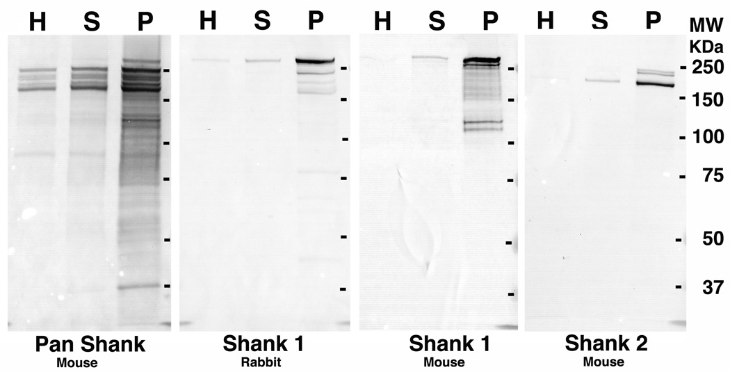
Figure 1.
Western immunoblots with Shank antibodies of homogenate (H), synaptosome (S) and PSD (P) fractions from cerebral cortex show significant enrichment of all Shank sub-types in the PSD fraction. The same amount of protein (10 µg) was loaded into each lane. Shank sub-families show further molecular diversity due to alternative splicing (reviewed in Boeckers et al., 2002). Reported isoforms of Shank1 in the UniProtKB database are in the 160–226 KDa range and for Shank 2 in the 134–200 KDa range.

