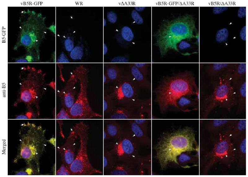
Fig. 2. B5-GFP is mis-targeted in the absence of A33.

HeLa cells were infected with the indicated viruses at a MOI of 1.0. At 24 h PI, fixed and permeabilized cells were stained with an anti-B5 MAb, followed by Texas Red-conjugated donkey anti-rat antibody (Red), and visualized using a fluorescent microscope. Localization of B5-GFP or B5 at the site of wrapping (concave arrowheads), at the vertices (arrows), and on virion-sized particles (arrowheads) is indicated. The DNA in nuclei and viral factories was stained with DAPI (Blue). B5-GFP is shown in green. The overlap of green and red is shown in yellow.
