Abstract
IL-4Rα-dependent responses are essential for granuloma formation and host survival during acute schistosomiasis. Previously, we demonstrated that mice deficient for macrophage-specific IL-4Rα (LysMcreIl4ra−/lox) developed increased hepatotoxicity and gut inflammation; whereas inflammation was restricted to the liver of mice lacking T cell-specific IL-4Rα expression (iLckcreIl4ra−/lox). In the study presented here we further investigated their role in liver granulomatous inflammation. Frequencies and numbers of macrophage, lymphocyte or granulocyte populations, as well as Th1/Th2 cytokine responses were similar in Schistosoma mansoni-infected LysMcreIl4ra−/lox liver granulomas, when compared to Il4ra−/lox control mice. In contrast, a shift to Th1 responses with high IFN-γ and low IL-4, IL-10 and IL-13 was observed in the severely disrupted granulomas of iLckcreIl4ra−/lox and Il4ra−/− mice. As expected, alternative macrophage activation was reduced in both LysMcreIl4ra−/lox and iLckcreIl4ra−/lox granulomas with low arginase 1 and heightened nitric oxide synthase RNA expression in granuloma macrophages of both mouse strains. Interestingly, a discrete subpopulation of SSChighCD11b+I-A/I-EhighCD204+ macrophages retained expression of mannose receptor (MMR) and Ym1 in LysMcreIl4ra−/lox but not in iLckcreIl4ra−/lox granulomas. While aaMφ were in close proximity to the parasite eggs in Il4ra−/lox control mice, MMR+Ym1+ macrophages in LysMcreIl4ra−/lox mice were restricted to the periphery of the granuloma, indicating that they might have different functions. In vivo IL-10 neutralisation resulted in the disappearance of MMR+Ym1+ macrophages in LysMcreIl4ra−/lox mice. Together, these results show that IL-4Rα-responsive T cells are essential to drive alternative macrophage activation and to control granulomatous inflammation in the liver. The data further suggest that in the absence of macrophage-specific IL-4Rα signalling, IL-10 is able to drive mannose receptor- and Ym1-positive macrophages, associated with control of hepatic granulomatous inflammation.
Author Summary
Schistosomiasis is a tropical disease caused by one of the species of the parasitic worm Schistosoma which infects over 200 million people worldwide. Signalling via the IL-4 receptor alpha (IL-4Rα), which is the common receptor chain for the ligands IL-4 and IL-13, is essential for inducing protective Type 2 immune response and granuloma formation in response to the parasite eggs. In experimental Schistosoma mansoni infection and egg-induced inflammation studies with cell type-specific IL-4Rα deficient mice, the role of IL-4Rα-activated alternative macrophages (aaMφ) and IL-4Rα-responsive T cells was investigated with focus on the control of hepatic inflammation and granuloma formation. Interestingly, aaMφ were not essential for the cellular composition or the Th1/Th2 cytokine profile in liver granulomas. In contrast, IL-4Rα-dependent T cell responses were important for predominant Th2 and IL-10 responses, as well as the presence of aaMφ in the granulomas, avoiding major disruption in the granuloma cell composition. Moreover, a macrophage subpopulation was identified and those cells expressed the two aaMφ markers, mannose receptor- and Ym1 in an IL-4Rα-independent but IL-10-dependent manner. These cells might be involved in the control of inflammation.
Introduction
Schistosomiasis is a severe parasitic disease with more than 200 million people infected worldwide with an estimated 280,000 deaths per annum in sub-Saharan Africa alone [1], [2]. In the murine model, mice infected with Schistosoma mansoni develop a severe liver pathology with granulomatous inflammatory responses directed towards the parasite eggs. During chronic infections, Th2-type inflammation in the liver results in fibrosis, which leads to portal hypertension, bleeding from collateral vessels and ultimately death [3], [4]. At peak egg excretion, Th2-mediated granuloma formation appears to be indispensable for host protection and control of egg-induced inflammation.
Previous studies have demonstrated signalling via interleukin 4 receptor α-chain (IL-4Rα) to be essential for granuloma formation and host survival [5], [6], [7], [8], [9]. The cellular contributions of IL-4Rα to the mechanisms conferring protection to the host can be dissected using mice with IL-4Rα expression disrupted on specific cell types. Infection of macrophage/neutrophil-specific (LysMcreIl4ra−/lox) and T cell-specific (iLckcreIl4ra−/lox) IL-4Rα-deficient mice showed these mice to have high mortality during acute schistosomiasis irrespective of granuloma formation [6], [10]. Eight weeks after infection, the absence of IL-4Rα-dependent T cell responses resulted in severe hepatotoxicity while an absence of IL-4Rα-activated macrophages was responsible for the development of severe inflammation and damage in both intestine and liver, with a subsequent increase in serum lipopolysaccharide (LPS) levels and aspartate transaminase levels in the serum, respectively [6]. Though the mechanism(s) explaining the high mortality rates observed in LysMcreIl4ra−/lox mice is(are) not fully understood, the observed increased Th1/type1 in presence of normal Th2/type 2 cytokine and antibody systemic responses could be detrimental for the host survival [6].
Tissue macrophages were recently classified into three major categories based on their functions [11]. “Classical” macrophage activation occurring during Th1 immune responses, essentially via IFN-γ signalling is involved in cellular immunity against intracellular pathogens via the production of pro-inflammatory molecules and nitric oxide (NO). Macrophages can also develop a “deactivated” phenotype in association with IL-10 and TGF-β signalling. “Alternative” macrophage activation occurs via IL-4 and IL-13 signalling through their heterodimeric IL-4R during Th2 immune responses [11], [12], [13]. Alternative activation of macrophages results in the downstream activation of various molecules/markers among which arginase 1 (Arg-1) has been considered to be decisive for their functional activities [11]. Earlier studies suggested that alternatively-activated macrophages (aaMφ) may be important regulators of wound healing via arginase-1 activation. Arg-1 catalyses L-arginine to produce polyamines and proline, an important factor for collagen deposition [14], [15], [16]. More recent studies in LysMcreIl4ra−/lox mice showed that aaMφ are involved in immuno-modulation, -suppression or -pathology in infectious diseases such as schistosomiasis, cutaneous leishmaniasis and cryptococcosis, as well as in non-infectious diseases such as insulin resistance, EAE, and tissue repair. In these studies, IL-4Rα-dependent aaMφ have been shown to influence innate and adaptive immune responses with both beneficial or detrifmental outcomes, depending on the nature of the disease [6], [17], [18], [19], [20], [21], [22]. Arg-1 activation by IL-4Rα signalling is able to inhibit the activation of inducible nitric oxide synthase (iNOS) and therefore NO production, directly repressing classical macrophage activation. In a recent study, mice selectively lacking Arg1 expression in macrophages during schistosomiasis were less susceptible than LysMcreIl4ra−/lox mice and developed higher Th2 cytokine responses and antigen-specific Th2 cell proliferation [23].
Besides inducing Arg-1, IL-4Rα signalling induces a large number of markers in macrophages [11], [24]. Of these, macrophage mannose receptor (MMR, Mrc1), “resistin-like” (Relm) (also referred as “found in inflammatory zone” (FIZZ)) molecules and chitinase-like molecules (also referred as “Ym”) have come to be considered as markers of the aaMφ phenotype [11], [25], [26]. MMR, a C-type lectin, is induced in granulomas during schistosomiasis and is involved in the uptake of soluble antigens by antigen presenting cells [25], [27], [28]. Resistin-like molecule alpha (Retnla/Relma/FIZZ1) upregulation has been recently shown to play essential roles in suppression of helminth-induced Th2 inflammation [29], [30]. Finally, though the chitinase-3-like 3 molecule (Ym1, Chi3l3) is strongly upregulated in macrophages during Th2-type immunity, its role remains unclear during helminth infection [31], [32]. It is also apparent that these markers can be upregulated independently of IL-4 and IL-13, with IL-10 in particular being able to induce the expression of MMR along with other factors including glucocorticoids [33], [34]. In addition, innate alternative activation of macrophages in response to chitin has also been reported [35]. Expression of these markers might therefore not be exclusively dependent on IL-4/IL-13 responsiveness of macrophages.
Although the absence of IL-4Rα-dependent aaMφ and T cell responses during schistosomiasis has been mainly studied at the systemic level [6], [10], little is known of their specific role(s) in the liver granuloma microenvironment. As the major alterations of the immune response depending on IL-4Rα might be restricted to the direct environment surrounding the parasite eggs, we focus in this study on the liver granuloma level to investigate how macrophage- or T cell-specific IL-4Rα responses affect the local responses directed against Schistosoma eggs. Our results demonstrate that the Th1/Th2 balance and the cellular composition of liver granulomas are not affected during infection of LysMcreIl4ra−/lox mice; but are both severely modified in iLckcreIl4ra−/lox mice developing granuloma cytokine and cellular responses very similar to the global Il4ra−/− mice. Although LysMcreIl4ra−/lox and iLckcreIl4ra−/lox granuloma macrophages show a global reduction of expression of aaMφ markers, we demonstrate that MMR and Ym1 protein expressions remain high in LysMcreIl4ra−/lox granuloma macrophages. This IL-4Rα-independent MMR+Ym1+ subpopulation was restricted at the periphery of LysMcreIl4ra−/lox granulomas, in contrast to Il4ra−/lox control mice where those cells were in close proximity to the parasite eggs. We finally used a parasite egg model with IL-10R blockade, and demonstrate that in absence of macrophage-specific IL-4Rα expression, IL-10 signalling can induce the expression of MMR and Ym1 in macrophages.
Methods
Mice
Macrophage/neutrophil-specific IL-4Rα-deficient mice (LysMcreIl4ra−/lox) and T cell specific IL-4Rα-deficient mice (iLckcreIl4ra−/lox) were generated with hemizygous Il4ra−/lox mice and homozygous Il4ra−/− mice were used as controls, as previously described [6], [10]. All mice used were on a BALB/c background. 8–12 week old sex-matched mice were obtained from the University of Cape Town specific-pathogen-free animal facility. All experiments were approved by the University of Cape Town Animal Ethics Committee.
Parasite infections and antigen preparation
Mice were infected percutaneously with 100 cercariae of a Puerto Rican strain of S. mansoni obtained from infected Biomphalaria glabrata snails as described [10]. Snails and S. mansoni eggs isolated from livers of infected mice were obtained from Dr A.P. Mountford (University of York, UK). In some experiments, mice were injected intraperitoneally (i.p.) with 3,000 liver eggs in PBS. Soluble egg antigen (SEA) was prepared from purified eggs (liver) as described [36] and used at 20µg/ml.
Liver granuloma-associated leukocyte purification
Granulomatous livers were finely cut in small pieces and digested in culture media containing 50µg/ml collagenase type IV (Sigma) at 37°C for 1.5h. Single cell suspensions were further passed through a 100-µm nylon mesh before leukocytes were isolated through a 30% Percoll cushion at 600×g during 15 min. Liver granuloma cells were further treated for 2 min with NH4Cl lysis buffer to lyse erythrocytes. Purified granuloma cells were used for ex vivo restimulation or flow cytometry analysis.
Anti-IL-10 receptor treatment
Mice were inoculated intraperitoneally with PBS or PBS containing 4 µg anti-mouse IL-10Rα (R&D Systems, goat IgG) at day 0, 4 and 6 after injection of eggs.
Antibodies and flow cytometry
mAbs targeting the following cell surface markers were used (the respective mAb clone names are given in italic): CD11b (APC-, FITC- or PE-conjugated, M1/70), I-A/I-E (MHC-II, FITC-conjugated or biotinylated, M5/114), F4/80 (PE-conjugated, A3-1, Caltag), Gr-1 (FITC- or PE-conjugated, RB6-8C5), Siglec-F (PE-conjugated, E50-2440), CD204 (Scavenger receptor-A, FITC-conjugated, 2F8, Serotec), CD206 (MMR, PE-conjugated or biotinylated, 5D3, Serotec), CD68 (macrosialin, FITC-conjugated, FA-11, Serotec), Dectin-1 (biotinylated, 2A11, gift from Dr. G. Brown), CD80 (B7-1, PE-conjugated, 16-10A1), CD86 (B7-2, PE-conjugated GL1), CD4 (PE-, FITC-conjugated, GK1.5, PerCP- conjugated, RM4-5), CD3 (PE- or FITC-conjugated, 145-2C11), CD8 (FITC-conjugated or biotinylated, 53-6.7), TCRβ (FITC-conjugated, H57-597), TCRγδ (biotinylated, GL3), CD49b (pan-NK, biotinylated, DX5), CD19 (FITC-conjugated, 1D3), CD124 (IL-4Rα, PE-conjugated or biotinylated, M-1). Staining specificity was verified with the appropriate isotype-matched antibody controls and compensation performed with single-stained samples before acquiring the multi-coloured samples. Incubations with antibodies were performed in washing buffer (PBS containing 0.1% BSA, 5mM EDTA and 2mM NaN3) supplemented with heat-inactivated rat serum (2%) and rat anti-mouse FcRII/III mAb (2.4G2, 10µg/ml). PerCP or APC-conjugated-streptavidin was used to detect biotinylated mAbs. For detection of intracellular Ym1 liver cells were fixed for 20 min on ice in 2% (wt/vol) paraformaldehyde and permeabilized during 30 min with 0.5% saponin buffer and further stained with biotinylated goat anti-Ym1/ECF-L (R&D systems). mAbs were from BD Pharmingen except where noted otherwise. Acquisition was performed using a FACSCalibur (BD Immunocytometry Systems), and data were analyzed by FlowJo software (Treestar). In some experiments, liver granuloma cells were sorted to high purity (≥98%) using a FACSVantage (BD Immunocytometry Systems) cell sorter. Sorted cell populations (SSChighCD11b+I-/I-E−F4/80+/−Gr-1int/high granulocytes or CD11b+I-A/I-EhighCD204+F4/80+ macrophages) were either used for RNA extraction or stained with Diff-Quick (Rapidiff set - Clinical Science Diagnostics) after cytospin for direct microscopic examination.
Ex vivo restimulation and cytokine detection
Single-cell suspensions of mesenteric lymph node (mLN) cells or liver granuloma-associated leukocytes were cultured overnight in Iscove's modified Dulbecco medium (IMDM) containing 10% FCS, 2mM L-glutamine, 0.1 mM non essential amino acids, 1 mM Na Pyruvate (Invitrogen) and 50 µM 2-mercaptoethanol (Sigma) in presence or absence of 20 µg/ml SEA before cytokine detection by flow cytometry. IL-4, IL-10 and IFN-γ secreting cells were detected with the cytokine-specific mouse secretion assay detection kits (PE) (Miltenyi) according to the manufacturer's instructions. Dead cells were excluded from analysis by using 7-amino-actinomycin D (7-AAD, Sigma). For detection of intracellular IL-13, cells were further incubated 4h with monensin, fixed for 20 min on ice in 2% (wt/ol) paraformaldehyde and permeabilized during 30 min with 1× permeabilization buffer (eBioscience) before being stained with anti-IL-13-PE (eBioscience, eBio13a).
Immunohistology
Liver tissue was embedded in OCT (Tissue-Tek, Sakura) before cryopreservation at −80°C. Seven-µm cryosections were cut and mounted onto 3-aminopropyltriethoxysilane (APES) coated slides, dehydrated overnight at 4°C before being fixed in ice-cold acetone. Washes were performed in 0.05% Tween-20 in PBS and staining steps in PBS containing 0.1% BSA. We used the primary antibodies to the following: CD206-Biotin (MMR, 5D3, Serotec), Ym1/ECF-L-Biotin (goat IgG, R&D Systems), CD204-FITC (Scavenger receptor-A, 2F8, Serotec), or iNOS (rabbit IgG, provided by Dr J.Pheilschifter, Germany). PE-conjugated streptavidin (BD Biosciences) was used to detect biotinylated primary antibodies after an avidin/biotin blocking step (Vector). FITC-conjugated goat anti-rabbit IgG secondary antibody (Abcam) was used for the detection of iNOS staining. Sections were washed in PBS before coverslipped in anti-fade fluorescent mounting medium (Dako). Staining specificity was verified by using irrelevant antibody controls. Images were taken with a Nikon Eclipse E400 microscope (Nikon), control by a NIS-Elements Basic Research 3.0 imaging system. Exposure times and fluorescence intensities were normalized to appropriate control images. Photomicrographs of liver granuloma focusing on autofluorescent parasite eggs were captured using a Nikon 5.0 mega pixels color digital camera (Digital SIGHT DS-SMc). Images were photographed separately for each fluorescent channels and were merged using Adobe Photoshop 7.0.
Quantitative RT-PCR
Total RNA was extracted and purified with RNeasy Microprep kit (Qiagen) and cDNA synthesised with Transcriptor First Strand cDNA synthesis kit™ (Roche). Real-time PCR was performed using Lightcycler® FastStart DNA MasterPLUS SYBR Green I reaction mix (Roche) and the reactions run on a Lightcycler® carousel-based system (Roche). Primers used are listed in Table 1.
Table 1. List of gene-specific primer pairs used for quantitative real-time PCR.
| Gene Product | Forward primer | Reverse primer |
| Il4ra | 5′-TGACCTCACAGGAACCCAGGC-3′ | 5′-GAACAGGCAAAACAACGGGAT-3′ |
| Nos2 | 5′-AGCTCCTCCCAGGACCACAC -3′ | 5′-ACGCTGAGTACCTCATTGGC -3′ |
| Arg1 | 5′-CAGAAGAATGGAAGAGTCAG -3′ | 5′-CAGATATGCAGGGAGTCACC-3′ |
| Retnla | 5′-TCCCAGTGAATACTGATGAGA-3′ | 5′-CCACTCTGGATCTCCCAAGA-3′ |
| Mrc1 | 5′-CTCGTGGATCTCCGTGACAC-3′ | 5′-GCAAATGGAGCCGTCTGTGC-3′ |
| Chi3l3 | 5′-GGGCATACCTTTATCCTGAG-3′ | 5′-CCACTGAAGTCATCCATGTC–3′ |
| r12S | 5′-GGAAGGCATAGCTGCTGGAGGT-3′ | 5′-CGATGACATCCTTGGCCTGA-3′ |
Statistical analysis
Statistical analyses were conducted using GraphPad Prism 4 software (www.graphpad.com). One-way ANOVA test was used to determine significant differences, with Bonferroni's multiple comparison post test applied to calculate significance values between samples.
Results
Characterization of macrophagic/granulocytic cell populations within liver granulomas in schistosomiasis
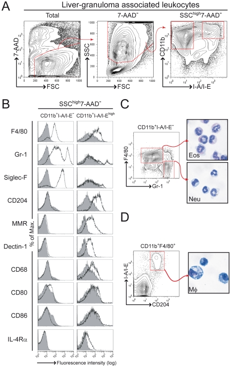
The major leukocytic cell types associated with the liver granuloma in S. mansoni infected wild-type mice are T cells, eosinophils and macrophages [3]. IL-4/IL-13-activated macrophages have been shown to play a crucial role in host survival during acute schistosomiasis [6]. Studies to define tissue macrophages are however complex as these cells can express different markers depending on the organ or tissue, in particular eosinophils are known to express high levels of both CD11b and F4/80 [37]. Here, we used a 4-colour flow cytometry strategy to detect and isolate liver granuloma-associated macrophages and granulocytes. Following exclusion of dead cells, a first gate was placed on CD11b+I-A/I-E− cells for granulocytes. As opposed to inflammatory macrophages, Küpffer cells express no/low levels of CD11b [38], [39]. Therefore, a second gate was placed on CD11b+I-A/I-Ehigh for detection of granuloma macrophages (Fig. 1A). Further analysis of additional markers expression demonstrated that a large proportion of CD11b+I-A/I-E− cells were also F4/80+, Gr-1int or Gr-1high (Ly-6G) and Siglec-F+ (sialic-acid binding Ig-like lectin, a CD33-related Siglec family member) (Fig. 1B). F4/80 and Siglec-F are both expressed on eosinophils but not on neutrophils [37], [38], [39], [40]. Most of the CD11b+I-A/I-E− cells did not express macrophage-specific surface markers, such as macrophage scavenger receptor-A (CD204), MMR, Dectin-1 (a β-glucan receptor), macrosialin (CD68); nor did they express members of the B7 co-stimulatory molecules (CD80 and CD86) and expressed low levels of IL-4Rα (Fig. 1B). Subsequently, we identified a four colour staining with CD11b, I-A/I-E, F4/80, and Gr-1 as a clear definition and discrimination of CD11b+I-A/I-E−F4/80+Gr-1int (and Siglec-F+) eosinophils and CD11b+I-A/I-E−F4/80−Gr-1high (and Siglec-F−) neutrophils, confirmed by morphologic analysis of cytospins (Fig. 1C). Most of CD11b+I-A/I-Ehigh cells however did not express Siglec-F or Gr-1, but expressed F4/80, IL-4Rα and macrophage-specific markers, such as CD204, MMR, Dectin-1 and CD68 (Fig. 1B). Granuloma macrophages were therefore defined as CD11b+F4/80+I-A/I-EhighCD204+ cells (with F4/80 not essential), confirmed by morphologic analysis of cytospins obtained from FACS-sorted CD11b+I-A/I-EhighCD204+ cells (Fig. 1D). Thus, granuloma macrophages within the liver of S. mansoni infected mice are SSChighCD11b+I-A/I-EhighCD204+.
Figure 1. Flow cytometry analysis of liver granuloma during acute schistosomiasis.
BALB/c mice were infected with 100 S. mansoni cercariae, killed 8 weeks later and liver granuloma-associated leukocytes isolated before 4-colour flow cytometry analysis. (A) Gating strategy for analysis of surface expression of CD11b and I-A/I-E on SSChigh and live (7-AAD−) liver granuloma-associated leukocytes. Outlined regions define CD11b+I-A/I-E− and CD11b+I-A/I-Ehigh, respectively. (B) CD11b+I-A/I-E−, and CD11b+I-A/I-Ehigh gated populations in A were analyzed for F4/80, Gr-1, Siglec-F, CD204, MMR, Dectin-1, CD68, CD80, CD86 and IL-4Rα expression, respectively. Greyscale histograms show relevant isotype control. (C) 4-colour flow cytometry analysis of liver granuloma-associated leukocytes gated on CD11b+I-A/I-E− cells as described in A. F4/80 and Gr-1 double staining contour plot is shown. Outlined regions were sorted for cytospin and morphological analysis. (D) 4-colour flow cytometry analysis of liver granuloma-associated leukocytes gated on CD11b+F4/80+ cells as described in A. MHC-II (I-A/I-E) and scavenger receptor-A (CD204) double staining contour plot is shown. Outlined region was sorted for cytospin and morphological analysis. Data are representative of three independent experiments with similar results.
Cellular composition and cell-specific cytokine responses of liver granulomas in LysMcreIl4ra−/lox and iLckcreIl4ra−/lox mice
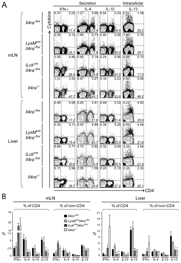
We previously reported that both S. mansoni-infected LysMcreIl4ra−/lox and iLckcreIl4ra−/lox mice developed hepatotoxicity and increased granuloma sizes [6], [10]. It is however unknown whether macrophage/neutrophil-specific or T cell-specific deletion of IL-4Rα affects the cellular composition or cytokine responses within liver granulomas. At 8 weeks post-infection (p.i.), corresponding to the peak of Th2 responses, leukocytes were isolated from granulomas of infected Il4ra−/lox, LysMcreIl4ra−/lox, iLckcreIl4ra−/lox,and global Il4ra−/− mice and analyzed by flow cytometry. Il4ra−/lox littermates were used as controls. Results obtained in Table 2 show that the relative frequencies of macrophages, neutrophils, eosinophils and lymphocyte subpopulations in granulomas were not affected by the specific impairment of IL-4Rα on macrophages, with eosinophils (CD11b+F4/80+Gr-1intSiglec-F+) dominating granulomatous cell composition in LysMcreIl4ra−/lox and infected Il4ra−/lox control mice. We observed however dramatically disrupted granuloma cell populations in iLckcreIl4ra−/lox mice with increased frequencies of macrophages (CD11b+I-A/I-EhighCD204+), neutrophils (CD11b+F4/80−Gr-1high), and γδ-T cells. Though we previously showed that these mice developed bigger granulomas with reduced numbers of eosinophils per granuloma [10], iLckcreIl4ra−/lox mice showed high numbers of eosinophils in their liver (143.1×10⁵ cells in iLckcreIl4ra−/lox mice vs. 110.5×10⁵ cells in Il4ra−/lox mice). This apparent discrepancy could be explained by the fact that these mice had globally increased cell numbers per liver (data not shown). The cellular changes in iLckcreIl4ra−/lox mice were very similar to the disruption of the granuloma cell frequencies observed in Il4ra−/− mice, also showing increased CD4+ T cells and B cells. The disruption of the granulomas in Il4ra−/− mice could be explained by the 4-fold-reduced numbers of eosinophils (25.4×10⁵ cells in Il4ra−/− mice vs. 115.9×10⁵ and 110.6×10⁵ cells in LysMcreIl4ra−/lox and Il4ra−/lox mice, respectively) (Table 2). CD4+CD62Lhigh naive T cell numbers however changed with a 4-fold increase in infected iLckcreIl4ra−/lox mice and a 10-fold increase in infected Il4ra−/− mice compared to Il4ra−/lox control mice over-proportionally (28.3×10⁴ cells in Il4ra−/− mice and 11.9×10⁴ cells in iLckcreIl4ra−/lox mice vs. 4.3×10⁴ and 2.9×10⁴ cells in LysMcreIl4ra−/lox and Il4ra−/lox mice, respectively). In contrast, NK (CD11b−SSClowTCRαβ −CD49b+) and NK-T (CD11b−SSClowTCRαβ +CD49b+) cells showed significant lower frequencies in the liver of infected Il4ra−/− mice compared to Il4ra−/lox control mice (10.3% vs 17.3% and 8.2% vs 25.7%, respectively), resulting in a 2-fold cell number reduction of NK-T cells compared to control mice (2.9×10⁵ cells in Il4ra−/− mice vs. 5.8×10⁵ cells in Il4ra−/lox mice) (Table 2). Following ex vivo SEA restimulation of mesenteric lymph node (mLN) cells or liver granuloma-associated leukocytes, cytokine productions of CD4+ T cells and non-CD4+ cells were analyzed by multicolour flow cytometry, either by catch secretion assay (IFN-γ, IL-4, IL-10) or intracellular staining (IL-13) (Fig. 2). Cytokine catch secretion assays give information on cytokines secreted by a specific cell-type, gathering therefore advantages of both ELISA and intracellular staining. In both tissues, lymphocytes from LysMcreIl4ra−/lox mice produced similar levels of IL-13, IL-10 and IFN-γ cytokines compared to Il4ra−/lox control mice with slightly but significant higher levels of IL-4 in liver CD4+ cells compared to Il4ra−/lox control mice (Fig. 2B). In contrast, lymphocytes from both iLckcreIl4ra−/lox and Il4ra−/− mice produced less Th2 cytokines but more IFN-γ revealing a shift towards Th1-type responses within the granulomas, explaining the observed differences in cellular composition.
Table 2. Cellular composition of liver granulomas depending on macrophage/neutrophil or T cell-specific IL-4Rα deletion.
| Gated populationa | Marker (cell population) | Proportion of gated populationb | Cell number per liver (×10−5)c | ||||||
| Il4ra−/lox | LysMcre Il4ra−/lox | iLckcre IL4ra−/lox | Il4ra−/− | Il4ra−/lox | LysMcre Il4ra−/lox | iLckcre IL4ra−/lox | Il4ra−/− | ||
| CD11b+SSChigh | I-A/I-EhighCD204+ (MΦ) | 21.8±1.4 | 21.9±3.7 | 35.8±5.4d | 39.2±3.4d | 42.3±8.2 | 48.3±14.1 | 134.9±29.1e | 65.9±22.7d |
| F4/80−Gr-1high (Neu) | 14.0±4.6 | 16.6±2.9 | 24.2±1.8d | 28.0±2.1d | 27.3±5.3 | 36.6±10.6 | 91.2±19.7e | 47.2±16.2 | |
| F4/80+Gr-1intSiglec-F+ (Eos) | 56.8±1.4 | 52.4±0.8 | 57.0±7.5 | 15.1±0.2d | 110.5±21.3 | 115.9±33.6 | 143.1±30.9 | 25.3±8.7e | |
| CD11b−SSClow | CD4+ (Th) | 23.2±3.9 | 21.1±5.3 | 21.5±0.3 | 31.1±4.6d | 19.5±3.8 | 16.3±4.7 | 27.7±6.0 | 32.5±11.2d |
| CD8+ (CTL) | 6.7±0.9 | 5.0±1.4 | 3.4±0.4d | 6.3±1.4 | 5.6±1.1 | 3.9±1.1 | 4.4±0.9 | 6.5±2.3 | |
| CD19+ (B) | 18.7±0.1 | 21.2±0.4 | 15.1±0.2 | 33.0±9.8d | 15.7±3.0 | 16.4±4.7 | 19.5±4.2 | 34.5±11.9e | |
| CD11b−SSClowTCRβ − | CD49b+ (NK) | 17.3±4.0 | 13.2±0.4 | 16.9±1.0 | 10.3±0.3d | 9.4±1.8 | 6.9±2.0 | 13.3±2.9d | 7.4±2.6 |
| CD11b−SSClowTCRβ + | CD49b+ (NK-T) | 25.7±2.5 | 23.3±6.8 | 15.4±2.9d | 8.2±0.3d | 5.8±1.1 | 4.5±1.3 | 4.7±1.0 | 2.9±1.0d |
| CD11b−SSClowCD4+ | CD62L high (Th naive) | 1.5±0.2 | 2.5±0.2 | 4.0±0.1d | 8.1±0.9d | 0.3±0.1 | 0.4±0.1 | 1.2±0.3d | 2.8±1.0e |
| CD44+ (Th effector/memory) | 73.2±5.4 | 73.2±8.2 | 69.8±5.6 | 63.0±7.9 | 14.3±2.8 | 12.7±3.7 | 21.0±4.5 | 22.0±7.5 | |
| CD11b−SSClowCD3+CD4− | TCRγδ + (γδT) | 5.0±1.4 | 6.7±3.0 | 10.4±3.9d | 10.7±2.3d | 0.5±0.1 | 0.5±0.2 | 1.5±0.3d | 0.7±0.2 |
Figure 2. IL-4Rα-expressing macrophages do not affect cytokine responses in granulomas.
Il4ra−/lox, LysMcreIl4ra−/lox, iLckcreIl4ra−/lox and Il4ra−/− mice were infected with 100 S. mansoni cercariae and killed 8 weeks later. Mesenteric lymph node (mLN) cells or liver granuloma-associated leukocytes (Liver) were then isolated and restimulated overnight with 20 µg/ml SEA before analyzed for ex vivo cytokine production capability. Staining of surface CD4 was followed by detection of IL-4, IL-10 and IFN-γ secretion in a cytokine catch assay or detection of intracellular levels of IL-13 after 4h of monensin treatment, as described in Methods. Live gate was placed on lymphocytes according to their forward- and side-scatter by flow cytometry. (A) Representative contour plots of IFN-γ, IL-4, IL-10 and IL-13-producing lymphocytes. Quadrant bars were set up on unstimulated cells and numbers represent percent cells in each quadrant. Data represent one out of three independent experiments with similar results. (B) Percentages of IFN-γ, IL-4, IL-10 or IL-13-producing cells were determined in gated CD4+ or non-CD4+ cell populations from mLN or liver granuloma-associated lymphocytes (Liver) (mean±SEM, n = 4). Data are representative of three independent experiments. *p<0.05; **p<0.001; ***p<0.001 compared to control Il4ra−/lox mice.
Mannose receptor and Ym1 expression in liver granuloma-associated macrophages of LysMcreIl4ra−/lox mice
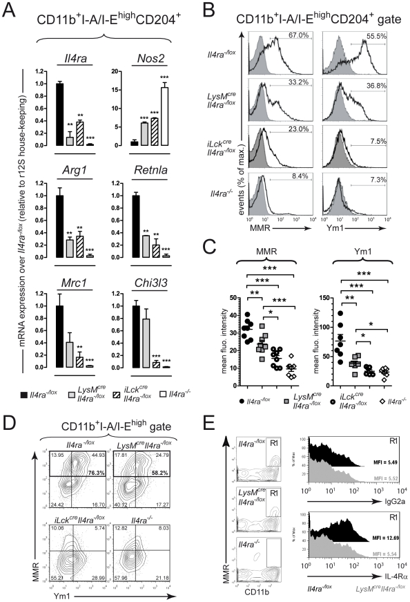
We further compared gene expression levels of markers used to distinguish classical from alternative macrophage activation within liver granulomas during acute S. mansoni infection. Liver granuloma macrophages (SSChighCD11b+I-A/I-EhighCD204+) were FACS-sorted to high-purity at 8 weeks p.i. (≥98%), RNA extraction performed, and gene expression profile analyzed (Fig. 3A). Il4ra (IL-4Rα) mRNA expression, observed in Il4ra−/lox control macrophages, was impaired in macrophages from LysMcreIl4ra−/lox mice and global Il4ra−/− mice, confirming the efficiency of Cre-loxP mediated DNA deletion in granuloma macrophages. Interestingly, Il4ra expression was also reduced in iLckcreIl4ra−/lox macrophages, suggesting a predominant classical activation in the granulomas of this mouse strain. Consistent with the IL-4/IL-13/IL-4Rα- and IFN-γ/IFN-γR-mediated macrophage activation by direct control of expression of the enzymes catalysing L-arginine, LysMcreIl4ra−/lox, iLckcreIl4ra−/lox, and global Il4ra−/− mice presented classically activated macrophages with heightened Nos2 but reduced Arg1 mRNA expression levels, compared to Il4ra−/lox control mice. This was in line with the expression pattern of the known alternative activation marker gene Retnla (FIZZ1), significantly reduced in macrophages from LysMcreIl4ra−/lox, iLckcreIl4ra−/lox and global Il4ra−/− mice, compared to Il4ra−/lox control mice. Of importance, the aaMφ marker Chi3l3 and Mrc1 gene expression levels were present although reduced for Mrc1 in LysMcreIl4ra−/lox macrophages, resulting in Ym1 and MMR surface expression in about 30% of SSChighCD11b+I-A/I-EhighCD204+ macrophages from LysMcreIl4ra−/lox mice. The expression of these two markers was however strongly reduced in both iLckcreIl4ra−/lox and global Il4ra−/− mice, compared to control Il4ra−/lox mice (Fig. 3B). Mean fluorescent intensities (MFI) values of MMR and Ym1 stainings were also significantly higher in macrophages from LysMcreIl4ra−/lox compared to both iLckcreIl4ra−/lox and global Il4ra−/− mice (Fig. 3C). The majority (76.3%) of MMR-expressing CD11b+I-A/I-Ehigh granuloma macrophages also expressed Ym1 in the Il4ra−/lox control mice (Fig. 3D). Absence of surface IL-4Rα expression was further confirmed by 4-colour staining on granuloma macrophages (Fig. 3E). MFI of IL-4Rα staining on gated CD11b+MMR+ cells revealed similar expression levels of the receptor between LysMcreIl4ra−/lox mice and isotype control (MFI = 5.54 and 5.52, respectively), whereas granuloma CD11b+MMR+ cells from Il4ra−/lox mice showed 2-fold increased expression of the IL-4Rα compared to LysMcreIl4ra−/lox mice and isotype control (MFI = 12.69) (Fig. 3E). Together, these data suggest that MMR and Ym1 can be expressed independently of macrophage-specific IL-4Rα signalling, whereas IL-4Rα-dependent T-cell responses are necessary for driving expression of aaMφ markers, including MMR and Ym1.
Figure 3. Mannose receptor and Ym1 expression in granuloma macrophages of LysMcreIl4ra−/lox mice.
Il4ra−/lox, LysMcreIl4ra−/lox, iLckcreIl4ra−/lox and Il4ra−/− mice were infected with 100 S. mansoni cercariae, killed 8 weeks later and liver granuloma-associated leukocytes purified. (A) CD11b+I-A/I-EhighCD204+ granuloma macrophages were sorted to high purity (>98%) and RNA extracted before analysis of expression levels of genes associated with macrophage activation by real time RT-PCR: IL-4Rα (Il4ra), nitric oxide synthase (Nos2), arginase 1 (Arg1), FIZZ1 (Retnla), macrophage mannose receptor (Mrc1), and Ym1 (Chi3l3). Relative expression levels normalized to r12S house-keeping gene are shown as fold-change over Il4ra−/lox mice values. Data is representative of two independent experiments with analysis of pooled samples (n = 4). Bars show mean±SD of analyses performed in triplicates. (B) 4-colour flow cytometry analysis of granuloma leukocytes gated on CD11b+I-A/I-EhighCD204+ cells. Representative monoparametric histograms show stainings of surface macrophage mannose receptor (MMR) or intracellular Ym1 expression. Data is representative of four independent experiments of pooled samples (n = 4). Greyscale histograms show relevant isotype control. (C) Mean fluorescent intensities of MMR and Ym1 stainings based on analysis by flow cytometry as presented in B. Data show results of individual mice for 2 independent experiments. (D) 4-colour flow cytometry analysis of granuloma leukocytes gated on CD11b+I-A/I-Ehigh cells. Representative contour plots show MMR/Ym1 double staining and outlined regions show MMR-positive staining. Data is representative of two independent experiments of pooled samples (n = 4). (E) 4-colour flow cytometry analysis of granuloma leukocytes. Analysis gate (R1) was placed on SSChighCD11b+MMR+7-AAD− and histograms show isotype IgG2a (upper panel) and anti-IL-4Rα (lower panel) stainings. Data is representative of two independent experiments of pooled samples (n = 4). Black, Il4ra−/lox; grey, LysMcreIl4ra−/lox. * p<0.05, **p<0.01; ***p<0.001.
Peripheral localisation of IL-4Rα-independent MMR- and Ym1-positive macrophages within egg-induced liver granulomas of LysMcreIl4ra−/lox mice
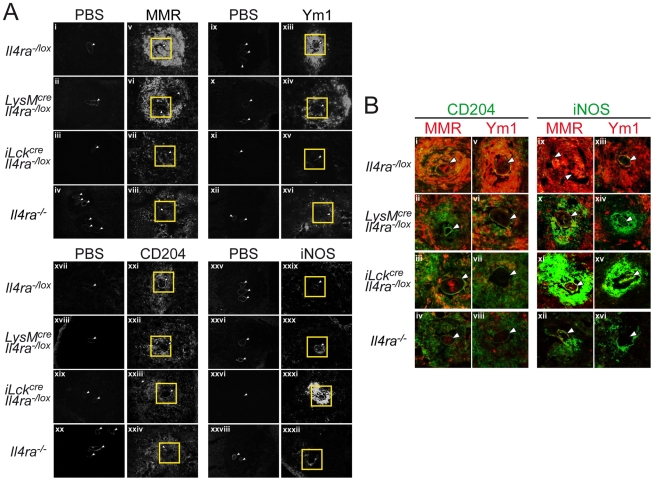
The results detailed in Figure 3 demonstrated that LysMcreIl4ra−/lox granuloma macrophages have retained expression of MMR and Ym1. This was surprising as we previously showed that liver granuloma from LysMcreIl4ra−/lox mice had low levels of MMR expression in close proximity to the eggs compared to Il4ra−/lox control mice [6]. To further determine the localisation of the MMR- and Ym1-positive macrophage subpopulation within the liver granuloma, immunofluorescent stainings were performed on liver cryosections at 8 weeks p.i. (Fig. 4). As expected from the expression data, MMR and Ym1 were highly expressed in granulomas from Il4ra−/lox control mice; and these cells co-expressing scavenger receptor A (CD204) were in close proximity to the parasite eggs in the centre of the granuloma (Fig. 4A and B). MMR- and Ym1-positive macrophages were restricted to the periphery of the granuloma in LysMcreIl4ra−/lox mice and nearly undetectable in both iLckcreIl4ra−/lox and Il4ra−/− mice. Instead, iNOS-positive, MMR- and Ym1-negative macrophages were in close proximity around the egg in the granuloma of LysMcreIl4ra−/lox,iLckcreIl4ra−/lox and Il4ra−/− mice (Fig. 4B), confirming previous observations [6], [10], [41]. These results show that a peripheral IL-4Rα-independent MMR- and Ym1-positive macrophage subpopulation surrounds the iNOS-producing classically activated macrophages present in close proximity to the egg within the liver granuloma of LysMcreIl4ra−/lox mice.
Figure 4. Localisation of mannose receptor and Ym1-expressing granuloma macrophages in close contact with S. mansoni eggs depends on their IL-4Rα signalling.
Livers were collected at 8 weeks p.i. from Il4ra−/lox, LysMcreIl4ra−/lox, iLckcreIl4ra−/lox and Il4ra−/− mice and immunofluorescent stainings performed on cryosections. (A) Representative micrographs of MMR (panels v–viii), Ym1 (panels xiii–xvi), CD204 (panels xxi–xxiv) and iNOS (panels xxix–xxxii) stainings of liver cryosections as described in Methods. Stainings with secondary antibody (MMR, iNOS), streptavidin alone (Ym1) or isotype-control (CD204) is shown for each corresponding staining (MMR = i–iv, Ym1 = ix–xii, CD204 = xvii–xx, iNOS = xxv–xxviii). Note that MMR+ and Ym1+ cells are restricted to the periphery of LysMcreIl4ra−/lox granulomas. Outlined regions represent the areas magnified in B. (B) Representative micrographs of liver cryosections stained with scavenger receptor (CD204) for macrophages detection (panels i–viii, green) or iNOS for classically activated macrophages detection (panels ix–xvi, green); and co-stained for MMR (panels i–iv and ix–xii, red) or Ym1 (panels v–viii and xiii–xvi, red). Note the low frequency of CD204+ macrophages co-expressing MMR+ or Ym1+ cells around the parasite eggs (panels ii and vi) but the high levels of iNOS+ cells (panels x and xiv) in LysMcreIl4ra−/lox mice, suggesting these macrophages to be classically activated. White arrows indicate the parasite eggs. Original magnification: 400×. Data represent one of three independent experiments.
IL-4Rα-independent MMR- and Ym1- positive macrophages depend on IL-10 signalling
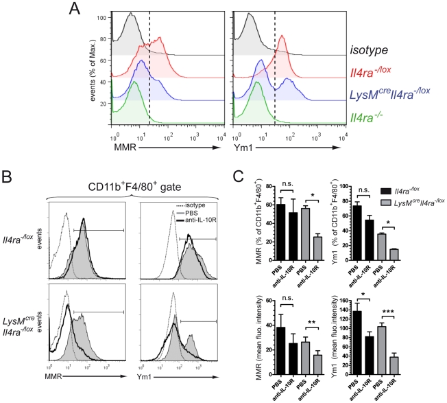
IL-10 signalling can induce MMR expression in macrophages [34]. We therefore investigated whether IL-10 could drive the MMR and Ym1 expression in macrophages of LysMcreIl4ra−/lox mice. To test this hypothesis we used a local egg model by injecting purified S. mansoni eggs intraperitoneally to induce Th2 responses and elicit macrophage activation in Il4ra−/lox, LysMcreIl4ra−/lox and Il4ra−/− mice. At 7 days p.i., corresponding to the peak of Th2 responses [42], MMR and Ym1 were highly expressed in peritoneal macrophages of Il4ra−/lox control mice (Fig. 5A). To investigate peritoneal macrophages, we placed a live gate on FSChighSSClowCD11b+F4/80+ cells. Here the association of the CD11b, F4/80 markers and side/forward scatter allows discrimination between eosinophils and macrophages, as previously described [37]. Similar to that observed in macrophages from liver granuloma, a subpopulation of cells expressing MMR and Ym1 was present in peritoneal macrophages of LysMcreIl4ra−/lox mice but absent in peritoneal macrophages from Il4ra−/− mice (Fig. 5A). In vivo blockade of IL-10 signalling via anti-IL-10 receptor antibody treatment was sufficient to significantly reduce the protein expression of both MMR and Ym1 in peritoneal macrophages of LysMcreIl4ra−/lox mice (Fig. 5B and C). These results demonstrate that IL-10 drives expression of MMR and Ym1 in macrophages independently of macrophage IL-4Rα.
Figure 5. IL-10 signalling drives mannose receptor and Ym1 expression in macrophages independently of their IL-4Rα expression.
Peritoneal cells were harvested 7 days after injection of 3000 S. mansoni purified eggs in the peritoneum of Il4ra−/lox, LysMcreIl4ra−/loxor Il4ra−/− mice. (A) Representative histograms of macrophage mannose receptor (MMR) or Ym1 expression by peritoneal macrophages gated on FSChighSSClowCD11b+F4/80+ cells after exclusion of peritoneal eosinophils (FSClowSSChighCD11b+F4/80+) [37]. Black, isotype control; red, Il4ra−/lox; blue, LysMcreIl4ra−/lox; green, Il4ra−/−. Broken lines show threshold for positive signal. Data are representative of two independent experiments of pooled samples (n = 3). (B) Anti-IL-10 receptor treatment. Four mice out of eight in each group received 4µg of anti-IL-10R i.p. at day 0, 4 and 6 post-injection. Overlays of representative monoparametric histograms of macrophage mannose receptor (MMR) or Ym1 expression by peritoneal macrophages are shown as described in A. Bracketed line indicates positive signal. Dotted line, isotype control; greyscale, untreated mice; bold line, mice treated with anti-IL-10R. (C) Percent and mean fluorescent intensities of peritoneal macrophages expressing MMR or Ym1 based on flow cytometry analysis in B. Data is representative of two independent experiments with mean±SEM (n = 3). *p<0.05, ** p<0.01, *** p<0.001.
Discussion
Recent S. mansoni infection studies demonstrated that LysMcreIl4ra−/lox mice, deficient for IL-4Rα-responsive aaMφ succumb to acute S. mansoni infection, due to the development of egg-induced gut inflammation, severe liver damage, and despite the formation of egg-induced granulomas and unaffected fibrosis in the liver [6]. These observations were very similar to the pathology developed by infected global Il4ra−/− mice [6], or Il4−/− and Il4−/−Il13−/− mice which developed hepatotoxicity and endotoxaemia due to the degradation of the intestinal barrier [5], [9]. Antibiotic treatment in infected LysMcreIl4ra−/lox mice resulted only in partial protection [6], suggesting that in addition to gut inflammation and systemic leakage of the intestinal content, hepatotoxic lesions could also explain the increased mortality. In addition to these findings, we also recently demonstrated, using iLckcreIl4ra−/lox mice, that IL-4Rα responsiveness by T cells was required for host survival mainly for controlling liver damage caused by the parasites eggs [10]. As iLckcreIl4ra−/lox mice did not develop gut injury or endotoxaemia, we concluded that IL-4Rα responsiveness by T cells is not essential for the control of intestine-derived sepsis. In this study we focused on the liver granuloma microenvironment and the consequences of impaired IL-4Rα signalling in macrophages or T cells on the local cellular responses directed against the parasite eggs.
Granulomas induced by S. mansoni contain both myeloid cells (mainly eosinophils and macrophages) and lymphoid cells (mainly T cells) [3]. Several markers are readily available for staining murine tissue macrophages but their expression can be highly tissue-dependent [43]. Most of the studies using such markers have been performed with blood, spleen or bone-marrow but little is known on granuloma macrophage markers during schistosomiasis. Liver granulomas are tightly organized, which renders their disruption difficult without affecting cell viability, especially macrophages. Though collagenase can in some conditions cleave surface proteins, comparison between collagenase-treated or untreated organs gave similar results concerning the expression levels of standard surface markers (including CD11b, F4/80, CD11c, Gr-1, and I-A/I-E), suggesting that collagenase treatment had no major influence on surface marker detection (data not shown). The combination of CD11b and/or F4/80 and MHC-II (I-A/I-E) was sufficient to specifically detect and isolate macrophages from granulomas. These macrophages were defined as SSChighCD11b+(F4/80+)I-A/I-EhighCD204+ cells, showed a macrophage-like morphology and expressed high levels of macrophage-specific markers such as scavenger receptor-A (CD204), mannose receptor (MMR), β-glucan receptor (Dectin-1) and macrosialin (CD68). In parallel, we defined granuloma eosinophils as SSChighCD11b+F4/80+I-A/I-E−Gr-1intSiglec-F+ and neutrophils as SSChighCD11b+F4/80−I-A/I-E−Gr-1highSiglec-F−.
Despite increased liver granuloma sizes, previously reported in infected LysMcreIl4ra−/lox mice [6], neither the cellular composition nor the Th1/Th2 cytokine profiles were affected. In contrast, we observed severe changes in granuloma cell composition and morphology of infected iLckcreIl4ra−/lox and Il4ra−/− mice. Interestingly, a recent report showed that infected LysMcreArg1−/lox or Tie2creArg1lox/lox mice (deficient for Arg1-expressing macrophages) did not develop lethal endotoxaemia or hepatotoxicity but showed increased Th2 responses and increased chronic disease [23]. Further, Arg1-expressing aaMφ were able to suppress schistosome-specific T cell proliferation, suggesting a possible mechanism for downmodulating granulomatous inflammation and slow the progression of Th2-driven fibrosis during chronic infection [23]. Similarly, studies in Retnla-deficient mice showed that absence of resistin-like molecule alpha (Retnla/FIZZ1) exacerbated Th2 cytokine responses in S. mansoni-egg-induced lung inflammation, suggesting that Retnla/FIZZ1 is able to control effector Th2 responses in S. mansoni egg-induced inflammation and infection [29], [30]. As IL-4Rα-sinalling is upstream from the induction of Arg1 and Retnla in aaMφ, it might be not surprising that LysMcreIl4ra−/lox mice have a more severe disease phenotype already detrimental in the acute phase. The relative normal granuloma morphology, cellular composition and Th1/Th2 cytokine responses in the liver of LysMcreIl4ra−/lox mice but more severe liver pathology in iLckcreIl4ra−/lox mice, suggest that abrogation of IL-4-promoted Th2 responses in combination with impairment of aaMφ negatively affect the control of liver pathology in mice. Together with the chronic phenotype and aaMφ-mediated T cell suppression observed in LysMcreArg1−/lox mice, aaMφ seem to control T helper cell responses in an optimal balance. Future studies need to further investigate the molecular mechanisms of how and which T cell subpopulations are controlled by aaMφ.
Interestingly, LysMcreIl4ra−/lox liver granulomas retained expression of MMR and Ym1 in a subpopulation of macrophages, confirmed in mRNA and protein (Chi3l3) or protein (Mrc1) expression analyses. Interestingly, MMR+Ym1+ macrophages were not observed in iLckcreIl4ra−/lox mice, suggesting that IL-4Rα-dependent Th2 responses drive MMR and Ym1 expression independently of IL-4Rα-responsive macrophages in LysMcreIl4ra−/lox mice. After staining of scavenger receptor-A (CD204) in situ to specifically detect macrophages [44], [45], we showed that macrophages were in close proximity to the parasite eggs within the granuloma in all mouse strains. However, macrophages expressing MMR or Ym1 were only found at the periphery of the LysMcreIl4ra−/lox granulomas, while present in close proximity to the eggs in the Il4ra−/lox control granulomas. As previously demonstrated, MMR- or Ym1-positive cells were absent of global Il4ra−/− granulomas [25] and we observed nearly undetectable MMR+ or Ym1+ cells in iLckcreIl4ra−/lox granulomas. Instead classically activated macrophages, defined by their iNOS expression were centred on the eggs in LysMcreIl4ra−/lox, iLckcreIl4ra−/lox and global Il4ra−/− granulomas (Fig. 4B). Increased iNOS activity in these mice strains could explain the heightened hepatocellular damage observed after S. mansoni infection [6], [10]. Together, these results suggest that IL-4Rα-responsiveness in aaMφ regulates their ability to migrate in close proximity and/or interact with the parasite eggs. The factors that could be responsible for the interaction with the eggs are not known, but a possible candidate could be MMR. This receptor encodes a C-type lectin, which has been involved in antigen uptake by antigen-presenting cells and components of S. mansoni eggs as well as a fraction of their egg-secreted molecules are ligands of MMR [25]. Furthermore, a study demonstrated that SEA can be internalized by DCs through the C-type lectins dendritic cell specific ICAM-3 grabbing non integrin (DC-SIGN), macrophage galactose-type lectin (MGL) and MMR [28]. We did not look at the expression of other C-type lectins, but the presence of macrophages expressing MMR in the proximity of the parasite eggs could allow IL-4Rα-responsive aaMφ to interact with antigen-specific T cells and also play regulatory functions on effector T cells as previously suggested [6], [23], [29], [30], [46]. Although Ym1 was described as a chitinase-like secreted protein, it lacks any chitinase activity and S. mansoni eggs do not contain chitin [47], [48]. A recent study described Ym1 to digest glycosaminoglycans and might therefore play a role in egg killing and/or antigen processing [49]. It has previously been suggested that Ym1 could encapsulate pathogens [31] and our observation of Ym1-expressing macrophages in the proximity of the parasite eggs supports this hypothesis. Encapsulation of the parasite eggs may serve to protect the host from pro-inflammatory molecules and harmful factors released by the eggs. Macrophages isolated from granulomas or macrophages elicited with S. mansoni egg components have previously been proposed to act as “myeloid suppressive cells (MSCs) by inducing clonal anergy in egg-antigen-specific Th1 cells by unknown mechanisms [46], [50], [51], [52], [53], [54]. Although we did not detect changes in cellular composition or cytokine production in the liver granulomas of LysMcreIl4ra−/lox mice, these mice quickly die from increased systemic Th1-type responses and acute infection due to uncontrolled gut inflammation [6]. Recently established MMR-deficient mice [55], generation of Ym1-deficient mice should allow further studies to address the role of such aaMφ biomarkers.
Previous studies proposed that MMR and Ym1 expression by macrophages could directly be driven by bacterial endotoxin or chitin [35], and independently of IL-4Rα signalling [56]. Linehan and colleagues (2003) previously described a complete absence of MMR expression in the liver granulomas of global Il4ra−/− mice [25], demonstrating that IL-4Rα responsiveness is essential for MMR expression in macrophages recruited to the granulomas. These authors however described resident liver macrophages to retain MMR expression and we also observed MMR expression in resident Küpffer cells (Fig. 4A). As Küpffer cells express no/low levels of CD11b in contrast to inflammatory macrophages [38], [39], we can exclude any contamination with these cells in our gating strategy targeting SSChighCD11b+(F4/80+)I-A/I-EhighCD204+ granuloma macrophages.
Our findings demonstrate that direct IL-4Rα signalling in a subpopulation of granuloma macrophages is not essential for MMR and Ym1 expression and these cells are restricted to the periphery of LysMcreIl4ra−/lox granulomas. IL-10 signalling in macrophages can induce MMR expression [34] and Ym1 expression is downregulated in Il10−/− and Il10−/−Il4−/− deficient mice [32]. These observations, together with reduced IL-10 production and impaired aaMφ markers (including MMR and Ym1) in iLckcreIl4ra−/lox and Il4ra−/− liver granulomas, prompted us to consider IL-10 as a good candidate to explain the IL-4Rα-independent expression of MMR and/or Ym1 in macrophages. In order to focus on the egg-induced inflammation in the LysMcreIl4ra−/lox mice, we chose to use an egg model to induce acute Th2 responses following challenge in the peritoneum. Here, we showed evidence by in vivo IL-10R signalling blockade that IL-10 signalling was responsible for the retained expression of MMR and Ym1 expression in LysMcreIl4ra−/lox peritoneal macrophages following S. mansoni egg exposure and induction of Th2 cytokine responses, while no significant effect could be observed in macrophages of Il4ra−/lox control mice. Although elicitation of macrophages in the peritoneal cavity by S. mansoni eggs might not exactly reflect what occurs in the liver granuloma during live infection, data generated with peritoneal macrophages gave similar results as those obtained with granuloma macrophages. Control Il4ra−/lox mice over-expressed both MMR and Ym1, whereas expression of those markers in peritoneal macrophages from Il4ra−/− mice was strongly impaired. Moreover, LysMcreIl4ra−/lox peritoneal macrophages also retained expression of MMR and Ym1 after injection of eggs and anti-IL-10R treatment blocked the expression of both markers. These results indicate that IL-10 plays a key role in the retained expression of MMR and Ym1 in the liver granulomas of LysMcreIl4ra−/lox mice. The macrophages retaining expression of MMR and Ym1 in infected LysMcreIl4ra−/lox mice could therefore represent a cellular subpopulation with IL-4Rα-independent aaMφ-related regulatory activities, driven by IL-10. The presence of such macrophage population could explain why LysMcreIl4ra−/lox liver granulomas do not have disrupted cellular or cytokine responses, in contrast to mice having impaired Th2 responses and showing strongly reduced IL-10 production such as iLckcreIl4ra−/lox and Il4ra−/− mice. Supporting these results, Herbert et al. recently showed that in vivo IL-10R neutralization results in increased hepatocellular damage without affecting gut inflammation in schistosomiasis [57]. Hepatotoxicity was however observed in infected LysMcreIl4ra−/lox mice [6], suggesting that IL-10 without IL-4/IL-13 signalling in macrophages might not be sufficient to control liver tissue damage. Future work on IL-4Rα-independent MMR+Ym1+ macrophages from liver granulomas may define whether those cells have an “alternative” or “deactivated” phenotype, which was previously described to be mediated by IL-10 signalling [11].
In conclusion, we demonstrated that IL-4Rα-responsive macrophages are dispensable for the control of cellular and cytokine responses in liver granulomas during acute schistosomiasis, whereas IL-4Rα-responsive T cells are necessary to sustain cellular composition and morphology of the liver granuloma. Though our results suggest that macrophage-specific IL-4Rα expression is necessary for an adequate interaction between aaMφ and the parasite eggs, we showed that MMR and Ym1 expression may be induced via IL-10R signalling in absence of IL-4Rα signalling in macrophages. Taken together, our results suggest that IL-4Rα-derived Th2 cytokine responses are essential to drive aaMφ in the liver and demonstrate that in absence of macrophage-specific IL-4Rα, IL-10 signalling induces MMR+Ym1+ macrophages. Future investigation may clarify whether IL-10-driven IL-4Rα-independent MMR+Ym1+ macrophages have important functions in the control of liver granulomatous inflammation.
Acknowledgments
B.D. is a Postdoctoral Researcher of the «Fonds National Belge de la Recherche Scientifique» (FNRS). We are thankful to Dr William Horsnell for critical reading of the manuscript. For technical assistance we would like to thank Mr Ronnie Dreyer, Mrs Lizette Fick, Mrs Wendy Green, Mrs Zenaria Abbas and Mr Reagan Peterson.
Footnotes
The authors have declared that no competing interests exist.
This work was supported by The Wellcome Trust (080921/Z/06/Z) and Royal Society, UK; South African Research Chair initiative of the Department of Science and Technology (DST), National Research Foundation (NRF), and Medical Research Council (MRC), South Africa, and Medical Research Council (MRC), South Africa. The funders had no role in study design, data collection and analysis, decision to publish, or preparation of the manuscript.
References
- 1.Gryseels B, Polman K, Clerinx J, Kestens L. Human schistosomiasis. Lancet. 2006;368:1106–1118. doi: 10.1016/S0140-6736(06)69440-3. [DOI] [PubMed] [Google Scholar]
- 2.King CH. Toward the elimination of schistosomiasis. N Engl J Med. 2009;360:106–109. doi: 10.1056/NEJMp0808041. [DOI] [PubMed] [Google Scholar]
- 3.Pearce EJ, MacDonald AS. The immunobiology of schistosomiasis. Nat Rev Immunol. 2002;2:499–511. doi: 10.1038/nri843. [DOI] [PubMed] [Google Scholar]
- 4.Wynn TA, Thompson RW, Cheever AW, Mentink-Kane MM. Immunopathogenesis of schistosomiasis. Immunol Rev. 2004;201:156–167. doi: 10.1111/j.0105-2896.2004.00176.x. [DOI] [PubMed] [Google Scholar]
- 5.Fallon PG, Richardson EJ, McKenzie GJ, McKenzie AN. Schistosome infection of transgenic mice defines distinct and contrasting pathogenic roles for IL-4 and IL-13: IL-13 is a profibrotic agent. J Immunol. 2000;164:2585–2591. doi: 10.4049/jimmunol.164.5.2585. [DOI] [PubMed] [Google Scholar]
- 6.Herbert DR, Holscher C, Mohrs M, Arendse B, Schwegmann A, et al. Alternative macrophage activation is essential for survival during schistosomiasis and downmodulates T helper 1 responses and immunopathology. Immunity. 2004;20:623–635. doi: 10.1016/s1074-7613(04)00107-4. [DOI] [PubMed] [Google Scholar]
- 7.Herbert DR, Orekov T, Perkins C, Rothenberg ME, Finkelman FD. IL-4R alpha expression by bone marrow-derived cells is necessary and sufficient for host protection against acute schistosomiasis. J Immunol. 2008;180:4948–4955. doi: 10.4049/jimmunol.180.7.4948. [DOI] [PMC free article] [PubMed] [Google Scholar]
- 8.Jankovic D, Kullberg MC, Noben-Trauth N, Caspar P, Ward JM, et al. Schistosome-infected IL-4 receptor knockout (KO) mice, in contrast to IL-4 KO mice, fail to develop granulomatous pathology while maintaining the same lymphokine expression profile. J Immunol. 1999;163:337–342. [PubMed] [Google Scholar]
- 9.McKenzie GJ, Fallon PG, Emson CL, Grencis RK, McKenzie AN. Simultaneous disruption of interleukin (IL)-4 and IL-13 defines individual roles in T helper cell type 2-mediated responses. J Exp Med. 1999;189:1565–1572. doi: 10.1084/jem.189.10.1565. [DOI] [PMC free article] [PubMed] [Google Scholar]
- 10.Dewals B, Hoving JC, Leeto M, Marillier RG, Govender U, et al. IL-4Ralpha responsiveness of non-CD4 T cells contributes to resistance in schistosoma mansoni infection in pan-T cell-specific IL-4Ralpha-deficient mice. Am J Pathol. 2009;175:706–716. doi: 10.2353/ajpath.2009.090137. [DOI] [PMC free article] [PubMed] [Google Scholar]
- 11.Gordon S. Alternative activation of macrophages. Nat Rev Immunol. 2003;3:23–35. doi: 10.1038/nri978. [DOI] [PubMed] [Google Scholar]
- 12.Ramalingam TR, Pesce JT, Sheikh F, Cheever AW, Mentink-Kane MM, et al. Unique functions of the type II interleukin 4 receptor identified in mice lacking the interleukin 13 receptor alpha1 chain. Nat Immunol. 2008;9:25–33. doi: 10.1038/ni1544. [DOI] [PMC free article] [PubMed] [Google Scholar]
- 13.LaPorte SL, Juo ZS, Vaclavikova J, Colf LA, Qi X, et al. Molecular and structural basis of cytokine receptor pleiotropy in the interleukin-4/13 system. Cell. 2008;132:259–272. doi: 10.1016/j.cell.2007.12.030. [DOI] [PMC free article] [PubMed] [Google Scholar]
- 14.Bronte V, Serafini P, Mazzoni A, Segal DM, Zanovello P. L-arginine metabolism in myeloid cells controls T-lymphocyte functions. Trends Immunol. 2003;24:302–306. doi: 10.1016/s1471-4906(03)00132-7. [DOI] [PubMed] [Google Scholar]
- 15.Bronte V, Zanovello P. Regulation of immune responses by L-arginine metabolism. Nat Rev Immunol. 2005;5:641–654. doi: 10.1038/nri1668. [DOI] [PubMed] [Google Scholar]
- 16.Noel W, Raes G, Hassanzadeh Ghassabeh G, De Baetselier P, Beschin A. Alternatively activated macrophages during parasite infections. Trends Parasitol. 2004;20:126–133. doi: 10.1016/j.pt.2004.01.004. [DOI] [PubMed] [Google Scholar]
- 17.Gallina G, Dolcetti L, Serafini P, De Santo C, Marigo I, et al. Tumors induce a subset of inflammatory monocytes with immunosuppressive activity on CD8+ T cells. J Clin Invest. 2006;116:2777–2790. doi: 10.1172/JCI28828. [DOI] [PMC free article] [PubMed] [Google Scholar]
- 18.Holscher C, Arendse B, Schwegmann A, Myburgh E, Brombacher F. Impairment of alternative macrophage activation delays cutaneous leishmaniasis in nonhealing BALB/c mice. J Immunol. 2006;176:1115–1121. doi: 10.4049/jimmunol.176.2.1115. [DOI] [PubMed] [Google Scholar]
- 19.Keating P, O'Sullivan D, Tierney JB, Kenwright D, Miromoeini S, et al. Protection from EAE by IL-4Ralpha(−/−) macrophages depends upon T regulatory cell involvement. Immunol Cell Biol. 2009;87:534–545. doi: 10.1038/icb.2009.37. [DOI] [PubMed] [Google Scholar]
- 20.Loke P, Gallagher I, Nair MG, Zang X, Brombacher F, et al. Alternative activation is an innate response to injury that requires CD4+ T cells to be sustained during chronic infection. J Immunol. 2007;179:3926–3936. doi: 10.4049/jimmunol.179.6.3926. [DOI] [PubMed] [Google Scholar]
- 21.Muller U, Stenzel W, Kohler G, Werner C, Polte T, et al. IL-13 induces disease-promoting type 2 cytokines, alternatively activated macrophages and allergic inflammation during pulmonary infection of mice with Cryptococcus neoformans. J Immunol. 2007;179:5367–5377. doi: 10.4049/jimmunol.179.8.5367. [DOI] [PubMed] [Google Scholar]
- 22.Odegaard JI, Ricardo-Gonzalez RR, Goforth MH, Morel CR, Subramanian V, et al. Macrophage-specific PPARgamma controls alternative activation and improves insulin resistance. Nature. 2007;447:1116–1120. doi: 10.1038/nature05894. [DOI] [PMC free article] [PubMed] [Google Scholar]
- 23.Pesce JT, Ramalingam TR, Mentink-Kane MM, Wilson MS, El Kasmi KC, et al. Arginase-1-expressing macrophages suppress Th2 cytokine-driven inflammation and fibrosis. PLoS Pathog. 2009;5:e1000371. doi: 10.1371/journal.ppat.1000371. [DOI] [PMC free article] [PubMed] [Google Scholar]
- 24.Martinez FO, Helming L, Gordon S. Alternative activation of macrophages: an immunologic functional perspective. Annu Rev Immunol. 2009;27:451–483. doi: 10.1146/annurev.immunol.021908.132532. [DOI] [PubMed] [Google Scholar]
- 25.Linehan SA, Coulson PS, Wilson RA, Mountford AP, Brombacher F, et al. IL-4 receptor signaling is required for mannose receptor expression by macrophages recruited to granulomata but not resident cells in mice infected with Schistosoma mansoni. Lab Invest. 2003;83:1223–1231. doi: 10.1097/01.lab.0000081392.93701.6f. [DOI] [PubMed] [Google Scholar]
- 26.Stein M, Keshav S, Harris N, Gordon S. Interleukin 4 potently enhances murine macrophage mannose receptor activity: a marker of alternative immunologic macrophage activation. J Exp Med. 1992;176:287–292. doi: 10.1084/jem.176.1.287. [DOI] [PMC free article] [PubMed] [Google Scholar]
- 27.Linehan SA. The mannose receptor is expressed by subsets of APC in non-lymphoid organs. BMC Immunol. 2005;6:4. doi: 10.1186/1471-2172-6-4. [DOI] [PMC free article] [PubMed] [Google Scholar]
- 28.van Liempt E, van Vliet SJ, Engering A, Garcia Vallejo JJ, Bank CM, et al. Schistosoma mansoni soluble egg antigens are internalized by human dendritic cells through multiple C-type lectins and suppress TLR-induced dendritic cell activation. Mol Immunol. 2007;44:2605–2615. doi: 10.1016/j.molimm.2006.12.012. [DOI] [PubMed] [Google Scholar]
- 29.Nair MG, Du Y, Perrigoue JG, Zaph C, Taylor JJ, et al. Alternatively activated macrophage-derived RELM-{alpha} is a negative regulator of type 2 inflammation in the lung. J Exp Med. 2009;206:937–952. doi: 10.1084/jem.20082048. [DOI] [PMC free article] [PubMed] [Google Scholar]
- 30.Pesce JT, Ramalingam TR, Wilson MS, Mentink-Kane MM, Thompson RW, et al. Retnla (relmalpha/fizz1) suppresses helminth-induced th2-type immunity. PLoS Pathog. 2009;5:e1000393. doi: 10.1371/journal.ppat.1000393. [DOI] [PMC free article] [PubMed] [Google Scholar]
- 31.Nair MG, Cochrane DW, Allen JE. Macrophages in chronic type 2 inflammation have a novel phenotype characterized by the abundant expression of Ym1 and Fizz1 that can be partly replicated in vitro. Immunol Lett. 2003;85:173–180. doi: 10.1016/s0165-2478(02)00225-0. [DOI] [PubMed] [Google Scholar]
- 32.Sandler NG, Mentink-Kane MM, Cheever AW, Wynn TA. Global gene expression profiles during acute pathogen-induced pulmonary inflammation reveal divergent roles for Th1 and Th2 responses in tissue repair. J Immunol. 2003;171:3655–3667. doi: 10.4049/jimmunol.171.7.3655. [DOI] [PubMed] [Google Scholar]
- 33.Goerdt S, Orfanos CE. Other functions, other genes: alternative activation of antigen-presenting cells. Immunity. 1999;10:137–142. doi: 10.1016/s1074-7613(00)80014-x. [DOI] [PubMed] [Google Scholar]
- 34.Martinez-Pomares L, Reid DM, Brown GD, Taylor PR, Stillion RJ, et al. Analysis of mannose receptor regulation by IL-4, IL-10, and proteolytic processing using novel monoclonal antibodies. J Leukoc Biol. 2003;73:604–613. doi: 10.1189/jlb.0902450. [DOI] [PubMed] [Google Scholar]
- 35.Reese TA, Liang HE, Tager AM, Luster AD, Van Rooijen N, et al. Chitin induces accumulation in tissue of innate immune cells associated with allergy. Nature. 2007;447:92–96. doi: 10.1038/nature05746. [DOI] [PMC free article] [PubMed] [Google Scholar]
- 36.Boros DL, Warren KS. Delayed hypersensitivity-type granuloma formation and dermal reaction induced and elicited by a soluble factor isolated from Schistosoma mansoni eggs. J Exp Med. 1970;132:488–507. doi: 10.1084/jem.132.3.488. [DOI] [PMC free article] [PubMed] [Google Scholar]
- 37.Taylor PR, Brown GD, Geldhof AB, Martinez-Pomares L, Gordon S. Pattern recognition receptors and differentiation antigens define murine myeloid cell heterogeneity ex vivo. Eur J Immunol. 2003;33:2090–2097. doi: 10.1002/eji.200324003. [DOI] [PubMed] [Google Scholar]
- 38.Gordon S, Hirsch S. Differentiation antigens and macrophage heterogeneity. Adv Exp Med Biol. 1982;155:391–400. doi: 10.1007/978-1-4684-4394-3_40. [DOI] [PubMed] [Google Scholar]
- 39.Gordon S, Keshav S, Chung LP. Mononuclear phagocytes: tissue distribution and functional heterogeneity. Curr Opin Immunol. 1988;1:26–35. doi: 10.1016/0952-7915(88)90047-7. [DOI] [PubMed] [Google Scholar]
- 40.Zhang JQ, Biedermann B, Nitschke L, Crocker PR. The murine inhibitory receptor mSiglec-E is expressed broadly on cells of the innate immune system whereas mSiglec-F is restricted to eosinophils. Eur J Immunol. 2004;34:1175–1184. doi: 10.1002/eji.200324723. [DOI] [PubMed] [Google Scholar]
- 41.Leeto M, Herbert DR, Marillier R, Schwegmann A, Fick L, et al. TH1-dominant granulomatous pathology does not inhibit fibrosis or cause lethality during murine schistosomiasis. Am J Pathol. 2006;169:1701–1712. doi: 10.2353/ajpath.2006.060346. [DOI] [PMC free article] [PubMed] [Google Scholar]
- 42.Baumgart M, Tompkins F, Leng J, Hesse M. Naturally occurring CD4+Foxp3+ regulatory T cells are an essential, IL-10-independent part of the immunoregulatory network in Schistosoma mansoni egg-induced inflammation. J Immunol. 2006;176:5374–5387. doi: 10.4049/jimmunol.176.9.5374. [DOI] [PubMed] [Google Scholar]
- 43.Gordon S, Taylor PR. Monocyte and macrophage heterogeneity. Nat Rev Immunol. 2005;5:953–964. doi: 10.1038/nri1733. [DOI] [PubMed] [Google Scholar]
- 44.de Villiers WJ, Fraser IP, Gordon S. Cytokine and growth factor regulation of macrophage scavenger receptor expression and function. Immunol Lett. 1994;43:73–79. doi: 10.1016/0165-2478(94)00148-0. [DOI] [PubMed] [Google Scholar]
- 45.Tomokiyo R, Jinnouchi K, Honda M, Wada Y, Hanada N, et al. Production, characterization, and interspecies reactivities of monoclonal antibodies against human class A macrophage scavenger receptors. Atherosclerosis. 2002;161:123–132. doi: 10.1016/s0021-9150(01)00624-4. [DOI] [PubMed] [Google Scholar]
- 46.Flores Villanueva PO, Harris TS, Ricklan DE, Stadecker MJ. Macrophages from schistosomal egg granulomas induce unresponsiveness in specific cloned Th-1 lymphocytes in vitro and down-regulate schistosomal granulomatous disease in vivo. J Immunol. 1994;152:1847–1855. [PubMed] [Google Scholar]
- 47.Chang NC, Hung SI, Hwa KY, Kato I, Chen JE, et al. A macrophage protein, Ym1, transiently expressed during inflammation is a novel mammalian lectin. J Biol Chem. 2001;276:17497–17506. doi: 10.1074/jbc.M010417200. [DOI] [PubMed] [Google Scholar]
- 48.Sun YJ, Chang NC, Hung SI, Chang AC, Chou CC, et al. The crystal structure of a novel mammalian lectin, Ym1, suggests a saccharide binding site. J Biol Chem. 2001;276:17507–17514. doi: 10.1074/jbc.M010416200. [DOI] [PubMed] [Google Scholar]
- 49.Harbord M, Novelli M, Canas B, Power D, Davis C, et al. Ym1 is a neutrophil granule protein that crystallizes in p47phox-deficient mice. J Biol Chem. 2002;277:5468–5475. doi: 10.1074/jbc.M110635200. [DOI] [PubMed] [Google Scholar]
- 50.Atochina O, Daly-Engel T, Piskorska D, McGuire E, Harn DA. A schistosome-expressed immunomodulatory glycoconjugate expands peritoneal Gr1(+) macrophages that suppress naive CD4(+) T cell proliferation via an IFN-gamma and nitric oxide-dependent mechanism. J Immunol. 2001;167:4293–4302. doi: 10.4049/jimmunol.167.8.4293. [DOI] [PubMed] [Google Scholar]
- 51.Elliott DE, Ragheb S, Wellhausen SR, Boros DL. Interactions between adherent mononuclear cells and lymphocytes from granulomas of mice with schistosomiasis mansoni. Infect Immun. 1990;58:1577–1583. doi: 10.1128/iai.58.6.1577-1583.1990. [DOI] [PMC free article] [PubMed] [Google Scholar]
- 52.McKee AS, MacLeod M, White J, Crawford F, Kappler JW, et al. Gr1+IL-4-producing innate cells are induced in response to Th2 stimuli and suppress Th1-dependent antibody responses. Int Immunol. 2008;20:659–669. doi: 10.1093/intimm/dxn025. [DOI] [PMC free article] [PubMed] [Google Scholar]
- 53.Smith P, Walsh CM, Mangan NE, Fallon RE, Sayers JR, et al. Schistosoma mansoni worms induce anergy of T cells via selective up-regulation of programmed death ligand 1 on macrophages. J Immunol. 2004;173:1240–1248. doi: 10.4049/jimmunol.173.2.1240. [DOI] [PubMed] [Google Scholar]
- 54.Terrazas LI, Walsh KL, Piskorska D, McGuire E, Harn DA., Jr The schistosome oligosaccharide lacto-N-neotetraose expands Gr1(+) cells that secrete anti-inflammatory cytokines and inhibit proliferation of naive CD4(+) cells: a potential mechanism for immune polarization in helminth infections. J Immunol. 2001;167:5294–5303. doi: 10.4049/jimmunol.167.9.5294. [DOI] [PubMed] [Google Scholar]
- 55.Lee SJ, Evers S, Roeder D, Parlow AF, Risteli J, et al. Mannose receptor-mediated regulation of serum glycoprotein homeostasis. Science. 2002;295:1898–1901. doi: 10.1126/science.1069540. [DOI] [PubMed] [Google Scholar]
- 56.Atochina O, Da'dara AA, Walker M, Harn DA. The immunomodulatory glycan LNFPIII initiates alternative activation of murine macrophages in vivo. Immunology. 2008 doi: 10.1111/j.1365-2567.2008.02826.x. [DOI] [PMC free article] [PubMed] [Google Scholar]
- 57.Herbert DR, Orekov T, Perkins C, Finkelman FD. IL-10 and TGF-beta redundantly protect against severe liver injury and mortality during acute schistosomiasis. J Immunol. 2008;181:7214–7220. doi: 10.4049/jimmunol.181.10.7214. [DOI] [PMC free article] [PubMed] [Google Scholar]


