Botulinum neurotoxin's nontoxic HA protein binds E-cadherin to disrupt cell–cell adhesion in a species-specific manner.
Abstract
Botulinum neurotoxin is produced by Clostridium botulinum and forms large protein complexes through associations with nontoxic components. We recently found that hemagglutinin (HA), one of the nontoxic components, disrupts the intercellular epithelial barrier; however, the mechanism underlying this phenomenon is not known. In this study, we identified epithelial cadherin (E-cadherin) as a target molecule for HA. HA directly binds E-cadherin and disrupts E-cadherin–mediated cell to cell adhesion. Although HA binds human, bovine, and mouse E-cadherin, it does not bind rat or chicken E-cadherin homologues. HA does not interact with other members of the classical cadherin family such as neural and vascular endothelial cadherin. Expression of rat E-cadherin but not mouse rescues Madin–Darby canine kidney cells from HA-induced tight junction (TJ) disruptions. These data demonstrate that botulinum HA directly binds E-cadherin and disrupts E-cadherin–mediated cell to cell adhesion in a species-specific manner and that the HA–E-cadherin interaction is essential for the disruption of TJ function.
Introduction
Botulinum neurotoxin (BoNT) is among the most toxic proteins, which is produced by the gram-positive bacterium Clostridium botulinum, and it is the etiological agent of botulism (Schiavo et al., 2000). BoNTs are classified into seven types (A–G) based on their serological specificity. Types A, B, E, and F cause botulism in both humans and animals. In contrast, types C and D cause botulism mainly in animals, and clinically significant disease associated with types C and D is rarely seen in humans (Arnon et al., 2001). BoNTs target peripheral nerve endings where they proteolytically cleave SNARE proteins inhibiting neurotransmitter release (Schiavo et al., 2000). In food-borne botulism, the orally ingested toxin must pass through the intestinal epithelial barrier to enter the systemic circulation from the gut lumen. Multiple studies have examined the mechanism of toxin transit across the intestinal epithelial barrier (Maksymowych and Simpson, 1998; Couesnon et al., 2008), but this remains controversial (Simpson, 2004; Poulain et al., 2008).
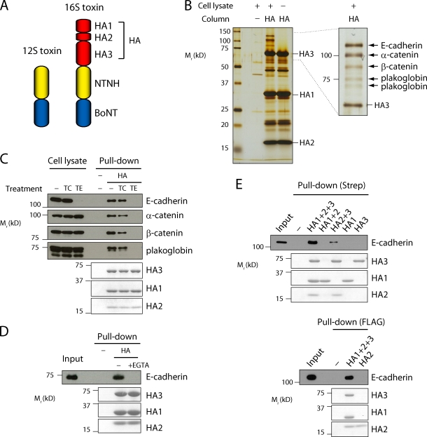
There are two forms of the type B BoNTs consisting of large protein complexes (BoNT/B complexes) referred to as the 12S and 16S toxins, in which nontoxic components are attached to BoNT (Sakaguchi, 1982; Oguma et al., 1999). BoNT with nontoxic non-HA (NTNH) constitutes the 12S toxin, and the 16S toxin is comprised of BoNT, NTNH, and HA, which itself is made up of three proteins, HA1, -2, and -3 (Fig. 1 A). These nontoxic components protect BoNT from the low pH and enzymes encountered in the digestive tract after oral ingestion, and these nontoxic factors greatly contribute to the oral toxicity of the BoNT complex (Ohishi et al., 1977; Sugii et al., 1977). We recently identified another role for the HA component of type B 16S toxin: this protein complex disrupts the intestinal epithelial intercellular junction and facilitates the transepithelial delivery of BoNT. We proposed that this activity contributes to the pathogenesis of food-borne botulism (Matsumura et al., 2008). In this study, we show that type B HA directly binds epithelial cadherin (E-cadherin) and thereby disrupts E-cadherin–mediated cell to cell adhesion and, in turn, the epithelial barrier.
Figure 1.
Botulinum HA binds to E-cadherin. (A) Schematic representation of the BoNT complexes. (B) Purification of HA binding proteins. Caco-2 cell lysates were loaded on a Strep-Tactin column prebound with HA. As a control, cell lysates were passed over a bare Strep-Tactin column, or a buffer was passed over a column containing HA. HA and associated proteins were eluted, and the eluates were further purified using anti-Flag M2 gel. HA and HA-binding proteins were eluted with low pH buffer (pH 3.5), separated by SDS-PAGE, and detected by silver stain (left). Alternatively, HA-binding proteins were eluted with EDTA and NaCl (right). Detected bands were excised and processed for mass spectrometric analysis, and the identified proteins are indicated (arrows). (C) Caco-2 cells were treated with trypsin in the presence of Ca2+ (TC) or EDTA (TE) before the extraction, and cell lysates were subjected to a HA pull-down assay followed by immunoblotting with antibodies for the indicated proteins. The HA proteins were detected by Coomassie blue stain. (D) The E-cadherin extracellular domain protein was pulled down with Strep-Tactin gel loaded with HA, but Ca2+ chelation by 5 mM EGTA prevented this interaction. (E) E-cadherin was pulled down from Caco-2 cell lysates with Strep-Tactin gel loaded with Strep-tagged HA1, Strep-tagged HA3, or the indicated combination of HA subunits (top). Flag-tagged HA2 was coupled to anti-Flag M2 gel and used for pull-down assay (bottom). HA1 and -2 do not form stable complexes.
Results and discussion
We generated recombinant type B HA comprised of Strep-tagged HA1, Flag-tagged HA2, and Flag-tagged HA3, and these proteins formed functional complexes comparable to native 16S toxin when assembled in vitro (unpublished data). We then purified HA-binding proteins using an affinity column in which recombinant HA was immobilized via its Strep tag. Using this approach, we reproducibly purified five proteins with molecular masses of ∼100 kD from the human intestinal epithelial cell line Caco-2 (Fig. 1 B). Mass spectrometric analysis identified these proteins as E-cadherin, a transmembrane protein that mediates Ca2+-dependent intercellular adhesion (Yoshida-Noro et al., 1984; Hatta et al., 1985), and its cytoplasmic binding proteins, α-catenin, β-catenin, and plakoglobin/γ-catenin (Ozawa et al., 1989). We confirmed these results using a HA pull-down assay followed by immunoblotting. Additionally, when E-cadherin was depleted by treating the cells with trypsin in the absence of Ca2+, the catenins no longer coprecipitated with HA, suggesting that HA directly interacts with the extracellular region of E-cadherin (Fig. 1 C). This interaction was also detected by preincubating cells with HA (Fig. S1, A and B). Furthermore, using a recombinant extracellular domain of E-cadherin, we confirmed the direct interaction of HA with E-cadherin and the dependence of this interaction on the presence of Ca2+ (Fig. 1 D). Although weak binding was observed between E-cadherin and a complex consisting of HA2 and -3, all three HA subunits were needed for full binding (Fig. 1 E).
We next examined the interaction of the native 16S toxin with E-cadherin. Both types B and A 16S toxins interacted with E-cadherin, but type C 16S toxin did not (Fig. S1 C). Interestingly, we previously showed that types A and B but not C 16S toxin disrupt the intercellular junction of Caco-2 cells (Jin et al., 2009). Collectively, these data suggest that the HA–E-cadherin interaction may be related to the disruption of intercellular junctions (see Fig. 5).
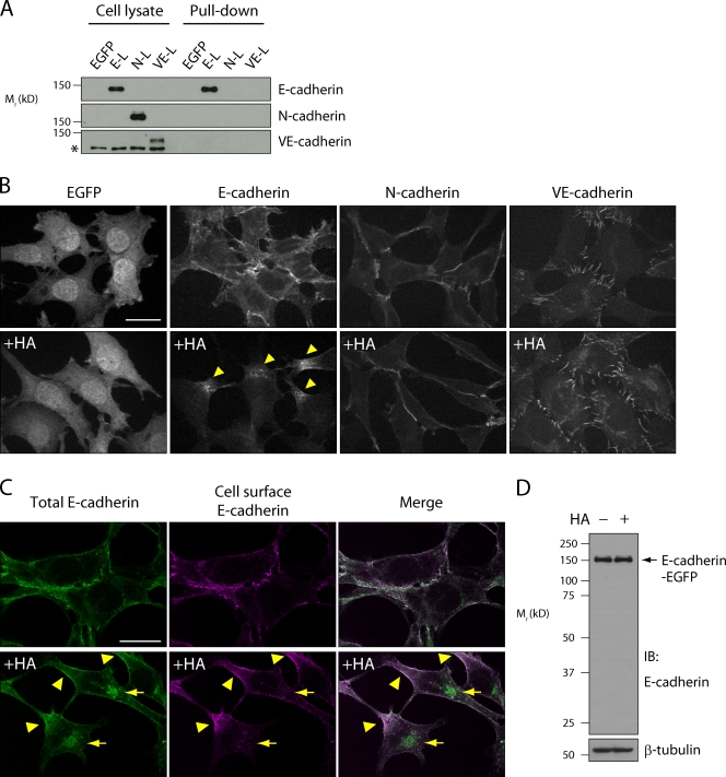
E-cadherin is a canonical member of the cadherin family, which has at least 80 members (Yagi and Takeichi, 2000). We examined the specificity of the HA interaction with cadherins by comparing HA binding to three classical cadherins, E-cadherin, neural cadherin (N-cadherin), and vascular endothelial cadherin (VE-cadherin), using L cells stably expressing EGFP-tagged cadherins. In a HA pull-down assay, the interaction was specific to E-cadherin (Fig. 2 A). When recombinant HA was applied to the E-cadherin–expressing cells, localization of E-cadherin around the cell boundaries was lost, E-cadherin clusters appeared on the cell surface, and E-cadherin was also internalized (Fig. 2, B and C). In contrast, HA had no effect on cadherin localization or internalization in cells expressing N- or VE-cadherin (Fig. 2 B). E-cadherin cleavage was not observed after HA treatment (Fig. 2 D). Thus, HA specifically disrupted E-cadherin–mediated cell to cell adhesion without proteolytic E-cadherin cleavage.
Figure 2.
Botulinum HA disrupts E-cadherin but not N-cadherin– and VE-cadherin–mediated cell adhesion. (A) Cell lysates prepared from L cells stably expressing EGFP, E-cadherin–EGFP (E-L), N-cadherin–EGFP (N-L), and VE-cadherin–EGFP (VE-L) were subjected to a HA pull-down assay followed by immunoblotting with specific antibodies for each protein. The asterisk denotes nonspecific bands. (B) Confocal images of EGFP fluorescence of the L cells pretreated with or without 100 nM HA for 6 h. E-cadherin–EGFP formed clusters (arrowheads). (C) E-L cells treated as in B were stained with the monoclonal antibody DECMA-1, which recognizes the extracellular region of E-cadherin (Ozawa et al., 1990), before permeabilization. After permeabilization, E-cadherin–EGFP was labeled with an anti–E-cadherin antibody that recognizes the intracellular domain of E-cadherin. E-cadherin–EGFP costained with these two antibodies is localized to the cell surface (arrowheads), and E-cadherin–EGFP stained only with the anti–intracellular domain antibody indicates internalized protein (arrows). (D) E-L cells treated as in B were lysed in SDS-PAGE sample buffer and analyzed by immunoblotting using anti–E-cadherin antibody. β-Tubulin was used as a loading control. IB, immunoblot. Bars, 20 µm.
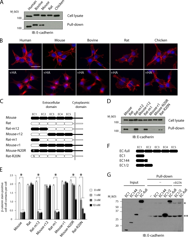
We then examined the ability of HA to interact with E-cadherin derived from different species using L cells stably expressing human, bovine, mouse, rat, or chicken E-cadherin (LCAM). HA bound to human, bovine, and mouse E-cadherin, but no binding was seen for rat and chicken E-cadherin (Fig. 3 A). Consistent with this, HA disrupted cell adhesion mediated by human-, bovine-, and mouse-derived E-cadherin (Fig. 3 B).
Figure 3.
E-cadherin EC1 and -2 domains are required for HA binding. (A) Human, mouse, and bovine but not rat and chicken E-cadherin bound HA. Cell lysates prepared from L cells stably expressing E-cadherin derived from each species were subjected to a HA pull-down assay followed by immunoblotting. (B) L cells were treated with or without 10 nM HA for 6 h, stained with an antibody against β-catenin (red) to demonstrate cadherin-dependent cell to cell boundary localization. Nuclei were stained with Hoechst (blue). (C) Schematic representation of E-cadherin chimeras between mouse (closed) and rat (open). (D) Cell lysates from the L cells expressing E-cadherin chimeras were subjected to a HA pull-down assay followed by immunoblotting. (E) The extent of the disruption of E-cadherin–mediated cell–cell adhesion was quantified as the mean number of β-catenin stain–positive cell–cell contacts per cell. A total of at least 250 cells were counted per condition in three independent experiments. Values are means ± SEM (*, P < 0.001). (F) Schematic representation of recombinant mouse EC domain proteins. (G) The recombinant EC domain proteins were subjected to a HA pull-down assay in the absence or presence of 5 mM EGTA followed by immunoblotting for E-cadherin. The asterisk and double asterisk denote HA3 and -1, respectively, which nonspecifically reacted with the antibody used. IB, immunoblot. Bar, 30 µm.
To identify the critical region of E-cadherin responsible for binding to HA, we generated chimeric constructs in which the extracellular cadherin (EC) domains of rat E-cadherin were sequentially replaced by the corresponding domains from mouse E-cadherin (Fig. 3 C). Replacement of the distal EC domain of rat E-cadherin with the corresponding domain from mouse E-cadherin (Rat-m1) was sufficient to mediate HA interaction and L cell dissociation (Fig. 3, D and E). Thus, the EC1 domain, which is known to mediate interactions between cells in trans (for review see Shapiro and Weis, 2009), appears to be critically involved in its interaction with HA. The mouse and rat EC1 domains differ by only 9 aa residues, and we generated single residue mutations in the mouse protein to identify residues that abrogate binding. Among these, replacement of Asn20 with Arg (Mouse-N20R) markedly reduced the interaction with HA in a pull-down assay (Fig. 3 D and Fig. S3). This residue is not directly involved in E-cadherin trans-dimerization, but it is located near the cadherin dimer interface (Boggon et al., 2002). Cells expressing Mouse-N20R remained susceptible to the effects of HA, but to a lesser extent than cells expressing wild-type mouse E-cadherin, and the reciprocal mutation (Rat-R20N) conferred only partial HA binding (Fig. 3, D and E). These results indicate that Asn20 is directly involved in the interaction of mouse E-cadherin with HA or located in close proximity to the interaction surface of them, but it seems likely that additional residues in EC1 contribute to this interaction.
We next examined whether the EC1 domain is sufficient for mediating the interaction of E-cadherin with HA using bacterially expressed EC domain proteins. In a HA pull-down assay, neither EC1 nor the extended construct EC144, which harbors EC1 and some residues of EC2, interacted with HA, but a construct containing the full EC1 and EC2 domains (EC1/2) was successfully pulled down with HA (Fig. 3, F and G). The interaction of EC1/2 with HA was comparable with that of the full-length EC domain protein expressed in mammalian cells (EC-full), and this interaction was Ca2+ dependent (Fig. 3 G). Thus, EC1 is necessary but not sufficient for the interaction of HA with E-cadherin, and both EC1 and -2 are required for full HA binding. Additionally, because the EC1/2 construct was produced in bacteria, it is clear that no posttranslational modifications of E-cadherin are needed to mediate its interaction with HA.
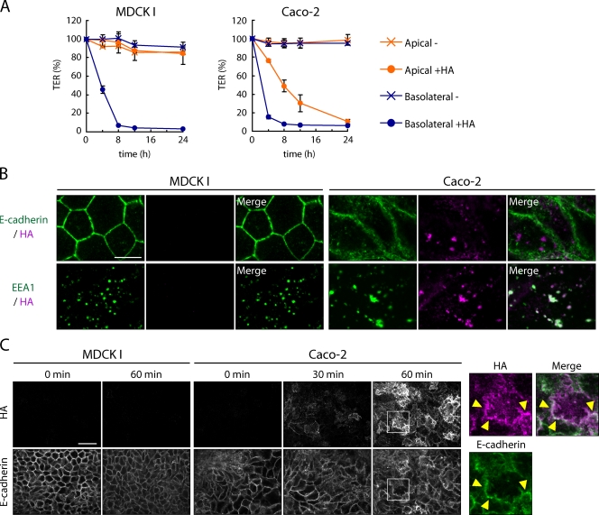
Epithelial cells form junctional complexes in which tight junctions (TJs), E-cadherin–based adherens junctions, and desmosomes are aligned from the apical surface at sites of cell to cell contact (Farquhar and Palade, 1963). Claudins are localized to the TJ and provide an important barrier function by preventing the paracellular influx of water, ions, and solutes (Furuse et al., 1998; Sonoda et al., 1999). Thus, HA would not have access to E-cadherin from the apical face of the cells, and this is consistent with our previous observation that the basolateral membrane is the site of HA action (Matsumura et al., 2008). When HA was added to the apical surface of Caco-2 cell monolayers, it was bound and internalized into early endosomes that costained with EEA1 (early endosome antigen 1; Fig. 4 B). 1 h after HA application, it reached the basal surface, presumably through transcytosis, and partially localized to the lateral cellular membrane with E-cadherin (Fig. 4 C). In contrast, in MDCK I cells, which were insensitive to apically applied HA but were sensitive when treated basolaterally, binding and internalization of HA was substantially reduced compared with Caco-2 cells, probably because of the lack of presumptive apical receptors for HA. Furthermore, large amounts of basolateral HA staining were not seen (Fig. 4, A–C). Although the detailed mechanism remains to be elucidated, HA transcytosis is required to achieve the proper colocalization of HA with E-cadherin in polarized epithelial cells. Under physiological conditions, intestinal M cells sample luminal antigens through high rates of transcytosis (Neutra et al., 2001), and these cells are thought to be the site of HA transcytosis in addition to enterocytes.
Figure 4.
HA is transcytosed in Caco-2 cells. (A) Caco-2 and MDCK I cells were grown in Transwell chambers, and TER was measured in the presence (•) or absence (x) of HA applied to the apical (1 µM) or basolateral (100 nM) chamber. Values are means ± SEM (n = 3). (B) Caco-2 and MDCK I cell monolayers were apically treated with 100 nM HA for 30 min at 4°C. Cells were washed and incubated for an additional 30 min at 37°C. HA was labeled with E-cadherin or EEA1 using specific antibodies against each molecule. (C) Caco-2 and MDCK I cell monolayers were treated with 1 µM HA from the apical side for the indicated times. The basolateral surface was labeled with anti-HA antibody before permeabilization. After permeabilization, cells were labeled with anti–E-cadherin antibody. Right panels show a higher magnification image of the boxed region. Partial colocalization of HA (magenta) and E-cadherin (green) was observed (arrowheads). Bars: (B) 10 µm; (C) 30 µm.
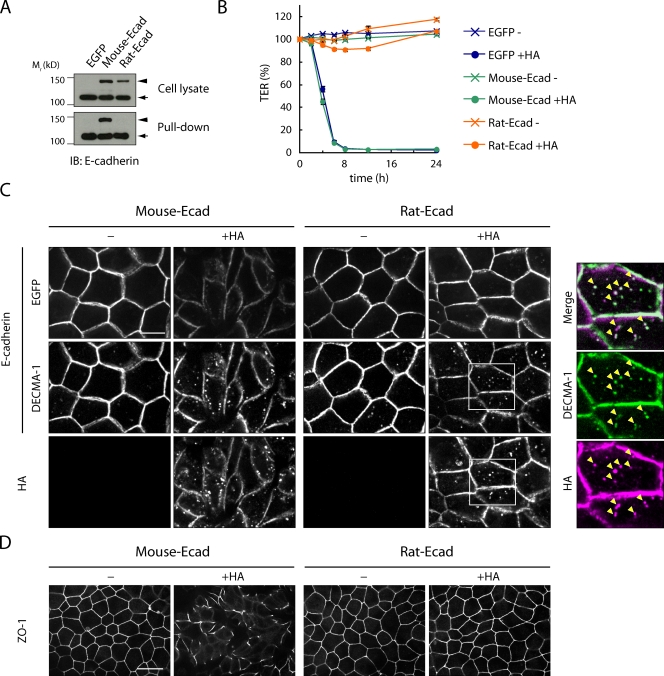
E-cadherin mediates the formation and maintenance of the entire epithelial junctional complex, including TJs (Behrens et al., 1985; Gumbiner and Simons, 1986; Gumbiner et al., 1988; Watabe et al., 1994; for review see Citi, 1993), although a recent study suggested that E-cadherin is not required for the maintenance of TJs (Capaldo and Macara, 2007). We performed the following experiments to confirm that HA disrupts TJs in an E-cadherin–dependent manner. We used MDCK I cell monolayers expressing EGFP alone or EGFP fused to mouse or rat E-cadherin. As described in the previous paragraph, MDCK I cells are susceptible to the effects of HA when treated basolaterally, and endogenous canine E-cadherin was pulled down by HA (Fig. 5 A). We applied HA to the basolateral surface of the cell monolayer and measured transepithelial electrical resistance (TER). Treatment with HA markedly reduced the TER of MDCK I cells expressing either EGFP or mouse E-cadherin–EGFP. However, rat E-cadherin–EGFP acted as a dominant negative, and cells expressing rat E-cadherin–EGFP maintained their TER even in the presence of HA (Fig. 5 B). The reduced TER induced by HA was partially inhibited by the expression of Mouse-N20R (Fig. S2 C). Endogenous canine E-cadherin in rat E-cadherin–expressing cells was pulled down by HA to a similar extent as in EGFP-expressing cells (Fig. 5 A). Moreover, in these cells, endogenous canine E-cadherin was cointernalized with HA, which is consistent with results seen in mouse E-cadherin–expressing cells (Fig. 5 C). Nonetheless, exogenous rat E-cadherin and ZO-1, a TJ protein, were localized normally at intercellular junctions (Fig. 5, C and D). These observations indicate that exogenous rat E-cadherin is capable of maintaining TJs despite HA-mediated disruption of endogenous canine E-cadherin function, and these data clearly demonstrate that E-cadherin plays a critical role in the disruption of TJs initiated by HA.
Figure 5.
The E-cadherin–HA interaction is critically involved in HA-mediated TJ disruption. (A) Cell lysates were prepared from MDCK I cells stably expressing EGFP, mouse E-cadherin–EGFP (Mouse-Ecad), or rat E-cadherin–EGFP (Rat-Ecad) and subjected to a HA pull-down assay followed by immunoblotting. Arrowheads and arrows denote E-cadherin–EGFP and endogenous canine E-cadherin, respectively. (B) MDCK transfectants were grown in Transwell chambers, and TER was measured in the presence (•) or absence (x) of 100 nM HA applied to the basolateral chamber. Values are means ± SEM (n = 3). (C) Confocal images of exogenous E-cadherin (EGFP), mouse and endogenous canine E-cadherin (DECMA-1), and HA of the cells treated with or without 100 nM HA for 24 h. Right panels show a higher magnification image of the boxed region. Endogenous E-cadherin (green) was cointernalized with HA (magenta; arrowheads). DECMA-1 recognizes mouse and canine but not rat E-cadherin. (D) Confocal images of ZO-1 of the cells treated with or without 100 nM HA for 24 h. Bars: (C) 10 µm; (D) 30 µm.
Previously, we showed that the nontoxic component of the 16S toxin (i.e., the complex of HA and NTNH) disrupts the intestinal epithelial barrier, leading to the absorption of the 12S toxin or soluble dextran in a mouse in situ loop assay (Matsumura et al., 2008). We performed this assay using rats and guinea pigs, and the disruption of the epithelial barrier was assessed by monitoring the influx of FITC-dextran in the serum. Like rat E-cadherin, guinea pig E-cadherin contains an Arg at its 20th aa (Lecuit et al., 1999), and it was unable to interact with HA (Fig. S3 A). In contrast to the results seen in mice, the nontoxic toxin component did not increase the absorption of FITC-dextran in rats or guinea pigs (Fig. S3). Thus, there is a strong in vivo correlate to our observations in vitro, and the interaction of HA with E-cadherin seems to play a pivotal role in inducing epithelial barrier disruption in vivo.
Several pathogens target E-cadherin to facilitate host invasion. Listeria monocytogenes and Candida albicans bind E-cadherin, leading to internalization (Mengaud et al., 1996; Phan et al., 2007). Alternatively, Bacteroides fragilis, Porphyromonas gingivalis, and C. albicans produce proteases that cleave E-cadherin, leading to epithelial barrier disruption and tissue invasion (Obiso et al., 1997; Wu et al., 1998; Katz et al., 2000, 2002; Frank and Hostetter, 2007; Villar et al., 2007). Although the mode of action of botulinum HA is more similar to that used by the latter pathogens, it does not proteolytically cleave E-cadherin. Disruption of the epithelial barrier by HA might facilitate the influx and dissemination of BoNT into the systemic circulation.
In this study, we showed that the HA–E-cadherin interaction is restricted to particular species, and, for example, HA of the BoNT/B complex did not interact with chicken E-cadherin. Interestingly, this observation is consistent with the rare reports of avian botulism caused by BoNT/B. Furthermore, birds were experimentally shown to be relatively resistant to BoNT/B complex, especially when administered orally (Gross and Smith, 1971; Notermans et al., 1980). Additionally, the inability of BoNT/C complex to associate with human E-cadherin is correlated with the fact that human type C botulism is rarely seen despite the ability of BoNT/C to block neuromuscular transmission in human tissue (Coffield et al., 1997). Collectively, these data suggest that the interaction of HA with E-cadherin is an important factor determining host susceptibility for orally ingested BoNT complexes. This hypothesis could be tested using a similar approach as that used in studies of L. monocytogenes infection (Lecuit et al., 2001; Disson et al., 2008), namely by comparing the oral toxicity of BoNT/B complex in wild-type and knockin mice in which mouse E-cadherin is replaced by rat E-cadherin.
Materials and methods
Antibodies
Antibodies for E-cadherin were purchased from BD, Sigma-Aldrich, Takara Bio Inc., and Invitrogen. An anti–VE-cadherin antibody was purchased from Santa Cruz Biotechnology, Inc. Anti–N-cadherin, anti–α-catenin, anti–β-catenin, antiplakoglobin (γ-catenin), and anti-EEA1 antibodies were purchased from BD. Anti–Pan-cadherin and anti–β-tubulin antibodies were purchased from Sigma-Aldrich. Rabbit anti–type A 16S, BoNT/A, type B 16S, and BoNT/B antisera were generated by immunizing rabbits with the respective forms of the toxins prepared as described previously (Matsumura et al., 2008; Jin et al., 2009). Rabbit anti–type C 16S and BoNT/C antisera were provided by S. Kozaki (Osaka Prefecture University, Nakaku, Sakai, Osaka, Japan).
Plasmid construction
Mouse E-, N-, and VE-cadherin cDNAs were described previously (Nagafuchi et al., 1987; Miyatani et al., 1989; Kametani and Takeichi, 2007). Human, bovine, rat, and chicken E-cadherin cDNA clones were amplified from the total RNA of Caco-2 (American Type Culture Collection), MDBK, NBT-T1 (RIKEN Cell Bank), and LMH cells (Japanese Collection of Research Bioresources Cell Bank), respectively, and subcloned into the pCA-IRES-hygromycin vector (Kametani and Takeichi, 2007).
Purified DNA from C. botulinum type B strain Lamanna was used as a template for the amplification of DNA encoding HA1, -2, and -3 by PCR. The amplified DNAs were inserted into pET-52b(+) (EMD) and pT7-Flag-1 (Sigma-Aldrich).
Cell culture and establishment of stably transfected cells
Caco-2 and MDCK I cells were cultured as previously described (Jin et al., 2009). IEC-18 (rat intestinal epithelial cells) and GPC-16 cells (guinea pig colon adenocarcinoma cells) were obtained from the American Type Culture Collection and cultured according to the supplier's protocol. CMT93-I cells (mouse rectal carcinoma cells; Inai et al., 2008) were provided by T. Inai (Kyushu University, Higashi-ku, Fukuoka, Japan).
L and MDCK I cells were transfected with vectors encoding cadherins using Lipofectamine 2000 (Invitrogen) according to the manufacturer's instructions. Stable transfectants were selected by limiting dilution in the presence of 1 mg/ml (L cells) or 0.25 mg/ml (MDCK I cells) hygromycin B (Invitrogen). Expression of exogenous cadherins was confirmed by immunoblotting.
Immunofluorescence
Cells grown on Transwell pore filters or glass coverslips were fixed with 4% paraformaldehyde in PBS for 20 min and permeabilized with 0.5% Triton X-100 in PBS for 5 min at room temperature. Monolayers or coverslips were incubated with primary antibodies, probed with secondary antibodies coupled to Alexa Fluor 405, Alexa Fluor 488 (Invitrogen), or Cy3 (Jackson ImmunoResearch Laboratories, Inc.), and mounted with ProLong Antifade kit (Invitrogen). Images were captured with a microscope (IX71; Olympus) equipped with UPlan-Apochromat 100× NA 1.35 oil or Plan-Apochromat 60× NA 1.40 oil objectives (Olympus) and a CSU-21 or CSU-X1 confocal scanner unit (Yokogawa). Images were analyzed with MetaMorph imaging software (version 6.3r5; Universal Imaging).
Protein expression and purification
Recombinant HAs were expressed as N-terminal Strep-tagged or Flag-tagged proteins in the Escherichia coli strains Rosetta (DE3) (EMD) or BL21-CodonPlus (DE3)-RIL (Agilent Technologies), and purified using the Strep-Tactin MacroPrep resin (EMD) and anti-Flag M2 resin (Sigma-Aldrich), respectively, according to the manufacturers' protocols. For the reconstitution of the HA complex, the recombinant HA1, -2, and -3 proteins were mixed at a molar ratio of 2:2:1 in PBS, pH 7.4, and incubated for 3 h at 37°C. The molarity of the HA complex was expressed as that of HA3. The extracellular domains (1–543 aa) of mouse E-cadherin were expressed in HEK293 cells as a C-terminally hexa-His–tagged protein and purified with a HisTrap column (GE Healthcare).
The recombinant mouse EC domain proteins (EC1, 1–104 aa; EC144, 1–144 aa; and EC1/2, 1–219 aa) were expressed in E. coli strain BL21-CodonPlus (DE3)-RIL as GST fusion proteins with a Factor Xa cleavage site adjacent to first Asp (Häussinger et al., 2004) and with a hexa-His tag at the C terminus. The proteins were purified with a GSTrap column (GE Healthcare), cleaved with Factor Xa (EMD), and further purified with the HisTrap column.
Purification of HA-binding proteins
Caco-2 cells were homogenized by being passed through a 24-gauge needle 10 times. The soluble fraction was removed by centrifugation (15,500 g for 10 min), and the resultant pellet was lysed in lysis buffer (10 mM Tris-HCl, pH 7.4, 1 mM MgCl2, 1 mM CaCl2, 150 mM NaCl, and 1% Triton X-100) supplemented with an EDTA-free protease inhibitor (Roche). Unsolubilized material was removed by ultracentrifugation (120,000 g for 1 h). The cell lysate was loaded on the Strep-Tactin MacroPrep column prebound with HA (a complex of Strep-HA1, Flag-HA2, and Flag-HA3), followed by extensive washing with lysis buffer. The HA and the HA-bound proteins were eluted from the resin with lysis buffer supplemented with 2.5 mM desthiobiotin and further purified using anti-Flag M2 resin.
HA pull-down assay
Cells stably expressing cadherins were solubilized in lysis buffer, and the cell lysates were incubated with the Strep-Tactin Superflow agarose (EMD) prebound with HA (a complex of Strep-HA1, Flag-HA2, and Strep-HA3). When the recombinant E-cadherin extracellular domain constructs were subjected to a HA pull-down assay, the protein concentration was adjusted to 50 nM in a Hepes buffer (20 mM Hepes-NaOH, pH 7.35, 2 mM CaCl2, 150 mM NaCl, and 0.01% Triton X-100).
In situ loop assay
In situ loop assay was performed as described previously (Kondoh et al., 2005; Matsumura et al., 2008) except that animals were anesthetized with pentobarbital Na. In brief, mice, rats, and guinea pigs were not fed for 24 h before surgery. A ligated intestinal loop (mice, 3.5–4.5 cm; and rats and guinea pigs, 4.5–5.5 cm of length) was made in the upper small intestine, and 10 mg/ml FITC-dextran 4k (Sigma-Aldrich) with or without 5 µM of the nontoxic component of B 16S toxin in PBS (mice, 100 µl; and rats and guinea pigs, 200 µl, pH 6.0) was injected into the loop with a 29-gauge needle. After 4 h, plasma was collected, and the FITC-dextran levels were determined with a fluorometer (Fluoroskan II; Labsystems). The experimental protocols were approved by the Ethics Review Committee for Animal Experimentation at Osaka University and were in accordance with its guidelines.
Statistical analysis
Data are representative of at least three independent experiments. In Fig. 3 E and Fig. S3 C, values were analyzed by the Student's t test.
Online supplemental material
Fig. S1 shows the interaction of E-cadherin with type B HA and native 16S toxins of types A to C. Fig. S2 shows the interaction of type B HA with the mouse to rat E-cadherin point mutants and the measurement of TER using MDCK cells expressing E-cadherin point mutants. Fig. S3 shows the differential effects of HA on cultured epithelial cells derived from mouse, rat, and guinea pig, the binding of HA with E-cadherin derived from these three species, and the in situ loop assay using mice, rats, and guinea pigs. Online supplemental material is available at http://www.jcb.org/cgi/content/full/jcb.200910119/DC1.
Acknowledgments
We thank Kazunobu Saito for mass spectrometric analysis, Shunji Kozaki for providing antibodies, Tetsuichiro Inai for providing cells, and Eisuke Mekada for insightful discussions and critical reading of the manuscript.
This work was supported in parts by grants from the Ministry of Education, Culture, Sports, Science and Technology of Japan to Y. Fujinaga and T. Matsumura.
Footnotes
Abbreviations used in this paper:
- BoNT
- botulinum neurotoxin
- EC
- extracellular cadherin
- E-cadherin
- epithelial cadherin
- N-cadherin
- neural cadherin
- NTNH
- nontoxic non-HA
- TER
- transepithelial electrical resistance
- TJ
- tight junction
- VE-cadherin
- vascular endothelial cadherin
References
- Arnon S.S., Schechter R., Inglesby T.V., Henderson D.A., Bartlett J.G., Ascher M.S., Eitzen E., Fine A.D., Hauer J., Layton M., et al. 2001. Botulinum toxin as a biological weapon: medical and public health management. JAMA. 285:1059–1070 10.1001/jama.285.8.1059 [DOI] [PubMed] [Google Scholar]
- Behrens J., Birchmeier W., Goodman S.L., Imhof B.A. 1985. Dissociation of Madin-Darby canine kidney epithelial cells by the monoclonal antibody anti-arc-1: mechanistic aspects and identification of the antigen as a component related to uvomorulin. J. Cell Biol. 101:1307–1315 10.1083/jcb.101.4.1307 [DOI] [PMC free article] [PubMed] [Google Scholar]
- Boggon T.J., Murray J., Chappuis-Flament S., Wong E., Gumbiner B.M., Shapiro L. 2002. C-cadherin ectodomain structure and implications for cell adhesion mechanisms. Science. 296:1308–1313 10.1126/science.1071559 [DOI] [PubMed] [Google Scholar]
- Capaldo C.T., Macara I.G. 2007. Depletion of E-cadherin disrupts establishment but not maintenance of cell junctions in Madin-Darby canine kidney epithelial cells. Mol. Biol. Cell. 18:189–200 10.1091/mbc.E06-05-0471 [DOI] [PMC free article] [PubMed] [Google Scholar]
- Citi S. 1993. The molecular organization of tight junctions. J. Cell Biol. 121:485–489 10.1083/jcb.121.3.485 [DOI] [PMC free article] [PubMed] [Google Scholar]
- Coffield J.A., Bakry N., Zhang R.D., Carlson J., Gomella L.G., Simpson L.L. 1997. In vitro characterization of botulinum toxin types A, C and D action on human tissues: combined electrophysiologic, pharmacologic and molecular biologic approaches. J. Pharmacol. Exp. Ther. 280:1489–1498 [PubMed] [Google Scholar]
- Couesnon A., Pereira Y., Popoff M.R. 2008. Receptor-mediated transcytosis of botulinum neurotoxin A through intestinal cell monolayers. Cell. Microbiol. 10:375–387 [DOI] [PubMed] [Google Scholar]
- Disson O., Grayo S., Huillet E., Nikitas G., Langa-Vives F., Dussurget O., Ragon M., Le Monnier A., Babinet C., Cossart P., Lecuit M. 2008. Conjugated action of two species-specific invasion proteins for fetoplacental listeriosis. Nature. 455:1114–1118 10.1038/nature07303 [DOI] [PubMed] [Google Scholar]
- Farquhar M.G., Palade G.E. 1963. Junctional complexes in various epithelia. J. Cell Biol. 17:375–412 10.1083/jcb.17.2.375 [DOI] [PMC free article] [PubMed] [Google Scholar]
- Frank C.F., Hostetter M.K. 2007. Cleavage of E-cadherin: a mechanism for disruption of the intestinal epithelial barrier by Candida albicans. Transl. Res. 149:211–222 10.1016/j.trsl.2006.11.006 [DOI] [PubMed] [Google Scholar]
- Furuse M., Fujita K., Hiiragi T., Fujimoto K., Tsukita S. 1998. Claudin-1 and -2: novel integral membrane proteins localizing at tight junctions with no sequence similarity to occludin. J. Cell Biol. 141:1539–1550 10.1083/jcb.141.7.1539 [DOI] [PMC free article] [PubMed] [Google Scholar]
- Gross W.B., Smith L.D.S. 1971. Experimental botulism in gallinaceous birds. Avian Dis. 15:716–722 10.2307/1588859 [DOI] [PubMed] [Google Scholar]
- Gumbiner B., Simons K. 1986. A functional assay for proteins involved in establishing an epithelial occluding barrier: identification of a uvomorulin-like polypeptide. J. Cell Biol. 102:457–468 10.1083/jcb.102.2.457 [DOI] [PMC free article] [PubMed] [Google Scholar]
- Gumbiner B., Stevenson B., Grimaldi A. 1988. The role of the cell adhesion molecule uvomorulin in the formation and maintenance of the epithelial junctional complex. J. Cell Biol. 107:1575–1587 10.1083/jcb.107.4.1575 [DOI] [PMC free article] [PubMed] [Google Scholar]
- Hatta K., Okada T.S., Takeichi M. 1985. A monoclonal antibody disrupting calcium-dependent cell-cell adhesion of brain tissues: possible role of its target antigen in animal pattern formation. Proc. Natl. Acad. Sci. USA. 82:2789–2793 10.1073/pnas.82.9.2789 [DOI] [PMC free article] [PubMed] [Google Scholar]
- Häussinger D., Ahrens T., Aberle T., Engel J., Stetefeld J., Grzesiek S. 2004. Proteolytic E-cadherin activation followed by solution NMR and X-ray crystallography. EMBO J. 23:1699–1708 10.1038/sj.emboj.7600192 [DOI] [PMC free article] [PubMed] [Google Scholar]
- Inai T., Sengoku A., Hirose E., Iida H., Shibata Y. 2008. Comparative characterization of mouse rectum CMT93-I and -II cells by expression of claudin isoforms and tight junction morphology and function. Histochem. Cell Biol. 129:223–232 10.1007/s00418-007-0360-0 [DOI] [PubMed] [Google Scholar]
- Jin Y., Takegahara Y., Sugawara Y., Matsumura T., Fujinaga Y. 2009. Disruption of the epithelial barrier by botulinum haemagglutinin (HA) proteins - differences in cell tropism and the mechanism of action between HA proteins of types A or B, and HA proteins of type C. Microbiology. 155:35–45 10.1099/mic.0.021246-0 [DOI] [PubMed] [Google Scholar]
- Kametani Y., Takeichi M. 2007. Basal-to-apical cadherin flow at cell junctions. Nat. Cell Biol. 9:92–98 10.1038/ncb1520 [DOI] [PubMed] [Google Scholar]
- Katz J., Sambandam V., Wu J.H., Michalek S.M., Balkovetz D.F. 2000. Characterization of Porphyromonas gingivalis-induced degradation of epithelial cell junctional complexes. Infect. Immun. 68:1441–1449 10.1128/IAI.68.3.1441-1449.2000 [DOI] [PMC free article] [PubMed] [Google Scholar]
- Katz J., Yang Q.B., Zhang P., Potempa J., Travis J., Michalek S.M., Balkovetz D.F. 2002. Hydrolysis of epithelial junctional proteins by Porphyromonas gingivalis gingipains. Infect. Immun. 70:2512–2518 10.1128/IAI.70.5.2512-2518.2002 [DOI] [PMC free article] [PubMed] [Google Scholar]
- Kondoh M., Masuyama A., Takahashi A., Asano N., Mizuguchi H., Koizumi N., Fujii M., Hayakawa T., Horiguchi Y., Watanbe Y. 2005. A novel strategy for the enhancement of drug absorption using a claudin modulator. Mol. Pharmacol. 67:749–756 10.1124/mol.104.008375 [DOI] [PubMed] [Google Scholar]
- Lecuit M., Dramsi S., Gottardi C., Fedor-Chaiken M., Gumbiner B., Cossart P. 1999. A single amino acid in E-cadherin responsible for host specificity towards the human pathogen Listeria monocytogenes. EMBO J. 18:3956–3963 10.1093/emboj/18.14.3956 [DOI] [PMC free article] [PubMed] [Google Scholar]
- Lecuit M., Vandormael-Pournin S., Lefort J., Huerre M., Gounon P., Dupuy C., Babinet C., Cossart P. 2001. A transgenic model for listeriosis: role of internalin in crossing the intestinal barrier. Science. 292:1722–1725 10.1126/science.1059852 [DOI] [PubMed] [Google Scholar]
- Maksymowych A.B., Simpson L.L. 1998. Binding and transcytosis of botulinum neurotoxin by polarized human colon carcinoma cells. J. Biol. Chem. 273:21950–21957 10.1074/jbc.273.34.21950 [DOI] [PubMed] [Google Scholar]
- Matsumura T., Jin Y., Kabumoto Y., Takegahara Y., Oguma K., Lencer W.I., Fujinaga Y. 2008. The HA proteins of botulinum toxin disrupt intestinal epithelial intercellular junctions to increase toxin absorption. Cell. Microbiol. 10:355–364 [DOI] [PubMed] [Google Scholar]
- Mengaud J., Ohayon H., Gounon P., Cossart P., Mège R.-M., Cossart P. 1996. E-cadherin is the receptor for internalin, a surface protein required for entry of L. monocytogenes into epithelial cells. Cell. 84:923–932 10.1016/S0092-8674(00)81070-3 [DOI] [PubMed] [Google Scholar]
- Miyatani S., Shimamura K., Hatta M., Nagafuchi A., Nose A., Matsunaga M., Hatta K., Takeichi M. 1989. Neural cadherin: role in selective cell-cell adhesion. Science. 245:631–635 10.1126/science.2762814 [DOI] [PubMed] [Google Scholar]
- Nagafuchi A., Shirayoshi Y., Okazaki K., Yasuda K., Takeichi M. 1987. Transformation of cell adhesion properties by exogenously introduced E-cadherin cDNA. Nature. 329:341–343 10.1038/329341a0 [DOI] [PubMed] [Google Scholar]
- Neutra M.R., Mantis N.J., Kraehenbuhl J.P. 2001. Collaboration of epithelial cells with organized mucosal lymphoid tissues. Nat. Immunol. 2:1004–1009 10.1038/ni1101-1004 [DOI] [PubMed] [Google Scholar]
- Notermans S., Dufrenne J., Kozaki S. 1980. Experimental botulism in Pekin ducks. Avian Dis. 24:658–664 10.2307/1589803 [DOI] [PubMed] [Google Scholar]
- Obiso R.J., Jr., Azghani A.O., Wilkins T.D. 1997. The Bacteroides fragilis toxin fragilysin disrupts the paracellular barrier of epithelial cells. Infect. Immun. 65:1431–1439 [DOI] [PMC free article] [PubMed] [Google Scholar]
- Oguma K., Inoue K., Fujinaga Y., Yokota K., Watanabe T., Ohyama T., Takeshi K., Inoue K. 1999. Structure and function of Clostridium botulinum progenitor toxin. Journal of Toxicololgy: Toxin Reviews. 18:17–34 [Google Scholar]
- Ohishi I., Sugii S., Sakaguchi G. 1977. Oral toxicities of Clostridium botulinum toxins in response to molecular size. Infect. Immun. 16:107–109 [DOI] [PMC free article] [PubMed] [Google Scholar]
- Ozawa M., Baribault H., Kemler R. 1989. The cytoplasmic domain of the cell adhesion molecule uvomorulin associates with three independent proteins structurally related in different species. EMBO J. 8:1711–1717 [DOI] [PMC free article] [PubMed] [Google Scholar]
- Ozawa M., Hoschützky H., Herrenknecht K., Kemler R. 1990. A possible new adhesive site in the cell-adhesion molecule uvomorulin. Mech. Dev. 33:49–56 10.1016/0925-4773(90)90134-8 [DOI] [PubMed] [Google Scholar]
- Phan Q.T., Myers C.L., Fu Y., Sheppard D.C., Yeaman M.R., Welch W.H., Ibrahim A.S., Edwards J.E., Jr., Filler S.G. 2007. Als3 is a Candida albicans invasin that binds to cadherins and induces endocytosis by host cells. PLoS Biol. 5:e64 10.1371/journal.pbio.0050064 [DOI] [PMC free article] [PubMed] [Google Scholar]
- Poulain B., Popoff M.R., Molgó J. 2008. How do the botulinum neurotoxins block neurotransmitter release: from botulism to the molecular mechanism of action. The Botulinum J. 1:14–87 10.1504/TBJ.2008.018951 [DOI] [Google Scholar]
- Sakaguchi G. 1982. Clostridium botulinum toxins. Pharmacol. Ther. 19:165–194 10.1016/0163-7258(82)90061-4 [DOI] [PubMed] [Google Scholar]
- Schiavo G., Matteoli M., Montecucco C. 2000. Neurotoxins affecting neuroexocytosis. Physiol. Rev. 80:717–766 [DOI] [PubMed] [Google Scholar]
- Shapiro L., Weis W.I. 2009. Structure and biochemistry of cadherins and catenins. Cold Spring Harb. Perspect. Biol. 1:a003053 10.1101/cshperspect.a003053 [DOI] [PMC free article] [PubMed] [Google Scholar]
- Simpson L.L. 2004. Identification of the major steps in botulinum toxin action. Annu. Rev. Pharmacol. Toxicol. 44:167–193 10.1146/annurev.pharmtox.44.101802.121554 [DOI] [PubMed] [Google Scholar]
- Sonoda N., Furuse M., Sasaki H., Yonemura S., Katahira J., Horiguchi Y., Tsukita S. 1999. Clostridium perfringens enterotoxin fragment removes specific claudins from tight junction strands: evidence for direct involvement of claudins in tight junction barrier. J. Cell Biol. 147:195–204 10.1083/jcb.147.1.195 [DOI] [PMC free article] [PubMed] [Google Scholar]
- Sugii S., Ohishi I., Sakaguchi G. 1977. Correlation between oral toxicity and in vitro stability of Clostridium botulinum type A and B toxins of different molecular sizes. Infect. Immun. 16:910–914 [DOI] [PMC free article] [PubMed] [Google Scholar]
- Villar C.C., Kashleva H., Nobile C.J., Mitchell A.P., Dongari-Bagtzoglou A. 2007. Mucosal tissue invasion by Candida albicans is associated with E-cadherin degradation, mediated by transcription factor Rim101p and protease Sap5p. Infect. Immun. 75:2126–2135 10.1128/IAI.00054-07 [DOI] [PMC free article] [PubMed] [Google Scholar]
- Watabe M., Nagafuchi A., Tsukita S., Takeichi M. 1994. Induction of polarized cell-cell association and retardation of growth by activation of the E-cadherin-catenin adhesion system in a dispersed carcinoma line. J. Cell Biol. 127:247–256 10.1083/jcb.127.1.247 [DOI] [PMC free article] [PubMed] [Google Scholar]
- Wu S., Lim K.C., Huang J., Saidi R.F., Sears C.L. 1998. Bacteroides fragilis enterotoxin cleaves the zonula adherens protein, E-cadherin. Proc. Natl. Acad. Sci. USA. 95:14979–14984 10.1073/pnas.95.25.14979 [DOI] [PMC free article] [PubMed] [Google Scholar]
- Yagi T., Takeichi M. 2000. Cadherin superfamily genes: functions, genomic organization, and neurologic diversity. Genes Dev. 14:1169–1180 [PubMed] [Google Scholar]
- Yoshida-Noro C., Suzuki N., Takeichi M. 1984. Molecular nature of the calcium-dependent cell-cell adhesion system in mouse teratocarcinoma and embryonic cells studied with a monoclonal antibody. Dev. Biol. 101:19–27 10.1016/0012-1606(84)90112-X [DOI] [PubMed] [Google Scholar]


