

Abstract
During mineralization, unbound water within the collagen matrix is replaced by apatite. This study tested the null hypothesis that there is no difference in the status of in vitro biomimetic remineralization of hybrid layers, regardless of their moisture contents. Acid-etched dentin was bonded with One-Step with ethanol-wet-bonding, water-wet-bonding, and water-overwet-bonding protocols. Composite-dentin slabs were subjected to remineralization for 1-4 months in a medium containing dual biomimetic analogs, with set Portland cement as the calcium source and characterized by transmission electron microscopy. Remineralization was either non-existent or restricted to the intrafibrillar mode in ethanol-wet-bonded specimens. Extensive intrafibrillar and interfibrillar remineralization was observed in water-wet-bonded specimens. Water-overwet specimens demonstrated partial remineralization of hybrid layers and precipitation of mineralized plates within water channels. The use of ethanol-wet-bonding substantiates that biomimetic remineralization is a progressive dehydration process that replaces residual water in hybrid layers with apatite crystallites.
Keywords: biomimetics, dentin bonding, ethanol, hybrid layer, intrafibrillar, interfibrillar, remineralization, water
Introduction
During hard-tissue mineralization, bulk and weakly bound water molecules in the collagen matrix are gradually replaced by apatite crystallites (Magne et al., 2001; Wehrli and Fernández-Seara, 2005). Likewise, during intentional or pathological demineralization, dissolved intrafibrillar and interfibrillar apatites are replaced with a comparable volume of water (Ito et al., 2005; Toroian et al., 2007). In the context of dentin bonding, water that fills the intrafibrillar and interfibrillar spaces after apatite dissolution by acids should be totally displaced by resin monomers during bonding (Vaidyanathan and Vaidyanathan, 2009). Demineralized collagen is responsible for the creep behavior of hard tissues (Bowman et al., 1999; Pashley et al., 2003). Thus, incomplete replacement of water introduced during adhesive application and by perfusion from vital dental pulps leaves behind a hydrated collagen matrix that could adversely affect the resistance of resin-dentin bonds to cyclic fatigue. To date, complete displacement of water from the interfibrillar compartment by contemporary dentin adhesives has never been reported. Moreover, the ability of these adhesives to replace depleted minerals from the intrafibrillar compartment of the collagen matrix is unknown (Pashley and Nakabayashi, 1998).
Ethanol-wet-bonding (Pashley et al., 2007) is an in vitro technique developed for the application of etch-and-rinse adhesives. In this technique, ethanol, a polar solvent with less hydrogen bonding capacity than water (Becker et al., 2007), is used to chemically dehydrate the demineralized collagen matrix (Nishitani et al., 2006). The resultant shrinkage of the collagen fibrils in the lateral dimension and reduction in hydrophilicity of the collagen matrix create wider interfibrillar spaces for hydrophobic resins to infiltrate the matrix more completely (Tay et al., 2007) as a potential mechanism for extending the longevity of resin-dentin bonds (Hosaka et al., 2009).
Biomimetic remineralization of resin-dentin bonds (Tay and Pashley, 2009) is an in vitro technique that incorporates biomimetic analogs for sequestration of amorphous calcium phosphate nanoprecursors from a remineralization medium. The analogs also function as templates for guiding the deposition of apatite crystallites within the intrafibrillar and interfibrillar compartments of incompletely resin-infiltrated hybrid layers created by etch-and-rinse (Mai et al., 2009) and self-etching adhesives (Kim et al., 2009). As apatites are deposited in these compartments, water within the microfibrils of collagen fibrils and the interfibrillar spaces (Traore et al., 2000) is gradually replaced by minerals (Chesnick et al., 2008). Compression of the collagen fibrils (Bonar et al., 1985) caused by intrafibrillar remineralization may additionally contribute to displacement of water from resin-infiltrated collagen matrices.
The aforementioned in vitro techniques differ from the in vivo technique of using matrix metalloproteinase inhibitors to prevent collagen degradation (Pashley et al., 2004; Carrilho et al., 2007), in that they recognize the significance of water replacement by either resin or apatite as the means to prevent degradation of resin-dentin bonds. Theoretically, if water within the intrafibrillar and interfibrillar compartments of a collagen matrix is completely replaced by resins by means of ethanol-wet-bonding, intrafibrillar and interfibrillar remineralization of collagen fibrils should not occur with the biomimetic remineralization technique; but this hypothesis has never been substantiated. This study tested the validity of this hypothesis by examining the extent of biomimetic remineralization in hybrid layers created by an etch-and-rinse adhesive under a spectrum of moisture conditions. The null hypothesis tested was that there is no difference in the status of in vitro biomimetic remineralization in hybrid layers created by a two-step etch-and-rinse adhesive regardless of the moisture condition under which they are created.
Materials & Methods
Dentin Bonding
Fifteen recently extracted human third molars were collected after donors' informed consents were obtained under a protocol reviewed and approved by the Human Assurance Committee of the Medical College of Georgia. A flat dentin surface was prepared perpendicular to the longitudinal axis of each tooth by means of an Isomet saw (Buehler Ltd., Lake Bluff, IL, USA) under water-cooling. We made a second parallel cut along the cemento-enamel junction to expose and remove the contents from the pulp chamber. The bonding surface was polished with 320-grit silicon carbide paper, etched with 37% H3PO4 (Bisco, Schaumburg, IL, USA) for 15 sec and rinsed with water for 10 sec.
The specimens were randomly divided into 3 groups (N = 5) with an increasing spectrum of water content within the bonding interface: (1) ethanol-wet-bonding [minimal water content]— water-moist dentin surface rinsed with 2 mL of 100% ethanol for 1 min, and the ethanol-saturated collagen matrix was not allowed to evaporate to dryness to prevent surface tension forces from collapsing the demineralized collagen network; (2) water-wet-bonding [manufacturer’s recommended optimal water content] — Kimwipes (Kimberly Clark, Roswell, GA, USA) used to blot excess water that remained after rinsing of the etchant; and (3) water-overwet-bonding [excessive water content] — The tooth segment was connected to a Plexiglas platform assembly to deliver 20 cm of water pressure during bonding. In addition, a 20-µL quantity of water was placed over the surface of the acid-etched dentin. Two coats of One-Step (Bisco) were applied and light-cured (600 mW/cm2) for 20 sec. Two 2-mm-thick incremental layers of resin composite (EPIC-TMPT, Parkell, Farmington, NY, USA) were then applied and light-cured separately for 40 sec each. Each tooth was sectioned occluso-gingivally to produce four 1-mm-thick slabs (5 × 4 = 20 slabs/group).
Remineralization Medium
White Portland cement (Lehigh Cement Company, Allentown, PA, USA) was mixed with de-ionized water (water-to-powder ratio 0.35:1) and allowed to set in silicone molds for 1 wk before use. A biomimetic remineralization medium (Tay and Pashley, 2009) was prepared, consisting of a simulated body fluid (SBF) with 100-5000 µg/mL of polyacrylic acid (Sigma-Aldrich, St. Louis, MO, USA) and polyvinylphosphonic acid (Sigma-Aldrich) as biomimetic analogs and buffered to pH 7.4.
Biomimetic Remineralization
Each specimen slab was placed over a set Portland cement block (ca. 1 g) inside a glass scintillation vial. The latter was filled with 15 mL of biomimetic remineralization medium and incubated at 37°C. The medium was changed monthly, with its pH monitored weekly so that it was above 9.25 to facilitate formation of apatite instead of octacalcium phosphate (Meyer and Eanes, 1978).
Confocal Laser Scanning Microscopy (CLSM)
For each group, 4 slabs were immersed for 4 mos in the remineralization medium without Portland cement blocks as the calcium source to identify porous zones within hybrid layers that were permeable to a water-soluble fluorescent dye (Kim et al., 2009). Each slab was ultrasonicated for 5 min and immersed in 0.1 wt% Rhodamine B (Sigma-Aldrich) dissolved in phosphate-buffered saline (pH = 7.4). After 24 hrs, the dye-infiltrated slabs were rinsed with de-ionized water and examined by a CLSM (LSM 510 META; Carl Zeiss, Thornwood, NY, USA) that was coupled with a helium neon gas laser (80% of 543-nm excitation, 1.2 mW). Images were captured at 5 µm beneath the polished surface to avoid superficial specimen preparation artifacts.
Transmission Electron Microscopy (TEM)
For each group, we retrieved specimens after 1-4 mos (4 slabs/mo x 4 mos = 16 slabs) for transmission electron microscopy (TEM) to examine the status of remineralization within the resin-dentin interface. The slabs were fixed in Karnovsky’s fixative, post-fixed in 1% osmium tetroxide (1 hr), and rinsed with sodium cacodylate buffer. The slab was dehydrated in an ascending ethanol series (50-100%), immersed in propylene oxide, and embedded in epoxy resin (Tay and Pashley, 2009). We examined 90-nm-thick unstained sections using a JEM-1230 TEM (JEOL, Tokyo, Japan) at 110 kV.
Results
Under CLSM examination, fluorescence was completely quenched in hybrid layers from ethanol-wet-bonded specimens that were aged for 4 mos without the use of Portland cement as the calcium source (Fig. 1A). Under TEM examination, 62.5% of the specimens that were aged in the Portland cement-block-containing remineralization medium showed no signs of hybrid layer remineralization over the entire four-month period (Fig. 1B). The rest of the specimens exhibited minimal remineralization around dentinal tubular orifices (Fig. 1C), with the mode of remineralization being exclusively intrafibrillar in nature (Fig. 1D).
Figure 1.
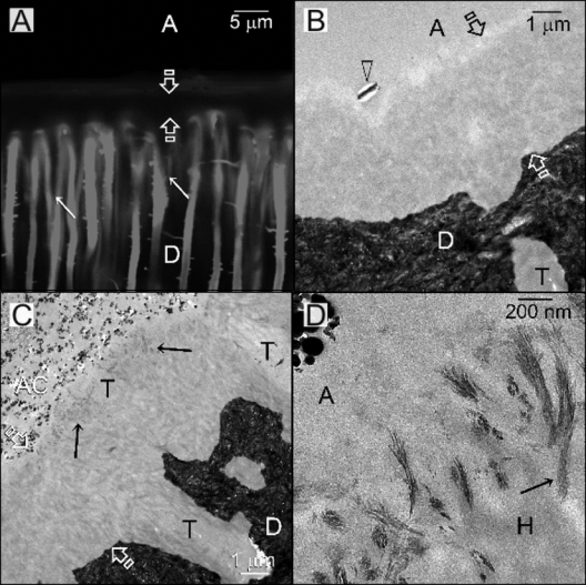
Specimens bonded with the ethanol-wet-bonding technique [minimal water content]. A, adhesive; between open arrows, hybrid layer; D, mineralized dentin base. (A) CLSM image from a specimen that had been immersed for 4 mos in the remineralization medium that was devoid of a Portland cement block (i.e., no remineralization). Fluorescence was absent from the entire hybrid layer. Resin tags (arrows) appeared to be longer than in the other groups. (B) TEM image of a specimen retrieved after 4 mos of biomimetic remineralization in which there was no remineralization of the hybrid layer. A large electron-dense crystal could be seen within a void (open arrowhead) along the surface of the hybrid layer. [Note: Absence of remineralization within the hybrid layer could be seen in 10 out of 16 specimens over the entire four-month period.] (C) TEM image of a specimen retrieved after 4 mos of biomimetic remineralization in which there was minimal remineralization of the hybrid layer along the dentin surface (arrows) in regions that were adjacent to a dentinal tubule (T). AC, composite that was blended with the oxygen inhibition layer of the adhesive. [Note: This feature was observed in 6 out of the 16 specimens over the entire four-month period.] (D) High-magnification TEM image showing exclusive intrafibrillar remineralization of the surface collagen fibrils depicted in (C). Electron-dense nanocrystallites could be observed within the remineralized fibrils (arrow).
Intense fluorescence throughout the hybrid layers could be detected from specimens prepared with water-wet-bonding (Fig. 2A). Remineralization of the hybrid layers was apparent beyond 10,000X magnification at 2 mos, but could be clearly discerned at 3000X after 3 mos. Remineralization was not uniform, being more extensive along the surface of the hybrid layer and around the periphery of the dentinal tubules (Fig. 2B). Both intrafibrillar and interfibrillar remineralization could be identified (Fig. 2C), with the mineral phase consistent with the electron diffraction patterns of poorly crystalline apatite (Fig. 2D).
Figure 2.
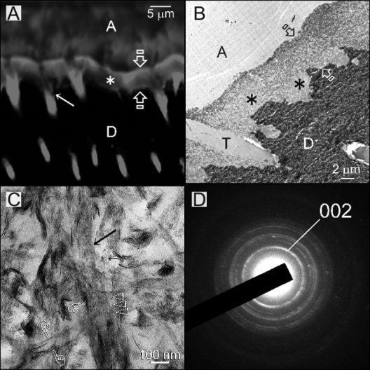
Specimens bonded by the water-wet-bonding technique [manufacturer’s recommended optimal water content]. A, adhesive; between open arrows, hybrid layer; D, mineralized dentin base. (A) CLSM image from a specimen that had been immersed for 4 mos in the remineralization medium that was devoid of a Portland cement block. Intense fluorescence present within the hybrid layer and dentinal tubules indicates that the polymerized adhesive was permeable to the water-soluble fluorescent dye after water sorption. (B) TEM image of a specimen retrieved after 4 mos of biomimetic remineralization, showing non-uniform remineralization of the hybrid layer. Regions that were more heavily remineralized are indicated by asterisks corresponding to the dark regions depicted in the CLSM image in (A). T, dentinal tubule. (C) High-magnification TEM image of (B) showing both intrafibrillar remineralization (arrow) and extrafibrillar remineralization (pointers) within the hybrid layer. Mineral nanocrystals were arranged in an ordered manner within the collagen fibril, giving a vague, banded appearance (open arrowheads). (D) Selected-area electron diffraction of those nanocrystals depicted in (C) yielded ring patterns that were consistent with the characteristics of poorly crystalline apatite.
Hybrid layers from the water-overwet bonding group also exhibited intense fluorescence. Vertically oriented water channels extended from the resin-dentin interface into the overlying adhesive (Fig. 3A). Fifty percent of the specimens examined at 3-4 mos after remineralization demonstrated filling of those vertical water channels with mineral plates (Fig. 3B). The rest of the specimens demonstrated remineralization of only the hybrid layers (not shown). Apart from partial remineralization of the collagen matrix (Fig. 3C), the smaller water channels within the hybrid layer (i.e., terminal branches of dentinal tubules that were continuous with the interfibrillar spaces) were filled with larger hexagonal mineral platelets (Fig. 3C), similar to those apatite crystals identified from the cap regions of the mineral-filled water channels (Fig. 3D).
Figure 3.
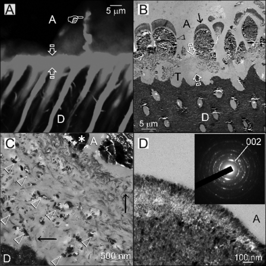
Specimens bonded with water-overwet-bonding [excessive water content]. A, adhesive; between open arrows, hybrid layer; D, mineralized dentin base. (A) CLSM image from a specimen that had been immersed for 4 mos in the remineralization medium that was devoid of a Portland cement block. Intense fluorescence could be observed within the entire hybrid layer, the dentinal tubules beneath the hybrid layer, and the vertically oriented water channel (pointer). (B) TEM image of a specimen retrieved after 4 mos of biomimetic remineralization. Numerous mineral-filled water channels could be seen on top of the partially remineralized hybrid layer. Each water channel consisted of a basal region that was filled with large mineral plates (open arrowhead) and a cap region (arrow) that was filled with very fine nanocrystals. (C) High-magnification TEM image of a similar remineralized hybrid layer in this group. Two modes of mineralization could be identified within the hybrid layer: (a) remineralization of the collagen fibrils (arrows) with nanocrystals similar to those depicted in Figs. 1D and 2C [This mode of remineralization occurred predominantly along the top of the hybrid layer surface, but could also be seen, albeit sparsely, from the base of the hybrid layer.]; and (b) the presence of electron-dense crystals in channels (open arrowheads) that were oriented roughly parallel to the mineralized dentin base. The large basal regions of the vertically oriented water channels above the hybrid layer were densely filled with much larger mineral plates (asterisk). (D) High-magnification TEM image of the cap region of a mineral-filled water channel revealed a dense conglomerate of 5-20 nm hexagonal nanocrystals within the cap region. Selected-area electron diffraction of those nanocrystals yielded spotted diffraction rings that were characteristic of apatites with a higher degree of crystallinity (inset).
Discussion
Water plays a pivotal role in maintaining the structural integrity of collagen molecules (Cameron et al., 2007) and mechanical properties of collagen fibrils (Zhang et al., 2007). Collagen molecules are surrounded by a highly ordered inner layer of tightly bound structural water and water bridges created by hydrogen bonding (Ramachandran and Chandrasekharan, 1968; Bella et al., 1995). It is unlikely that rinsing a demineralized collagen matrix with ethanol for 1 min will remove this innermost layer of structural water from the collagen fibrils, which in turn results in irreversible disruption of the triple-helical structure. (Magne et al., 2001).
In water-wet-bonding, the collagen matrix is suspended in water to prevent direct contact of the collagen fibrils via the development of interpeptide hydrogen bonds (Pashley et al., 2007). Conversely, suspension of the collagen matrix in a liquid chemical dehydrant (Nalla et al., 2006) removes bulk water from the dentinal tubule anastomosis and the loosely bound water layers from the intrafibrillar compartments and results in increased intramolecular hydrogen bonding among the collagen molecules (Mogilner et al., 2002). Intramolecular hydrogen bonds cause shrinkage of the fibrils by reduction in the lateral spacing of the collagen molecules (Miles and Ghelashvili, 1999). There was no biomimetic remineralization of the hybrid layers when ethanol wet-bonding was meticulously performed. However, in the absence of tubular occlusion (Sadek et al., 2007), ethanol-wet-bonding replaced water from the interfibrillar compartment effectively, but incompletely removed the loosely bound water from the intrafibrillar compartment of collagen fibrils around the tubular orifices. The exclusive intrafibrillar remineralization identified from those water-contaminated ethanol-wet-bonded specimens indirectly showed that low-molecular-weight ethanol-solvated resin monomers could infiltrate the intrafibrillar compartment of a dehydrated collagen network (Tay et al., 2007), despite the presence of a size exclusion effect (Toroian et al., 2007). By replacing most of the water from the collagen matrix, the ethanol-wet-bonding technique has the potential to create durable resin-dentin bonds (Hosaka et al., 2009).
The concept that biomimetic remineralization of hybrid layers can occur only in the presence of incomplete water replacement by adhesive resins was further fortified by the fairly extensive intrafibrillar and interfibrillar remineralization in the water-wet-bonding group. In our previous studies, we were surprised by the extent of intrafibrillar remineralization observed in hybrid layers created by both etch-and-rinse and self-etching adhesives (Mai et al., 2009; Tay and Pashley, 2009). Results from the ethanol-wet-bonding group in the present study helped to clarify our concern and implied that contemporary adhesives, when used according to the manufacturers’ instructions, are incapable of displacing water from the intrafibrillar compartment of the collagen matrix. Incomplete water replacement from the interfibrillar compartment, dilution of water-soluble monomers with water, or subsequent water sorption during aging could have additionally accounted for the susceptibility of hybrid layers to interfibrillar remineralization.
The inclusion of a water-overwet-bonding group, although far removed from clinical practice, broadens our understanding of the biomimetic remineralization scheme. Identification of large mineral plates in the basal region of the filled water channels signifies the absence of an appropriate scaffold for the deposition of nanocrystals. Conversely, the conglomerates of apatite nanocrystals within the cap region of the water channel probably represent hierarchical three-dimensional precipitation of the mineral phase within a water-entrapped polymer hydrogel matrix (Liu et al., 2009). The close adaptation of the large mineral plates to the conformation of the basal region of the water channel provides support for the involvement of a liquid amorphous precursor pathway as a biomineralization strategy (Gower, 2008). The fragmented plates are likely to be artifacts created during ultramicrotomy of a single brittle mineral sheet.
Identification of mineral platelets within the horizontally oriented water channels of the hybrid layer suggests that these channels were intricately connected to the vertical water channels on the hybrid layer surface, as confirmed in previous nanoleakage studies (Tay et al., 2005a,b). Mineral-filled water channels had never been identified within hybrid layers created with etch-and-rinse adhesives by the conventional water-wet-bonding technique. These channels represent the terminal branches of the dentinal tubule anastomosis and are continuous with the interfibrillar compartments of the collagen matrix. They were also identified when a non-resin- containing glass-ionomer cement was placed over moist acid-etched dentin (Kim et al., 2010). During adhesive application without perfusion, and with blot-drying, these terminal branches collapse due to shrinkage of the collagen matrix that is induced by the polar adhesive solvents with chemical dehydration potential (Nakajima et al., 2002; Nalla et al., 2006; Becker et al., 2007). During water-overwet-bonding, continuous replenishment of water via perfusion and an excessive amount of water enables these terminal branches to remain fully extended, even in the presence of the dehydrating adhesive solvents. On polymerization of the adhesive, these fully extended channels were stabilized in their fully extended conditions. Lacking an appropriate scaffold, only larger mineral platelets could be formed within these water channels.
The null hypothesis—that there is no difference in the status of in vitro biomimetic remineralization in hybrid layers created by a two-step etch-and-rinse adhesive, regardless of the moisture condition under which they are created—is rejected. Biomimetic remineralization of the hybrid layer and the ethanol wet-bonding technique achieved similar results, in that both led to less-porous hybrid layers by the elimination of denuded collagen matrices with progressive dehydration and water replacement (Fernández-Seara et al., 2004). Collectively, they represent an alternative philosophy to the use of matrix metalloproteinase inhibitors to prevent collagen degradation and may be combined with the latter to extend the longevity of resin-dentin bonds.
Acknowledgments
We thank Bob Smith for TEM technical assistance, Thomas Bryan for resin embedding, and Michelle Barnes for secretarial support.
Footnotes
This study was supported by Grant R21 DE019213-01 from the National Institute of Dental and Craniofacial Research (PI. Franklin R. Tay). The dentin adhesive used in this study was a generous gift from Bisco Inc.
References
- Becker TD, Agee KA, Joyce AP, Rueggeberg FA, Borke JL, Waller JL, et al. (2007). Infiltration/evaporation-induced shrinkage of demineralized dentin by solvated model adhesives. J Biomed Mater Res B Appl Biomater 80:156-165 [DOI] [PubMed] [Google Scholar]
- Bella J, Brodsky B, Berman HM. (1995). Hydration structure of a collagen peptide. Structure 3:893-906 [DOI] [PubMed] [Google Scholar]
- Bonar LC, Lees S, Mook HA. (1985). Neutron diffraction studies of collagen in fully mineralized bone. J Mol Biol 181:265-270 [DOI] [PubMed] [Google Scholar]
- Bowman SM, Gibson LJ, Hayes WC, McMahon TA. (1999). Results from demineralized bone creep tests suggest that collagen is responsible for the creep behavior of bone. J Biomech Eng 121:253-258 [DOI] [PubMed] [Google Scholar]
- Cameron IL, Short NJ, Fullerton GD. (2007). Verification of simple hydration/dehydration methods to characterize multiple water compartments on tendon type 1 collagen. Cell Biol Int 31:531-539 [DOI] [PubMed] [Google Scholar]
- Carrilho MR, Geraldeli S, Tay F, de Goes MF, Carvalho RM, Tjäderhane L, et al. (2007). In vivo preservation of the hybrid layer by chlorhexidine. J Dent Res 86:529-533 [DOI] [PubMed] [Google Scholar]
- Chesnick IE, Mason JT, Giuseppetti AA, Eidelman N, Potter K. (2008). Magnetic resonance microscopy of collagen mineralization. Biophys J 95:2017-2026 [DOI] [PMC free article] [PubMed] [Google Scholar]
- Fernández-Seara MA, Wehrli SL, Takahashi M, Wehrli FW. (2004). Water content measured by proton-deuteron exchange NMR predicts bone mineral density and mechanical properties. J Bone Miner Res 19:289-296 [DOI] [PubMed] [Google Scholar]
- Gower LB. (2008). Biomimetic model systems for investigating the amorphous precursor pathway and its role in biomineralization. Chem Rev 11:4551-4627 [DOI] [PMC free article] [PubMed] [Google Scholar]
- Hosaka K, Nishitani Y, Tagami J, Yoshiyama M, Brackett WW, Agee KA, et al. (2009). Durability of resin-dentin bonds to water- vs. ethanol-saturated dentin. J Dent Res 88:146-151 [DOI] [PMC free article] [PubMed] [Google Scholar]
- Ito S, Saito T, Tay FR, Carvalho RM, Yoshiyama M, Pashley DH. (2005). Water content and apparent stiffness of non-caries versus caries-affected human dentin. J Biomed Mater Res B Appl Biomater 72:109-116 [DOI] [PubMed] [Google Scholar]
- Kim J, Vaughn RM, Gu L, Rickman RA, Arola DD, Schafer TE, et al. (2009). Imperfect hybrid layers created by an aggressive one-step self-etch adhesive in primary dentin are amenable to biomimetic remineralization in vitro. J Biomed Mater Res A [Epub ahead of print: Sept 18, 2009]. (in press) [DOI] [PMC free article] [PubMed] [Google Scholar]
- Kim YK, Yiu CKY, Kim J, Gu L, Kim SK, Weller NR, et al. (2010). Failure of a glass ionomer to remineralize apatite-depleted dentin. J Dent Res. E-pub ahead of print, doi 10.1177/0022034510363665 [DOI] [PMC free article] [PubMed] [Google Scholar]
- Liu G, Zhao D, Tomsia AP, Minor AM, Song X, Saiz E. (2009). Three-dimensional biomimetic mineralization of dense hydrogel templates. J Am Chem Soc 131:9937-9939 [DOI] [PubMed] [Google Scholar]
- Magne D, Weiss P, Bouler JM, Laboux O, Daculsi G. (2001). Study of the maturation of the organic (type I collagen) and mineral (nonstoichiometric apatite) constituents of a calcified tissue (dentin) as a function of location: a Fourier transform infrared microspectroscopic investigation. J Bone Miner Res 16:750-757 [DOI] [PubMed] [Google Scholar]
- Mai S, Kim YK, Toledano M, Breschi L, Ling JQ, Pashley DH, et al. (2009). Phosphoric acid esters cannot replace polyvinylphosphonic acid as phosphoprotein analogs in biomimetic remineralization of resin-bonded dentin. Dent Mater 25:1230-1239 [DOI] [PMC free article] [PubMed] [Google Scholar]
- Meyer JL, Eanes ED. (1978). A thermodynamic analysis of the amorphous to crystalline calcium phosphate transformation. Caclif Tissue Res 25:59-68 [DOI] [PubMed] [Google Scholar]
- Miles CA, Ghelashvili M. (1999). Polymer-in-a-box mechanism for the thermal stabilization of collagen molecules in fibers. Biophys J 76:3243-3252 [DOI] [PMC free article] [PubMed] [Google Scholar]
- Mogilner IG, Ruderman G, Grigera JR. (2002). Collagen stability, hydration and native state. J Mol Graph Model 21:209-213 [DOI] [PubMed] [Google Scholar]
- Nakajima M, Okuda M, Pereira PN, Tagami J, Pashley DH. (2002). Dimensional changes and ultimate tensile strengths of wet decalcified dentin applied with one-bottle adhesives. Dent Mater 18:603-608 [DOI] [PubMed] [Google Scholar]
- Nalla RK, Kinney JH, Tomsia AP, Ritchie RO. (2006). Role of alcohol in the fracture resistance of teeth. J Dent Res 85:1022-1026 [DOI] [PMC free article] [PubMed] [Google Scholar]
- Nishitani Y, Yoshiyama M, Donnelly AM, Agee KA, Sword J, Tay FR, et al. (2006). Effects of resin hydrophilicity on dentin bond strength. J Dent Res 85:1016-1021 [DOI] [PMC free article] [PubMed] [Google Scholar]
- Pashley DH, Nakabayashi N. (1998). Hybridization of dental hard tissues. Tokyo: Quintessence Publishing Co. Ltd [Google Scholar]
- Pashley DH, Agee KA, Wataha JC, Rueggeberg F, Ceballos L, Itou K, et al. (2003). Viscoelastic properties of demineralized dentin matrix. Dent Mater 19:700-706 [DOI] [PubMed] [Google Scholar]
- Pashley DH, Tay FR, Yiu C, Hashimoto M, Breschi L, Carvalho RM, et al. (2004). Collagen degradation by host-derived enzymes during aging. J Dent Res 83:216-221 [DOI] [PubMed] [Google Scholar]
- Pashley DH, Tay FR, Carvalho RM, Rueggeberg FA, Agee KA, Carrilho M, et al. (2007). From dry bonding to water-wet bonding to ethanol-wet bonding. A review of the interactions between dentin matrix and solvated resins using a macromodel of the hybrid layer. Am J Dent 20:7-20 [PubMed] [Google Scholar]
- Ramachandran GN, Chandrasekharan R. (1968). Interchain hydrogen bonds via bound water molecules in the collagen triple helix. Biopolymers 6:1649-1658 [DOI] [PubMed] [Google Scholar]
- Sadek FT, Pashley DH, Ferrari M, Tay FR. (2007). Tubular occlusion optimizes bonding of hydrophobic resins to dentin. J Dent Res 86:524-528 [DOI] [PubMed] [Google Scholar]
- Tay FR, Pashley DH. (2009). Biomimetic remineralization of resin-bonded acid-etched dentin. J Dent Res 88:719-724 [DOI] [PMC free article] [PubMed] [Google Scholar]
- Tay FR, Pashley DH, Hiraishi N, Imazato S, Rueggeberg FA, Salz U, et al. (2005a). Tubular occlusion prevents water-treeing and through-and-through fluid movement in a single-bottle, one-step self-etch adhesive model. J Dent Res 84:891-896 [DOI] [PubMed] [Google Scholar]
- Tay FR, Pashley DH, Suh BI, Hiraishi N, Yiu CK. (2005b). Water treeing in simplified dentin adhesives—déjà vu? Oper Dent 30:561-579 [PubMed] [Google Scholar]
- Tay FR, Pashley DH, Kapur RR, Carrilho MR, Hur YB, Garrett LV, et al. (2007). Bonding BisGMA to dentin—a proof of concept for hydrophobic dentin bonding. J Dent Res 86:1034-1039 [DOI] [PubMed] [Google Scholar]
- Toroian D, Lim JE, Price PA. (2007). The size exclusion characteristics of type I collagen: implications for the role of noncollagenous bone constituents in mineralization. J Biol Chem 282:22437-22447 [DOI] [PubMed] [Google Scholar]
- Traore A, Foucat L, Renou JP. (2000). 1H-NMR study of water dynamics in hydrated collagen: transverse relaxation-time and diffusion analysis. Biopolymers 53:476-483 [DOI] [PubMed] [Google Scholar]
- Vaidyanathan TK, Vaidyanathan J. (2009). Recent advances in the theory and mechanism of adhesive resin bonding to dentin: a critical review. J Biomed Mater Res B Appl Biomater 88:558-578 [DOI] [PubMed] [Google Scholar]
- Wehrli FW, Fernández-Seara MA. (2005). Nuclear magnetic resonance studies of bone water. Ann Biomed Eng 33:79-86 [DOI] [PubMed] [Google Scholar]
- Zhang D, Chippada U, Jordan K. (2007). Effect of the structural water on the mechanical properties of collagen-like microfibrils: a molecular dynamics study. Ann Biomed Eng 35:1216-1230 [DOI] [PubMed] [Google Scholar]