Educational Objectives
After completing this program, participants should be able to:
▪ Discuss the role of epidermal growth factor receptor (EGFR)-targeted agents as therapy for patients with solid tumors.
▪ Identify the efficacy of EGFR-targeted agents in a variety of solid tumors, including colorectal, non–small-cell lung cancer, and pancreatic cancer.
▪ Recognize points of consideration for administration of EGFR inhibitors, including pharmacokinetic data and the significance of KRAS mutation testing.
▪ Describe the significance of anti-EGFR side effects on patients’ quality of life, optimizing adherence, and improving patients’ outcomes.
▪ Evaluate current strategies for preventing and managing side effects of EGFR-targeted therapies to ensure optimal patient care.
Accreditation Statement
Medical Education Resources (MER) is accredited by the Accreditation Council for Pharmacy Education (ACPE) as a provider of Continuing Pharmacy Education (CPE). MER designates this continuing education activity for 1.5 contact hours (0.15 CEUs) of the ACPE.
Universal Program No.: 816-999-10-005-H01-P
Abstract
Traditionally, patients receiving cancer treatment contend with the potentially life-threatening side effects of cytotoxic chemotherapy. The recent emergence of newer cancer therapies, such as the epidermal growth factor receptor (EGFR) inhibitors, present new management challenges for oncology pharmacists, nurses, and physicians. Even though EGFR inhibitors are generally considered to be “well tolerated,” this does not mean that they are devoid of side effects. Prior to the initiation of anti-EGFR therapy, it is imperative that patients be able to recognize the early signs of toxicity and seek prompt intervention to minimize such reactions. Patients should also understand that side-effect management may improve compliance with therapy and can lead to better outcomes. Importantly, pharmacists play a central role in such patient education.
Introduction
Conventional cytotoxic chemotherapy is an effective mode of therapy for the treatment of cancer. However, patients undergoing cytotoxic chemotherapy can experience life-threatening side effects, and the continued use of these agents is often limited by these toxicities. Furthermore, development of resistance may limit their effectiveness.1 In recent years, the advancement of molecular biology has led to the development of therapies that specifically target tumor cells, thus minimizing damage to normal tissues (Table 1). With molecularly targeted therapies, therapeutic agents are designed to influence the individual genetic and molecular signature of tumor cells. Accordingly, diagnosis, treatment, and monitoring can be tailored to meet the specific needs of each patient.
Table 1.
Comparison of Conventional Chemotherapy versus Molecular Therapies
| Conventional Chemotherapy | Molecular Targeted Treatment | |
|---|---|---|
| Mechanism | Forms covalent bonds or competes for metabolites normally incorporated in DNA/RNA; targets micro-tubules, resulting in impaired meiosis | Targets aberrant cell signaling pathways unique to cancer |
| Activity | Cytotoxic | Cytostatic/cytotoxic |
| Specificity | Low; generally targets rapidly proliferating cells | Is more specific to tumors |
Adapted from Harari PM, Huang SM. Epidermal growth factor receptor modulation of radiation response: preclinical and clinical development. Semin Radiat Oncol 2002;12(3 Suppl 2):21–26. Reproduced with permission of Elsevier.1
Tyrosine kinases are a family of proteins that play an important role in the normal regulation of many cellular processes. They are critical in capturing and transducing extracellular signals carried by peptide-based ligands, or growth factors.2 In their normal state, they regulate typical cellular processes associated with the cell cycle, including cellular proliferation and differentiation. However, when abnormalities in their expression occur, they can cause cells to divide uncontrollably and can contribute to the development of cancer.2
At present, there are approximately 60 known and characterized tyrosine kinase receptors that are divided into more than 20 different subfamilies based on similar characteristics, common ligands, or both.3 Recent research has focused on developing agents that can modify or inhibit these receptors.2
Epidermal Growth Factor Receptors
The epidermal growth factor receptor (EGFR) is a current promising molecular target for cancer therapeutics.1 EGFR is a tyrosine kinase receptor from a larger family of ErbB receptors that mediate cell survival, proliferation, invasion, and angiogenesis.6 Investigations in this area of cancer research have indicated that the ErbB subclass of tyrosine kinase receptors is abnormal in some cancers.2
Currently, there are four members of the ErbB subclass: Erb-B1 (or EGFR), Erb-B2 (or HER-2/neu), Erb-B3, and Erb-B4.
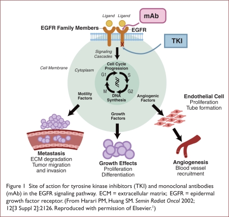
EGFR is a membrane-bound protein that is involved in signal transduction pathways, and it is critical in the regulation of cellular proliferation and survival. Although EGFR is expressed in many different cell types in normal tissue, EGFR over-expression and dysregulation can occur in neoplastic tissue (Table 2).4,5 The activation of tumor cell EGFR can trigger a series of intracellular events: cellular proliferation, the blocking of apoptosis, invasion and metastasis, and the commencement of tumor-induced neovascularization, all of which result in carcinogenesis (Figure 1).1,4
Table 2.
Epidermal Growth Factor Receptor (EGFR) Expression and Correlation With Poor Prognosis in Solid Tumors
| Tumor Type | EGFR Expression (%) |
|---|---|
| Squamous cell cancer of the head and neck | 95 |
| Prostate | 41–100 |
| Non–small-cell lung cancer | 40–80 |
| Glioblastoma | 40–60 |
| Esophageal | 35–88 |
| Ovarian | 35–70 |
| Gastric | 33–74 |
| Bladder | 31–72 |
| Pancreatic | 30–50 |
| Colorectal | 25–77 |
| Breast | 14–91 |
Adapted from Arteaga C. Semin Oncol 2003;30(Suppl 7):3–14, Elsevier.5
Figure 1.
Site of action for tyrosine kinase inhibitors (TKI) and monoclonal antibodies (mAb) in the EGFR signaling pathway. ECM = extracellular matrix; EGFR = epidermal growth factor receptor. (From Harari PM, Huang SM. Semin Radiat Oncol 2002; 12[3 Suppl 2]:2126. Reproduced with permission of Elsevier.1)
Anti-EGFR Strategies
There are various mechanisms by which the EGFR can be blocked. The major classes of currently available EGFR inhibitors include monoclonal antibodies (mAbs) and small-molecular-weight tyrosine kinase inhibitors (TKIs).6
EGFR Monoclonal Antibodies
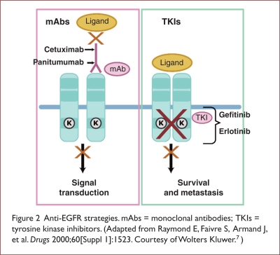
EGFR mAbs are large molecules that do not readily traverse the epithelial basement membrane. They are administered intravenously.1 Anti-EGFR mAbs bind extracellularly to the ligand-binding domain of the EGFR and prevent binding of the endogenous ligands, EGF and transforming growth factor–alpha (TGF-α). Extracellular blockade prevents receptor dimerization and autophosphorylation from occurring, thus inhibiting activation of the tyrosine kinase signaling pathways (Figure 2).7,8
Figure 2.
Anti-EGFR strategies. mAbs = monoclonal antibodies; TKIs = tyrosine kinase inhibitors. (Adapted from Raymond E, Faivre S, Armand J, et al. Drugs 2000;60[Suppl 1]:1523. Courtesy of Wolters Kluwer.7)
Cetuximab. Cetuximab (Erbitux, ImClone/Bristol-Myers Squibb) is a mouse-human chimeric EGFR mAb that was first approved by the FDA as either monotherapy or in combination with irinotecan (Camptosar, Pfizer) for the treatment of metastatic colorectal cancer (mCRC). The FDA granted cetuximab accelerated approval after the landmark Bowel Oncology with Cetuximab Antibody (BOND-1) trial showed objective response rates in 22.9% of patients who received cetuximab plus irinotecan and in 10.8% of patients with irinotecan-refractory mCRC treated with cetuximab monotherapy (P = 0.007).9 Cetuximab is also available for patients with advanced or metastatic/recurrent squamous cell carcinoma of the head and neck (SCCHN).6 This agent is indicated in combination with radiation therapy for the initial treatment of locally or regionally advanced SCCHN and as single-agent therapy for patients with recurrent or metastatic SCCHN for whom prior platinum-based therapy has failed.
In a randomized, multicenter, controlled trial of 424 patients with locally or regionally advanced SCCHN, Binner et al. administered cetuximab plus radiation therapy or radiation therapy alone.10 The median overall survival rates were 49 months for patients receiving cetuximab plus radiation and 29.3 months for those receiving radiation alone (P = 0.03).
In a single-arm, multicenter clinical trial conducted by Vermorken et al., the objective response rate for 103 patients with recurrent or metastatic SCCHN was 13% for those receiving single-agent cetuximab, and the median duration of response was determined to be 5.8 months (range, 1.2–5.8 months).11
Panitumumab. Another fully humanized EGFR mAb is panitumumab (Vectibix, Amgen/Abgenix). The evidence is still anecdotal, but it has been proposed that its properties may reduce the risk of infusion reactions and may decrease the likelihood of generating antibodies against the therapeutic antibody.12 Panitumumab is currently indicated for patients with EGFR-expressing mCRC that is refractory to chemotherapy.6
The approval of panitumumab was based on the findings of an open-label, multinational, randomized phase 3 clinical trial of 463 patients with EGFR-expressing colorectal cancer (CRC) who did not respond to treatment with a fluoropyrimidine, oxaliplatin (Eloxatin, Sanofi-Synthelabo), or irinotecan.13 In this trial, a significant increase in progression-free survival was observed in patients receiving panitumumab and best supportive care (96 days) compared with patients receiving best supportive care alone (60 days) (P < 0.0001).
Tyrosine Kinase Inhibitors
The small-molecule EGFR TKIs such as erlotinib (Tarceva, OSI/Genentech), gefitinib (Iressa, AstraZeneca), and lapatinib (Tykerb, Glaxo-SmithKline) are administered orally. EGFR TKIs act directly on the cytoplasmic domain of the EGFR, inhibiting the activity of the EGFR pathway.8
Gefitinib. Gefitinib inhibits EGFR tyrosine kinase by binding to the adenosine triphosphate (ATP)–binding site of the enzyme, thus functioning by deactivating the Ras signal transduction cascade and inhibiting malignant cells. Gefitinib was granted FDA approval in the U.S. in 2003 with an indication as third-line therapy for patients with non–small-cell lung cancer (NSCLC)—a setting without any other approved therapy—following the results of two phase 2 studies—Iressa Dose Evaluation in Advanced Lung Cancer (IDEAL-1 and IDEAL-2).14,15 However, upon extensive review of these data as well as subsequent studies, it was determined that the survival benefit was not significant in the overall populations studied. Interestingly, in a subsequent review of numerous clinical trials, a consistent clinical benefit was identified in patients of Asian ethnicity.16–18 Accordingly, in 2005, the FDA revised the labeling of gefitinib to be prescribed only for patients who had previously taken the drug and were benefiting from its use.2
Erlotinib. Like gefitinib, erlotinib specifically targets the EGFR by reversibly binding to the ATP binding site of this receptor.7 For the signal to be transmitted, two members of the EGFR family must come together to form a homodimer. Using the ATP molecule to undergo autophosphorylation with each other results in a conformational change in their intracellular structure; this further exposes a binding site for proteins that cause a signal cascade to the nucleus. If ATP binding is blocked, autophosphorylation is not possible and the signal is stopped.7
Erlotinib is approved in the U.S. as a second-line therapy in patients with locally advanced or metastatic NSCLC and as a first-line therapy when used along with gemcitabine in patients with locally advanced or metastatic pancreatic cancer.19
Lapatinib. Some malignancies, such as breast cancer, are associated with the overexpression of human EGFR type 2 (HER-2). Stimulation of HER-2 is associated with cell proliferation and with multiple processes involved in tumor progression and metastases.20
Lapatinib, another TKI, is associated with both the EGFR and HER-2/neu oncogenes and inhibits receptor signal processes by binding to the ATP-binding pocket of the EGFR/HER-2 protein kinase domain, thereby preventing autophosphorylation and subsequent activation of the signal mechanism.21 Following a randomized clinical trial of women with breast cancer that progressed after previous treatment with anthracyclines, taxanes, or trastuzumab (Herceptin, Genentech), the FDA approved the use of lapatinib in conjunction with capecitabine (Xeloda, Roche) when it was observed that the combination demonstrated an improved time to progression and delayed the time to further cancer growth compared with a regimen that used capecitabine alone.22
Application of EGFR Inhibitor Therapy: Practical Considerations
The clinical benefit associated with EGFR inhibitors can vary within patient populations and among different tumor types.23 Obviously, the goals of therapy with EGFR inhibitors are to optimize the clinical response for each patient and to avoid treating patients who are unlikely to derive any clinical benefit from these agents.
Skin Rash as Clinical Marker of Activity
The clinical sensitivity of a patient receiving treatment with EGFR inhibitors has been associated with the development of a number of dermatologic toxicities, including skin rash.24 Generally described as an acneiform rash because of its inflammatory follicular appearance, this frequently occurring skin toxicity mainly affects the face, scalp, neck, upper chest, and back.6 Interestingly, compared with the other dermatologic toxicities, skin rash may be a surrogate marker of clinical activity.
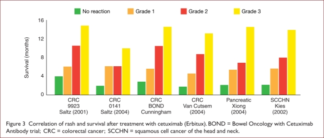
In several clinical studies, the development of a papulopustular rash was associated with improved responses to treatment and a trend toward improved survival (Figure 3).9,25–29 In a study published by Lievre and colleagues in 2008, the severity of skin toxicity correlated with improved survival.30 The reported median overall survival rates were 13.9 months for patients experiencing a grade 2 or 3 skin toxicity and 8.2 months for those experiencing a grade 0 or 1 skin toxicity (P = 0.029). Furthermore, progression-free survival rates were 28.8 weeks (grade 2 or 3) and 12 weeks (grade 0 or 1) (P = 0.146).
Figure 3.
Correlation of rash and survival after treatment with cetuximab (Erbitux). BOND = Bowel Oncology with Cetuximab Antibody trial; CRC = colorectal cancer; SCCHN = squamous cell cancer of the head and neck.
In another analysis published in 2009, the data from two large phase 3 studies (BR.21 and PA.3) were examined to characterize the correlation between the occurrence of rash during treatment with erlotinib and improved clinical outcomes.31 In this investigation, the presence of rash strongly correlated with overall survival in both trials. Data from BR.21 demonstrated an increased correlation with the grade of rash severity (grade 1 vs. no rash (hazard ratio [HR]), 0.41; P < 0.001) and grade 2 rash or higher vs. no rash (HR, 0.29; P < 0.001). Similar results were also observed for progression-free survival.
In the analysis of PA.3, grade 2 rash or greater strongly correlated with an improvement in overall survival (grade 2 rash or more vs. no rash [HR, 0.47]; P < 0.001).31 Similarly, grade 2 rash or greater was associated with improved progression-free survival and disease control. However, not every patient who develops a rash subsequently responds to treatment.32
KRAS Mutations and Clinical Efficacy of EGFR Inhibitors
The Raf–Ras–mitogen-activated protein (Ras–Raf–MAP) kinase pathway is activated in the EGFR cascade. Ras proteins (G proteins) cycle between active Ras–guanosine triphosphate (Ras–GTP) and inactive Ras–guanosine diphosphate (Ras–GDP) forms in response to stimulation from a cell–surface receptor EGFR and downstream signaling pathways important in cancer cell proliferation, invasion, metastasis, and stimulation of neovascularization. The KRAS gene (V-Ki-ras2 Kirsten rat sarcoma) of the Ras–Raf–MAP kinase pathway can harbor oncogenic mutations that result in a constitutively activated protein, independent of EGFR ligand binding, rendering EGFR-targeted therapeutics upstream of the EGFR ineffective.33
Mutations of the KRAS gene can be a significant predictor of resistance to anti-EGFR mAbs. Curiously, KRAS mutations are common and found in approximately 40% to 50% of CRC tumors.34 Several concordant clinical studies conducted in patients with CRC have shown that the presence of KRAS mutation can affect survival in patients receiving cetuximab or panitumumab (Table 3).
Table 3.
Retrospective Studies Supporting KRAS as a Biomarker for Epidermal Growth Factor Receptor Inhibitors (Panitumumab and Cetuximab)
| Treatment | No. of Patients (%) | Objective Response No. (%) | ||
|---|---|---|---|---|
| Mutant | Wild-Type | (Mutant) | ||
| Lievre et al.30 | Cetuximab ± chemo | 114 (32) | 34 (44) | 0 (0) |
| Benvenuti et al.35 | Panitumumab or cetuximab or cetuximab + chemo | 48 (33) | 10 (31) | 1 (6) |
| De Roock et al.36 | Cetuximab or cetuximab + irinotecan | 113 (41) | 27 (41) | 0 (0) |
| Capuzzo et al.37 | Cetuximab ± chemo | 81 (40) | 13 (26) | 2 (6) |
| DiFiore et al.38 | Cetuximab + chemo | 59 (27) | 12 (28) | 0 (0) |
| Khambata-For, et al.39 | Cetuximab | 80(38) | 5 (10) | 0 (0) |
| Amado et al.40 | Panitumumab | 208 (40) | 21 (17) | 0 (0) |
chemo = chemotherapy.
Lievre et al. reported that overall survival for patients with CRC who received cetuximab and who had a mutated form of KRAS was 10.1 months; for patients without KRAS mutations, the rate was 14.3 months (P = 0.0017).30 Progression-free survival rates were determined to be 9 weeks for patients with the mutation and 32 weeks for those without it (P = 0.0000001).
Amado and colleagues investigated the effect of KRAS mutations on overall survival in CRC patients with wild-type KRAS who were receiving best supportive care compared with panitumumab.40 Patients experienced overall survival at rates of 7.6 months with best supportive care and 8.1 months with panitumumab. However, panitumumab patients with mutated KRAS experienced an overall survival rate of 4.9 months, compared with 4.4 months for patients with mutated KRAS who received best supportive care.40
Not surprisingly, mutations in the EGFR tyrosine kinase domain have also been strongly associated with greater sensitivity of NSCLC to EGFR TKIs. Accordingly, screening patients for KRAS mutations before therapy is selected may be clinically beneficial, especially for those with CRC, because the cost and toxicity of ineffective therapy can be avoided. However, although current findings support avoiding EGFR inhibitors in patients with KRAS mutations, the data are not as clear regarding the use of EGFR inhibitors in patients with wild-type KRAS.41
Pharmacokinetics
Small-molecule EGFR TKIs are administered orally, whereas EGFR mAbs are administered via intravenous (IV) infusion. Therefore, some important pharmacokinetic distinctions should be mentioned.
Cetuximab. As demonstrated by a pharmacokinetic analysis, female patients with CRC had a 25% lower intrinsic clearance of cetuximab compared with their male counterparts.42 Smaller sex differences in cetuximab clearance were also observed in patients with SCCHN. Yet the sex difference in clearance does not necessitate any alteration in dosing as a result of a similar safety profile.
Panitumumab. In an analysis of special populations, baseline covariates such as body weight, cancer type, age, sex, and race were studied for their influence on panitumumab pharmacokinetics.43 Of the covariates studied, body weight was found to be the most influential on panitumumab exposure, affecting clearance, the maximum rate of metabolism for any reaction (Vmax), and the volume of the central or plasma compartment in a two-compartment model (V1). At present, however, no dosing adjustments are suggested.
Lapatinib. The bioavailability of lapatinib is greatly increased by food, especially a high-fat meal.44 In a randomized, crossover, food-effect study, peak concentration (Cmax) and an area-under-the-concentration (AUC) time curves were markedly increased when a single 1,500-mg dose of lapatinib was taken with food compared with fasting conditions. Moreover, this effect appeared to increase further when lapatinib was consumed with a high-fat meal. The resulting mean increase for the AUC time curve was 167% for low-fat meals and 325% for high-fat meals. The results suggest that patients should be counseled about the impact of high-fat foods and should be instructed to take lapatinib on an empty stomach.
Gefitinib and Erlotinib. These two agents are similar in many ways; both are extensively distributed throughout the tissues and highly protein-bound, resulting in a large volume of distribution. Both TKIs are predominantly excreted in the feces (more than 85%). Only a minor fraction is eliminated in urine (less than 1%). When analyzed in both healthy volunteers and in cancer patients, the absolute bioavailability of erlotinib and gefitinib was approximately 60%.45 However, some pharmacokinetic variations in these agents merit consideration. The Cmax of erlotinib is approximately 4 hours, whereas that of gefitinib is more in the range of 3 to 7 hours.45 In addition, although the effect of food can result in a moderate increase (about 35%) in gefitinib, it is not considered clinically relevant.32 However, food increases the bioavailability of erlotinib to almost 100%. Consequently, patients should be advised to take erlotinib on an empty stomach (one hour before or two hours after a meal).32
A large interpatient variability also exists for both gefitinib and erlotinib, although the cause is not completely understood. Jänne et al. explored the impact of inherited genetic variants and their effect on the pharmacokinetics of these two agents.23 They stated that germline polymorphisms had the potential to influence drug metabolism, drug toxicity, or drug efficacy. Interestingly, polymorphisms within intron-1 of EGFR have been associated with drug metabolism, toxicity, and clinical outcomes. Shorter cytosine–adenine (CA) dinucleotide repeats are also associated with increased EGFR expression and increased gefitinib sensitivity in wild-type EGFR cell lines.23 Although this finding has not yet been fully explained, ethnic variation may also be a factor contributing to these differences, as suggested by the observed advantage in clinical efficacy of EGFR inhibitors in Asian and Caucasian patients with lung cancer.
Gefitinib and erlotinib are also substrates for the cytochrome P450 (CYP) enzymes (CYP 3A4, CYP 3A5, and CYP 1A1). Although studies have not confirmed a significant association between CYP 3A4 and CYP 3A5 polymorphisms and the pharmacokinetics of TKIs, cigarette smoking (which induces the expression of CYP 1A1) does appear to influence erlotinib metabolism and decreases its exposure in smokers when compared with non-smokers.23
Toxicities and Management Strategies
EGFR inhibitors have a favorable toxicity profile when compared with conventional cytotoxic chemotherapy regimens. However, anti-EGFR therapy is not without complications. As previously mentioned, a number of dermatologic toxicities (skin rash, paronychia, and xerosis) are associated with EGFR inhibitors. Other adverse events can include trichomegaly, trichiasis, ocular changes, diarrhea, infusion reactions, interstitial lung disease, and hepatotoxicity.6
Dermatologic Effects
Approximately 60% to 90% of patients receiving anti-EGFR therapy experience skin toxicity.6,46 The skin rash associated with EGFR inhibitors occurs more commonly than other dermatologic toxicities, and it can be severe. Rash formation follows a fairly distinctive course: erythema and changes in sensation within one week of initiating therapy, development of the papulopustular rash during weeks 3 through 5, and hyper-pigmentation of the skin for weeks to months after therapy is discontinued.6 The rash can wax and wane over time with intermittent flare-ups, and many patients are unable to tolerate this toxicity, leading to premature discontinuation of therapy or a dose reduction. Accordingly, a preventative approach to the management of rash is recommended.
In a prospective study, Mitchell et al. evaluated the prophylactic management of skin rash associated with EGFR inhibitors.47 The Skin Toxicity Evaluation Protocol with Panitumumab (STEPP) trial was designed to compare prophylactic therapy with reactive skin treatment using panitumumab in patients with CRC.
In the prophylactic therapy arm, patients received daily skin treatment from 24 hours prior to their first dose of panitumumab through week 6. Study participants in this cohort received oral doxycycline 100 mg twice daily and were advised to apply a skin moisturizer each morning and hydrocortisone 1% cream each evening to their face, hands, feet, back, and chest. They were also instructed to use a sunscreen without para-aminobenzoic acid (PABA) at a sun-protective factor (SPF) of greater than 15 when exposed to sunlight. In the reactive skin-treatment group, patients received therapy based on skin toxicity at any time between weeks 1 and 6.
In this study, prophylactic therapy resulted in more than a 50% reduction in grade 2 toxicities or higher. Patients in the prophylactic group reported an improved quality of life, especially around week 3, when the median time to first grade 2 skin toxicity or higher was reached in the reactive skin-treatment group. This reduction allowed many patients to continue with therapy and to avoid dose reductions and delays.24,47
Xerosis occurs in approximately 35% of patients undergoing therapy with EGFR inhibitors.48 Characterized by extreme dryness and erythema of the tips of the fingers and toes, xerosis can cause fissuring of the skin and can make wearing shoes or performing physical tasks almost impossible.49 Because of the risk of superinfection, lesions arousing suspicion, if they appear, should be submitted for microbial culture and treated accordingly. Prophylactic management of xerosis includes the use of urea-containing emollients. For patients with skin fissures, liquid cyanoacrylate (e.g., Liquid Bandage) and a protective covering are recommended.46
The use of EGFR inhibitors can also result in alterations in the mucous membranes. Painful dryness and ulcerations of the nasal mucosa can cause epistaxis and dry anogenital mucosa.50 Over-the-counter nasal saline sprays, gels, and petroleum jelly can be used for nasal dryness; water-based personal lubricants are indicated to prevent and treat irritation of the anal and genital mucosa.
Paronychia occurs in up to 15% of patients receiving EGFR inhibitors.48 This inflammation and swelling of nail folds affect the fingernails and toenails (mainly the thumb and big toe) and can cause pain, erythema infectious granulomas, and abscesses. Paronychia usually develops about two months after treatment begins, and it can impair walking and the ability to perform fine-motor tasks. Patients should be advised to avoid trauma to the cuticles, such as biting their nails or cutting them too short. If paronychia develops, patients are advised to wear loose-fitting shoes and to soak their feet in aluminum acetate or Epsom salts.
Hair Abnormalities
Approximately 20% to 30% of patients receiving anti-EGFR therapies report changes such as hair growth, hair loss, or changes in the character or texture of the hair. These changes in hair growth or composition are typically a late effect of therapy, occurring approximately 100 days after treatment has begun. Additional changes have included frontal alopecia and slowed hair growth, hair growth in areas where it is usually not present, and hyperpigmentation of the hair. Currently, no management strategies are recommended, but patients should be informed that these changes typically resolve within one month after they discontinue EGFR inhibitor therapy.6
Trichomegaly and trichiasis have also occurred after months of treatment with EGFR inhibitors.6 Trichomegaly has occurred in about 20% of patients and typically presents after two months of treatment with EGFR inhibitors. In patients with trichomegaly, the eyelashes grow long and curly and may turn inward, brushing the cornea and leading to corneal erosions. In patients with trichiasis, the lashes grow inward and can irritate the conjunctiva, the cornea, or both. Patients should be advised not to cut the lashes; they should promptly be referred to an ophthalmologist to trim or remove the hairs if necessary.6,51
Ocular Changes
Internal and external ocular changes have been reported in approximately 30% of patients receiving anti-EGFR therapies.51 Common eye reactions include blepharitis, tear dysfunction, conjunctivitis, and corneal epithelial defects. Most ocular side effects do not threaten vision, but many do require prompt medical attention. The diagnosis of blepharitis is fairly straightforward. Dysfunctional meibomian glands of blepharitis can cause dry eyes, foreign-body sensation, pruritus, tearing, and crusting along the lash line.
In patients with blepharitis, warm compresses, topical antibiotic ointments, and gentle cleansing of the eyelids can be helpful.6,51 In addition to blepharitis, conjunctivitis, often accompanied by redness and ocular discharge, may develop in some patients receiving EGFR inhibitors. For conjunctivitis, over-the-counter lubricating eyedrops and artificial tears can be applied. Topical steroid preparations may be indicated for inflammation, erythema, and eyelid dermatitis.51
Diarrhea
Diarrhea has occurred in up to 75% of patients receiving EGFR inhibitors. Interestingly, it is more common with the use of oral EGFR TKIs than with infused EGFR mAbs. Although usually mild to moderate in nature, diarrhea induced by EGFR inhibitors can usually be controlled with high-dose loperamide (Imodium A-D, McNeil Consumer). However, patients should be careful to maintain adequate fluid intake to compensate for volume loss.50
Infusion Reactions
Nearly all systemic agents used in the treatment of cancer today areassociated with possible hypersensitivity reactions.52 These reactions can range in severity from mild flushing and itching to anaphylaxis and, in rare cases, death. Although the reactions are not common, severe infusion reactions occur in approximately 3% of patients who receive cetuximab (Erbitux). Ninety percent of reactions occur at first infusion, but it is crucial that each patient be monitored vigilantly with each infusion. Rescue resources should be readily available. Accordingly, prophylactic H1 antagonists should be administered prior to infusion. If infusion reactions occur, subsequent infusions for grade 1 and 2 adverse events should result in a permanent reduction in the infusion rate by 50%. For grade 3 and 4 adverse events, cetuximab should be discontinued immediately and permanently.42
Interestingly, anecdotal evidence suggests geographical location may affect the rate of infusion reactions; several medical centers in the southeastern U.S. reported a higher rate of infusion reactions than expected. In one report, O’Neil and colleagues revealed an alarming frequency of infusion-related hypersensitivity reactions at the three sites examined, including North Carolina and Tennessee.53 There was a strong relationship between prior allergy history and the chance of such a reaction.
The immediate and severe nature of the reactions suggested a pre-existing immunoglobulin E (IgE)-basedimmune reaction directed at the antibody itself. In fact, an IgE antibody directed against cetuximab has been identified in patients who experience hypersensitivity to cetuximab.54 It is thought that a cross-reactive response might be caused by increased exposure to mouse antigens or another antigen mimic of cetuximab that is regionally based, such as a particular plant or tree pollen. However, all reactions occurred with the first dose, and there is no reason to consider discontinuing therapy for patients living in this region who are already tolerating the drug.53
Given the similarities in homology, the use of panitumumab (Vectibix) could cause severe infusion reactions. However, infusion reactions with this medication are less common than those observed with cetuximab, with only about 1% of patients experiencing hypersensitivity. As of 2007, no fatal infusion reactions had been reported with panitumumab.55 Accordingly, no observation or premedications are currently required. Successful administration of panitumumab in patients who previously experienced severe infusion reactions with cetuximab has been reported.56,57
Electrolyte Imbalances
Electrolyte abnormalities have occurred in patients receiving EGFR mAbs. Most commonly, hypomagnesemia has occurred within days to months after the initiation of treatment, but it can sometimes be seen at six weeks or more following cessation of therapy. With cetuximab, hypomagnesemia reportedly occurs in approximately 50% of patients,42 whereas hypomagnesemia requiring oral or IV electrolyte repletion has occurred in only 2% of patients receiving panitumumab.58 In some patients, however, hypomagnesemia can be accompanied by hypocalcemia.59
Although there are currently no established guidelines for optimal repletion of magnesium for patients receiving EGFR inhibitors, magnesium and calcium levels should be closely monitored. When necessary, oral magnesium repletion should be given for patients with mild cases, whereas IV supplementation may be required for severe hypomagnesemia.42,60
Interstitial Lung Disease
Interstitial lung disease (ILD) refers to a type of inflammation of the interstitium of the lungs. Interstitial inflammation is typically a diffuse process that occurs throughout the lungs and is not confined to one location. ILD and pneumonitis have been reported to occur in 1% of patients worldwide who received EGFR TKIs.61
Symptoms include, but are not limited to, dyspnea, dry cough, fever, fatigue, muscle and joint pain, and rales and rhonchi.62 If new pulmonary symptoms develop or if existing pulmonary symptoms worsen during therapy, treatment should be discontinued immediately and corticosteroids should be administered empirically until EGFR TKI-induced ILD can be excluded as the cause.61,62
Hepatotoxicity
Drugs are an important cause of liver injury. As of 2007, more than 900 drugs, toxins, and herbs had been reported to cause hepatic damage.63 Liver function abnormalities have been noted with gefitinib and erlotinib, whereas lapatinib carries a black-box warning for hepatotoxicity. Patients are usually asymptomatic, but some individuals may present with increased liver transaminases.
Medical intervention is usually not necessary, but laboratory values (aspartate and alanine transaminases, bilirubin, and alkaline phosphatase) should be monitored regularly. In some cases, the dose may need to be interrupted or reduced to prevent worsening of the condition.64,65
Decreased Left Ventricular Ejection Fraction
Lapatinib has been shown to decrease left ventricular ejection fraction (LVEF).66 Symptoms of LVEF can include burning in the neck, lack of oxygen in the chest and shoulder, and heart pain. Clearly, normal LVEF values should be established before patients begin treatment with lapatinib. Patients should continue to be monitored as treatment persists. Caution should also be taken if lapatinib is to be administered to patients with pre-existing cardiac conditions, including uncontrolled or symptomatic angina, arrhythmias, or congestive heart failure.66
Conclusion
During the past decade, our understanding of the unique molecular pathways responsible for cancer cell proliferation, survival, and metastasis has increased dramatically. This knowledge has led to the development of numerous biologic and molecularly targeted therapies in oncology. EGFR inhibitors have become increasingly more common both in combination with conventional chemotherapy regimens and as monotherapy. Research has helped us to better understand the mechanism of action of EGFR inhibitors—in particular, the basis of resistance and the potential application in a variety of tumor types, including colorectal and pancreatic cancers.
As the use of EGFR inhibitor therapy develops in new directions and its integration evolves, pharmacists will continue to play an important role in the care of cancer patients and will need to be able to appropriately identify and manage adverse events in order to reduce the severity of symptoms, enhance adherence, and improve patient survival and quality of life.
Footnotes
Disclaimer: The clinical information presented in this manuscript is as current as possible at the time of manuscript preparation. Owing to regularly emerging data on developmental or newly approved drug therapies, this manuscript includes information published or presented in abstract form and available to the authors up until the time of manuscript submission. Participants have an implied responsibility to use the newly acquired information to enhance patient outcomes and their own professional development.
The information presented in this activity is meant to inform and not to serve as a guideline for patient management. Any procedures, medications, or other courses of diagnosis or treatment discussed or suggested in this activity should not be used by clinicians without evaluation of their patients’ conditions and possible contraindications or dangers of use, review of any applicable manufacturer’s product information, and comparison with recommendations of other authorities.
References
- 1.Harari PM, Huang SM. Epidermal growth factor receptor modulation of radiation response: preclinical and clinical development. Semin Radiat Oncol. 2002;12(3 Suppl 2):21–26. doi: 10.1053/srao.2002.34865. [DOI] [PubMed] [Google Scholar]
- 2.Culhane J, Li E.Targeted therapy with tyrosine kinase inhibitors US Pharmacist 20083310(Oncology Suppl):3–14. 23 [Google Scholar]
- 3.Pawson T. Regulation and targets of receptor tyrosine kinases. Eur J Cancer. 2002;38(Suppl 5):S3–S10. doi: 10.1016/s0959-8049(02)80597-4. [DOI] [PubMed] [Google Scholar]
- 4.Ciardiello F, Giampaolo T. EGFR antagonists in cancer treatment. N Engl J Med. 2008;358(11):1160–1174. doi: 10.1056/NEJMra0707704. [DOI] [PubMed] [Google Scholar]
- 5.Arteaga C. Targeting HER1/EGFR: A molecular approach to cancer therapy. Semin Oncol. 2003;30(Suppl 7):3–14. [PubMed] [Google Scholar]
- 6.Burtness B, Anadkat M, Basti S, et al. NCCN Task Force Report: Management of dermatologic and other toxicities associated with EGFR inhibition in patients with cancer. J Natl Compr Cancer Network. 2009;7(Suppl 1):S5–S21. doi: 10.6004/jnccn.2009.0074. [DOI] [PubMed] [Google Scholar]
- 7.Raymond E, Faivre S, Armand J. Epidermal growth factor receptor tyrosine kinase as a target for anticancer therapy. Drugs. 2000;60(Suppl 1):15–23. doi: 10.2165/00003495-200060001-00002. [DOI] [PubMed] [Google Scholar]
- 8.Baselga J. New technologies in epidermal growth factor receptor-targeted cancer therapy. Signal. 2000;1:12–21. [Google Scholar]
- 9.Cunningham D, Humblet Y, Siena S, et al. Cetuximab monotherapy and cetuximab plus irinotecan in irinotecan-refractory metastatic colorectal cancer. N Engl J Med. 2004;351(4):337–345. doi: 10.1056/NEJMoa033025. [DOI] [PubMed] [Google Scholar]
- 10.Bonner JA, Harari PM, Giralt J, et al. Radiotherapy plus cetuximab for squamous-cell carcinoma of the head and neck. N Engl J Med. 2006;354:567–578. doi: 10.1056/NEJMoa053422. [DOI] [PubMed] [Google Scholar]
- 11.Vermorken JB, Trigo J, Hitt R, et al. Open-label, uncontrolled, multicenter phase II study to evaluate the efficacy and toxicity of cetuximab as a single agent in patients with recurrent and/or metastatic squamous cell carcinoma of the head and neck who failed to respond to platinum-based therapy. J Clin Oncol. 2007;25:2171–2177. doi: 10.1200/JCO.2006.06.7447. [DOI] [PubMed] [Google Scholar]
- 12.Panitumumab pivotal trial completes patient enrollmentAbgenix, Inc., news release. March182005. Available at: www.amgen.com/pdfs/abgenix/2005-03-18.pdf Accessed January 27, 2010.
- 13.Van Cutsem E, Peeters M, Siena S, et al. Open-label phase III trial of panitumumab plus best supportive care compared with best supportive care alone in patients with chemotherapy-refractory metastatic colorectal cancer. J Clin Oncol. 2007;25(13):1658–1664. doi: 10.1200/JCO.2006.08.1620. [DOI] [PubMed] [Google Scholar]
- 14.Fukuoka M, Yano S, Giaccone G, et al. Multi-institutional randomized phase II trial of gefitinib for previously treated patients with advanced non-small-cell lung cancer. J Clin Oncol. 2003;21:2237–2246. doi: 10.1200/JCO.2003.10.038. [DOI] [PubMed] [Google Scholar]
- 15.Kris MG, Natale RB, Herbst RS, et al. Efficacy of gefitinib, an inhibitor of the epidermal growth factor receptor tyrosine kinase, in symptomatic patients with non–small cell lung cancer: A randomized trial. JAMA. 2003;290:2149–2158. doi: 10.1001/jama.290.16.2149. [DOI] [PubMed] [Google Scholar]
- 16.Jiang H. Overview of gefitinib in non-small cell lung cancer: An Asian perspective. Jpn J Clin Oncol. 2009;39(3):137–150. doi: 10.1093/jjco/hyn139. [DOI] [PubMed] [Google Scholar]
- 17.Armour A. Gefitinib in advanced non-small cell lung cancer: Clinical experience in patients of Asian origin. Asia Pac J Clin Oncol. 2007;3:66–78. [Google Scholar]
- 18.Park K, Goto K. A review of the benefit–risk profile of gefitinib in Asian patients with advanced non–small-cell lung cancer. Curr Med Res Opin. 2006;22:561–573. doi: 10.1185/030079906X89847. [DOI] [PubMed] [Google Scholar]
- 19.Tarceva (erlotinib), prescribing information. OSI Pharmaceuticals/Genentech, 2009. Available at: www.gene.com/gene/products/information/pdf/tarceva-prescribing.pdf Accessed on February 18, 2010.
- 20.Prat A, Baselga J. The role of hormonal therapy in the management of hormonal-receptor–positive breast cancer with co-expression of HER2. Nat Clin Pract Oncol. 2008;5:531–542. doi: 10.1038/ncponc1179. [DOI] [PubMed] [Google Scholar]
- 21.Nelson MH, Dolder CR. Lapatinib: A novel dual tyrosine kinase inhibitor with activity in solid tumors. Ann Pharmacother. 2006;40(2):261–269. doi: 10.1345/aph.1G387. [DOI] [PubMed] [Google Scholar]
- 22.Geyer CE, Forster J, Lindquist D, et al. Lapatinib plus capecitabine for HER2-positive advanced breast cancer. N Engl J Med. 2006;355(26):2733–2743. doi: 10.1056/NEJMoa064320. [DOI] [PubMed] [Google Scholar]
- 23.Jänne PA, Gray N, Settleman J. Factors underlying sensitivity of cancers to small-molecule kinase inhibitors. Nat Rev Drug Discovery. 2009;8:709–723. doi: 10.1038/nrd2871. [DOI] [PubMed] [Google Scholar]
- 24.Lacouture ME, Mitchell EP, Piperdi B, et al. Skin toxicity evaluation protocol with panitumumab (STEPP), a phase II, open-label, radomized trial evaluating the impact of a pre-emptive skin treatment regimen on skin toxicities and quality of life in patients with metastatic colorectal cancer. J Clin Oncol. 2010;28(8):1351–1357. doi: 10.1200/JCO.2008.21.7828. [DOI] [PubMed] [Google Scholar]
- 25.Saltz L, Rubin M, Hochster H, et al. Cetuximab (IMC-C225) plus irinotecan (CPT-11) is active in CPT-11–refractory colorectal cancer (CRC) that expresses epidermal growth factor receptor (EGFR) (Abstract 7) Proc Am Soc Clin Oncol. 2001;20:3a. [Google Scholar]
- 26.Saltz LB, Meropol NJ, Loehrer PJ. Phase II trial of cetuximab in patients with refractory colorectal cancer that expresses the epidermal growth factor receptor. J Clin Oncol. 2004;22:1201–1208. doi: 10.1200/JCO.2004.10.182. [DOI] [PubMed] [Google Scholar]
- 27.Van Cutsem E, Mayer R, Gold P, et al. Correlation of acne rash and tumor response with cetuximab monotherapy in patients with colorectal cancer refractory to both irinotecan and oxaliplatin (Abstract 279) Eur J Cancer. 2004;2(Suppl):85–86. [Google Scholar]
- 28.Xiong H, Rosenberg A, LoBuglio A, et al. Cetuximab, a monoclonal antibody targeting the epidermal growth factor receptor, in combination with gemcitabine for advanced pancreatic cancer: A multicenter phase II trial. J Clin Oncol. 2004;22:2610–2616. doi: 10.1200/JCO.2004.12.040. [DOI] [PubMed] [Google Scholar]
- 29.Kies MS, Arguette MA, Narbell L, et al. Final report of the efficacy and safety of the anti-epidermal growth factor antibody Erbitux (IMC-C225), in combination with cisplatin in patients with recurrent squamous cell carcinoma of the head and neck (SCCHN) refractory to cisplatin containing chemotherapy (Abstract) Proc Am Soc Clin Oncol. 2002;1:232a. [Google Scholar]
- 30.Lievre A, Bachet JB, Boige V, et al. KRAS mutations as an independent prognostic factor in patients with advanced colorectal cancer treated with cetuximab. J Clin Oncol. 2008;26:374–379. doi: 10.1200/JCO.2007.12.5906. [DOI] [PubMed] [Google Scholar]
- 31.Wacker B, Nagrani T, Weinberg J, et al. Correlation between development of rash and efficacy in patients treated with the epidermal growth factor receptor tyrosine kinase inhibitor erlotinib in two large phase III studies. Clin Cancer Res. 2007;13:3913–3921. doi: 10.1158/1078-0432.CCR-06-2610. [DOI] [PubMed] [Google Scholar]
- 32.Rukazenkov Y, Speake G, Marshall G, et al. Epidermal growth factor receptor tyrosine kinase inhibitors: Similar but different? Anticancer Drugs. 2009;20(10):856–866. doi: 10.1097/CAD.0b013e32833034e1. [DOI] [PubMed] [Google Scholar]
- 33.Zhu C-Q, da Cunha Santos G, Ding K, et al. Role of KRAS and EGFR as biomarkers of response to erlotinib in National Cancer Institute of Canada Clinical Trials Group Study BR.21. J Clin Oncol. 2008;26(26):4268–4275. doi: 10.1200/JCO.2007.14.8924. [DOI] [PubMed] [Google Scholar]
- 34.Olivier M, Eeles R, Hollstein M, et al. The IARC TP53 database: New online mutation analysis and recommendations to users. Hum Mutat. 2002;9:607–614. doi: 10.1002/humu.10081. [DOI] [PubMed] [Google Scholar]
- 35.Benvenuti S, Sartore-Bianchi A, Di Nicolantonio F, et al. Oncogenic activation of the RAS/RAF signaling pathway impairs the response of metastatic colorectal cancers to anti-epidermal growth factor receptor antibody therapies. Cancer Res. 2007;67(6):2643–2468. doi: 10.1158/0008-5472.CAN-06-4158. [DOI] [PubMed] [Google Scholar]
- 36.De Roock W, Piessevaux H, De Schutter J, et al. KRAS wild-type state predicts survival and is associated to early radiological response in metastatic colorectal cancer treated with cetuximab. Ann Oncol. 2008;19(3):508–515. doi: 10.1093/annonc/mdm496. [DOI] [PubMed] [Google Scholar]
- 37.Capuzzo F, Finocchiaro G, Rossi E, et al. EGFR FISH assay predicts for response to cetuximab in chemotherapy refractory colorectal cancer patients. Ann Oncol. 2008;19(4):717–723. doi: 10.1093/annonc/mdm492. [DOI] [PubMed] [Google Scholar]
- 38.Di Fiore F, Charbonnier F, Lefebure B, et al. Clinical relevance of KRAS mutation detection in metastatic colorectal cancer treated by cetuximab plus chemotherapy. Br J Cancer. 2007;96(8):1166–1169. doi: 10.1038/sj.bjc.6603685. [DOI] [PMC free article] [PubMed] [Google Scholar]
- 39.Khambata-Ford S, Garrett CR, Meropol NJ, et al. Expression of epiregulin and amphiregulin and K-ras mutation status predict disease control in metastatic colorectal cancer patients treated with cetuximab. J Clin Oncol. 2007;25:3230–3237. doi: 10.1200/JCO.2006.10.5437. [DOI] [PubMed] [Google Scholar]
- 40.Amado RG, Wolf M, Peeters M, et al. Wild-type Kras is required for panitumumab efficacy in patients with metastatic colorectal cancer. J Clin Oncol. 2008;26:1626–1634. doi: 10.1200/JCO.2007.14.7116. [DOI] [PubMed] [Google Scholar]
- 41.Mancl EE, Kolesar JM, Vermeulen LC, et al. Clinical and economic value of screening for Kras mutations as predictors of response to epidermal growth factor receptor inhibitors. Am J Health Syst Pharm. 2009;66(23):2105–2112. doi: 10.2146/ajhp090036. [DOI] [PubMed] [Google Scholar]
- 42.Erbitux (cetuximab), prescribing information. ImClone/Bristol-Myers Squibb. July 2009. Available at: http://packageinserts.bms.com/pi/pi_erbitux.pdf Accessed January 27, 2010.
- 43.Ma P, Yang B-B, Wang Y-M, et al. Population pharmacokinetic analysis of panitumumab in patients with advanced solid tumors. J Clin Pharm. 2009;49:1142–1156. doi: 10.1177/0091270009344989. [DOI] [PubMed] [Google Scholar]
- 44.Ratain MJ, Cohen EE. The value meal: How to save $1,700 per month or more on lapatinib. J Clin Oncol. 2007;25(23):3397–3398. doi: 10.1200/JCO.2007.12.0758. [DOI] [PubMed] [Google Scholar]
- 45.Van Erp N, Gelderblom H, Guchelaar H. Clinical pharmacokinetics of tyrosine kinase inhibitors. Cancer Treat Rev. 2009;35(8):692–706. doi: 10.1016/j.ctrv.2009.08.004. [DOI] [PubMed] [Google Scholar]
- 46.Agero AL, Dusza S, Benvenuto-Andrade C, et al. Dermatologic side effects associated with the epidermal growth factor receptor inhibitors. J Am Acad Dermatol. 2006;55:657–670. doi: 10.1016/j.jaad.2005.10.010. [DOI] [PubMed] [Google Scholar]
- 47.Mitchell EP, Lacouture M, Shearer H, et al. Final STEPP results of prophylacatic versus reactive skin toxicity (ST) treatment (tx) for panitumumab (pmab)-related ST in patients (pts) with metastatic colorectal cancer (mCRC) (Abstract CRA4027) J Clin Oncol 20092718SPost-ASCO meeting edition. [Google Scholar]
- 48.Bianchini D, Jayanth A, Chua YJ, et al. Epidermal growth factor receptor inhibitor-related skin toxicity: Mechanisms, treatment, and its potential role as a predictive marker. Clin Colorectal Cancer. 2008;7:33–43. doi: 10.3816/CCC.2008.n.005. [DOI] [PubMed] [Google Scholar]
- 49.Robert C, Soria JC, Spatz A, et al. Cutaneous side-effects of kinase inhibitors and blocking antibodies. Lancet Oncol. 2005;6:491–500. doi: 10.1016/S1470-2045(05)70243-6. [DOI] [PubMed] [Google Scholar]
- 50.Galimont-Collen AF, Vos LE, Lavrijsen AP, et al. Classification and management of skin, hair, nail and mucosal side-effects of epidermal growth factor receptor (EGFR) inhibitors. Eur J Cancer. 2007;43:845–851. doi: 10.1016/j.ejca.2006.11.016. [DOI] [PubMed] [Google Scholar]
- 51.Basti S. Ocular toxicities of epidermal growth factor receptor inhibitors and their management. Cancer Nurs. 2007;30(4 Suppl 1):S10–S16. doi: 10.1097/01.NCC.0000281759.23823.82. [DOI] [PubMed] [Google Scholar]
- 52.Zanotti KM, Markman M. Prevention and management of anti-neoplastic-induced hypersensitivity reactions. Drug Saf. 2001;24:767–779. doi: 10.2165/00002018-200124100-00005. [DOI] [PubMed] [Google Scholar]
- 53.O’Neil BH, Allen R, Spigel DR, et al. High incidence of cetuximab-related infusion reactions in Tennessee and North Carolina and the association with atopic history. J Clin Oncol. 2007;25:3644–3648. doi: 10.1200/JCO.2007.11.7812. [DOI] [PubMed] [Google Scholar]
- 54.Chung CH, Mirakhur B, Chan E, et al. Cetuximab-induced anaphylaxis and IgE specific for galactose-alpha-1,3-galactose. N Eng J Med. 2008;358:1109–1117. doi: 10.1056/NEJMoa074943. [DOI] [PMC free article] [PubMed] [Google Scholar]
- 55.Kang SP, Saif MW. Infusion-related and hypersensitivity reactions of monoclonal antibodies used to treat colorectal cancer: Identification, prevention, and management. J Support Oncol. 2007;5(9):451–457. [PubMed] [Google Scholar]
- 56.Cartwright TH, Genther R. Successful administration of panitumumab alone after severe infusion reaction to cetuximab in a patient with metastatic colorectal cancer. Clin Colorectal Cancer. 2008;7(3):202–203. doi: 10.3816/CCC.2008.n.027. [DOI] [PubMed] [Google Scholar]
- 57.Helbling D, Borner M. Successful challenge with the fully human EGFR antibody panitumumab following an infusion reaction with the chimeric EGFR antibody cetuximab. Ann Oncol. 2007;18(5):963–964. doi: 10.1093/annonc/mdm130. [DOI] [PubMed] [Google Scholar]
- 58.Vectibix (panitumumab), prescribing information. Amgen, 2008. Available at: www.vectibix.com/pdfs/misc/vectibix_pi.pdf Accessed February 18, 2010.
- 59.Giusti RM, Shastri KM, Cohen MH, et al. FDA Drug Approval Summary: Panitumumab (Vectibix) Oncologist. 2007;12(5):577–583. doi: 10.1634/theoncologist.12-5-577. [DOI] [PubMed] [Google Scholar]
- 60.Novello NP, Blumstein HA.Hypomagnesemia Emedicine 2007. Available at: http://emedicine.medscape.com/article/767546-overview Accessed March 15, 2010.
- 61.Liu V, WhiteDa, Zakowski MF, et al. Pulmonary toxicity associated with erlotinib. Chest. 2007;132:1042–1044. doi: 10.1378/chest.07-0050. [DOI] [PubMed] [Google Scholar]
- 62.Herbst RS, Prager D, Hermann R, et al. TRIBUTE: A phase II trial of erlotinib hydrochloride (OSI-774) combined with carboplatin and paclitaxel chemotherapy in advanced non–small-cell lung cancer. J Clin Oncol. 2005;23:5892–5899. doi: 10.1200/JCO.2005.02.840. [DOI] [PubMed] [Google Scholar]
- 63.Mehta N, Ozick L, Gbadehan E.Drug-induced hepatotoxicity Emedicine 2007. Available at: http://emedicine.medscape.com/article/169814-overview Accessed January 27, 2010.
- 64.Ramanarayanan J, Scarpace S. Acute drug induced hepatitis due to erlotinib. J Pancreas. 2007;8:1, 39–43. [PubMed] [Google Scholar]
- 65.Sandler AB. Nondermatologic adverse events associated with anti-EGFR therapy. Oncology. 2006;20(5 Suppl 2):35–40. [PubMed] [Google Scholar]
- 66.Tykerb (lapatinib), prescribing information. GlaxoSmithKline, 2008. Available at: http://us.gsk.com/products/assets/us_tykerb.pdf Accessed January 27, 2010.


