Abstract
Background:
Transtibial drilling techniques are widely used for arthroscopic reconstruction of the anterior cruciate ligament, most likely because they simplify femoral tunnel placement and reduce surgical time. Recently, however, there has been concern that this technique results in nonanatomically positioned bone tunnels, which may cause abnormal knee function. The purpose of this study was to use three-dimensional computed tomography models to visualize and quantify the positions of femoral and tibial tunnels in patients who underwent traditional transtibial single-bundle reconstruction of the anterior cruciate ligament and to compare these positions with reference data on anatomical tunnel positions.
Methods:
Computed tomography scans were performed on thirty-two knees that had undergone transtibial single-bundle reconstruction of the anterior cruciate ligament. Three-dimensional computed tomography models were aligned into an anatomical coordinate system. Tibial tunnel aperture centers were measured in the anterior-to-posterior and medial-to-lateral directions on the tibial plateau. Femoral tunnel aperture centers were measured in anatomic posterior-to-anterior and proximal-to-distal directions and with the quadrant method. These measurements were compared with reference data on anatomical tunnel positions.
Results:
Tibial tunnels were located at a mean (and standard deviation) of 48.0% ± 5.5% of the anterior-to-posterior plateau depth and a mean of 47.8% ± 2.4% of the medial-to-lateral plateau width. Femoral tunnels were measured at a mean of 54.3% ± 8.3% in the anatomic posterior-to-anterior direction and at a mean of 41.1% ± 10.3% in the proximal-to-distal direction. With the quadrant method, femoral tunnels were measured at a mean of 37.2% ± 5.5% from the proximal condylar surface (parallel to the Blumensaat line) and at a mean of 11.3% ± 6.6% from the notch roof (perpendicular to the Blumensaat line). Tibial tunnels were positioned medial to the anatomic posterolateral position (p < 0.001). Femoral tunnels were positioned anterior to both anteromedial and posterolateral anatomic tunnel locations (p < 0.001 for both).
Conclusions and Clinical Relevance:
Transtibial anterior cruciate ligament reconstruction failed to accurately place femoral and tibial tunnels within the native anterior cruciate ligament insertion site. If anatomical graft placement is desired, transtibial techniques should be performed only after careful identification of the native insertions. If anatomical positioning of the femoral tunnel cannot be achieved, then an alternative approach may be indicated.
Over the past four decades, anterior cruciate ligament surgery has transitioned from open to predominantly arthroscopic techniques, reducing trauma and improving cosmesis. The advent of transtibial drilling of femoral tunnels simplified the procedure, enabling further reductions in surgical time and trauma by means of a single-incision approach. As a result, the transtibial technique was seen as a major innovation, and it quickly became the most widely used arthroscopic technique for anterior cruciate ligament reconstruction.
Unfortunately, the transtibial technique fails to restore normal knee function and does not prevent osteoarthritis in a high percentage of patients1-3. This may be due in part to the failure of the transtibial technique to place graft tunnels within the footprint of the native anterior cruciate ligament.
The purpose of this study was to retrospectively evaluate the three-dimensional positions of the femoral and tibial tunnels in patients who had undergone a traditional transtibial single-bundle anterior cruciate ligament reconstruction. Two aims were addressed to achieve this goal. First, tibial and femoral tunnel positions were determined with use of three-dimensional computed tomography for a group of subjects who had undergone transtibial single-bundle anterior cruciate ligament reconstruction. These tunnel positions were then compared with reference data of established anatomic double-bundle (anteromedial and posterolateral) tunnel positions4.
Materials and Methods
Computed tomography scans were performed on thirty-two knees in thirty patients (twenty-two male and eight female patients, with a mean age [and standard deviation] of 35.1 ± 9.1 years; range, 17.1 to 59.6 years) who underwent an arthroscopic transtibial single-bundle anterior cruciate ligament reconstruction. Bilateral anterior cruciate ligament reconstructions were included for two subjects. Patients with reinjury (a partial or complete tear) of the reconstructed anterior cruciate ligament and/or increased knee laxity (a side-to-side difference of >3 mm on the KT-2000 arthrometer [MEDmetric, San Diego, California], a positive Lachman test, and/or a positive anterior drawer test) of the operatively treated knee were excluded. One femur was excluded because the femoral tunnel aperture could not be clearly identified in the three-dimensional computed tomography model. The surgical procedures were performed by nine different surgeons between 1989 and 2007. The mean time from surgery to the computed tomography scan was 7.6 ± 6.7 years (range, 0.2 to 19.3 years). Computed tomography scans were performed with use of a standard protocol. All research procedures were approved by the institutional review board.
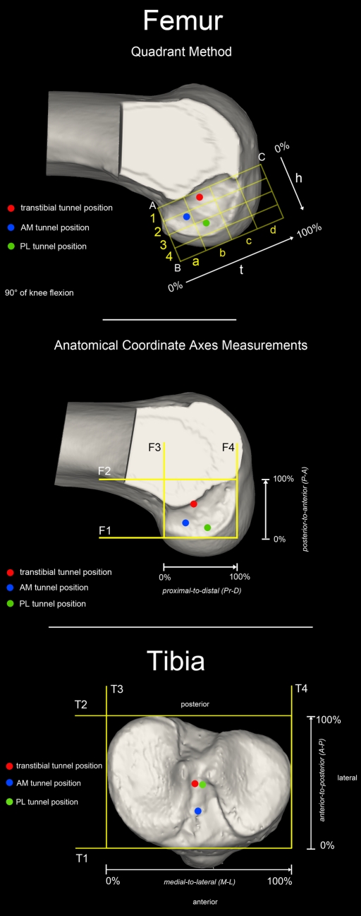
Tunnel measurements with use of three-dimensional computed tomography were performed as previously developed and described4. Femoral tunnel positions were determined with use of two different coordinate systems, as shown in Figure 1, to facilitate comparisons with other published studies. The quadrant method employed a grid defined parallel and perpendicular to the Blumensaat line (Fig. 1, A). The anatomical coordinate axes method defined tunnel positions relative to standard anatomical directions (proximal to distal and posterior to anterior), as shown in Figure 1, B. Tibial tunnel locations were presented in a grid aligned with the medial-to-lateral and anterior-to-posterior anatomical tibial axes (Fig. 1, C). All measurements were expressed as a percentage of the corresponding maximum dimension. Tunnel locations were presented as the mean and standard deviation, with the range (minimum to maximum) in parentheses. Anatomic comparison data were taken from our previous cadaver study, which evaluated tunnel positions after anatomic double-bundle anterior cruciate ligament reconstruction4. Independent t tests were performed to compare transtibial single-bundle tunnel position with both anatomic anteromedial and anatomic posterolateral tunnel positions. The significance level was set at p < 0.025 to account for the two comparisons of the femoral and tibial tunnels.
Fig. 1.
The mean results of the quadrant method measurements on the femur (A) and the anatomical coordinate axes measurements on both the femur (B) and tibia (C) are plotted visually. Traditional transtibial anterior cruciate ligament reconstruction results in anterior (“high”) tunnel placement on the femur and posterior tunnel placement, medial to the anatomic posterolateral (PL) position, on the tibia. AM = anteromedial, t = line parallel to Blumensaat line, and h = line perpendicular to Blumensaat line. F1 through F4 represent femoral line numbers 1 through 4, and T1 through T4 represent tibial line numbers 1 through 4.
Source of Funding
This work was partially supported by a grant from the National Institutes of Health, National Institute of Arthritis and Musculoskeletal and Skin Diseases AR46387.
Results
The tunnel positions are summarized in Tables I and II and displayed in Figure 1. On the tibia in the anterior-to-posterior direction, the transtibial tunnel positions were not significantly different from the anatomic posterolateral tunnel positions (p = 0.428) but were significantly different posterior to the anatomic anteromedial tunnel (p < 0.001). In the medial-to-lateral direction, the transtibial tunnel positions were significantly more medial than the anatomic anteromedial tunnels (p = 0.022) and the anatomic posterolateral tunnels (p < 0.001) (Fig. 1, C). On the femur, with use of the anatomical coordinate axes method (Fig. 1, B), the transtibial femoral tunnels were positioned anterior (“high”) relative to the anatomic anteromedial and posterolateral tunnel positions (p < 0.001 for both), distal to the anatomic anteromedial tunnels (p = 0.002), and proximal to the posterolateral tunnels (p < 0.001). Using the quadrant method (Fig. 1, A), the transtibial tunnels were positioned significantly closer to the Blumensaat line (more anterior) compared with the anatomic anteromedial and posterolateral tunnel positions (p < 0.001 for both). Parallel to the Blumensaat line (t), the position of the transtibial tunnels was significantly different from the anatomic anteromedial tunnels (p < 0.001), but closer to the anatomic posterolateral tunnels (p = 0.295). In the 4 × 4 grid (Fig. 1, A), the tunnels were located in box 1b (twenty-nine tunnels), box 1c (one tunnel), and box 1a (one tunnel).
TABLE I.
Results of Tibial and Femoral Bone-Tunnel Position Measurements and Significant Differences within the Anatomical Coordinate Axes
| Transtibial Position*(%) | Anatomic Anteromedial Position*(%) | Anatomic Posterolateral Position*(%) | |
| Femoral measurements | |||
| Posterior to anterior from L1 to L2 | 54.3 ± 8.3 (41.5-79.5)†‡ | 23.1 ± 6.1 (16.3-36.4)† | 15.3 ± 4.8 (8.9-24.3)‡ |
| Proximal to distal from L3 to L4 | 41.1 ± 10.3 (15.1-67.4)†‡ | 28.2 ± 5.4 (20.1-36.2)† | 58.1 ± 7.1 (50.2-73.1)‡ |
| Tibial measurements | |||
| Anterior to posterior from L1 to L2 | 48.0 ± 5.5 (35.6-59.5)† | 25.0 ± 2.8 (21.1-29.5)† | 46.4 ± 3.7 (40.1-51.5) |
| Medial to lateral from L3 to L4 | 47.8 ± 2.4 (42.7-51.6)†‡ | 50.5 ± 4.2 (44.1-54.7)† | 52.4 ± 2.5 (49.5-56.1)‡ |
The values are given as the mean and the standard deviation, with the range in parentheses.
The difference between transtibial and anteromedial tunnels was significant (p < 0.001).
The difference between transtibial and posterolateral tunnels was significant (p < 0.001).
TABLE II.
Results and Significant Differences of the Quadrant Method
| Femoral Measurements | Transtibial Position*(%) | Anatomic Anteromedial Position*(%) | Anatomic Posterolateral Position*(%) |
| Along line t from point A to point C | 37.2 ± 5.5 (24.9-50.6)† | 21.7 ± 2.5 (18.9-25.7)† | 35.1 ± 3.5 (31.2-40.0) |
| Along line h from point A to point B | 11.3 ± 6.6 (–5.5-22.9)†‡ | 33.2 ± 5.6 (24.1-42.1)† | 55.3 ± 5.3 (47.7-65.1)‡ |
The values are given as the mean and the standard deviation, with the range in parentheses.
The difference between transtibial and anteromedial tunnels was significant (p < 0.001).
The difference between transtibial and posterolateral tunnels was significant (p < 0.001).
Discussion
The results of this study show that the traditional transtibial technique for arthroscopic anterior cruciate ligament reconstruction results in bone tunnels that are positioned nonanatomically (Fig. 2). More specifically, the transtibially drilled femoral tunnels were consistently positioned anterior (“high”) to the anatomic anteromedial and posterolateral tunnels. Tibial tunnel apertures in this study were positioned medial to the anatomic posterolateral and anatomic anteromedial tunnels and posterior to the anteromedial tunnel (Fig. 1, C). These findings are consistent with prior cadaver studies that evaluated tunnel position in traditional transtibial anterior cruciate ligament reconstruction5-8. In those studies, the tunnels were drilled during arthroscopy or in an open fashion and were evaluated postoperatively during dissection. In contrast, two studies have found that it was possible to locate the femoral tunnel within the anatomic femoral insertion when a transtibial method was used to drill the tunnel9,10. However, the first study (a cadaver study) recommended use of the accessory medial portal for femoral tunnel drilling because the tibial bone tunnels were short and the anterior tibial tunnel entrances were close to the joint line9. The second investigation was a retrospective radiographic study of patients after transtibial anterior cruciate ligament reconstruction10. Only plain radiographs were utilized to evaluate tunnel position, revealing posteriorly positioned tibial tunnels measured along the tibial plateau and anatomically positioned femoral tunnels measured directly along the Blumensaat line. To our knowledge, there is no anatomical study in the literature that has effectively used the Blumensaat line alone to describe the anatomical location of the native femoral anterior cruciate ligament insertion. Thus, it is difficult to draw the conclusion that the femoral bone tunnels in the aforementioned study were anatomically positioned. Additionally, one-dimensional measurements do not define tunnel locations within the three-dimensional intercondylar notch. A recent in vivo study evaluated femoral tunnel position after transtibial drilling in eight patients and confirmed our results that the transtibial technique results in nonanatomical tunnel placement11. However, the sample size was small and the authors did not evaluate tibial tunnel positions. Furthermore, they used magnetic resonance imaging to evaluate bone-tunnel positions, whereas computed tomography is considered the gold standard to evaluate osseous structures.
Fig. 2.
This arthroscopic view of a knee after transtibial anterior cruciate ligament (ACL) reconstruction reveals posterior graft placement on the tibial side and anterior (“high”) graft placement on the femoral side. The vertical intra-articular orientation of the anterior cruciate ligament graft is secondary to nonanatomical tunnel placement, which is well demonstrated by the corresponding three-dimensional computed tomography image. AM = anteromedial, PL = posterolateral, and PCL = posterior cruciate ligament.
A strength of the current study is the fact that the anterior cruciate ligament reconstructions were performed by multiple surgeons from different institutions. This reduced the likelihood of bias due to the techniques of a single surgeon and should make the findings more generalizable to the wider orthopaedic community. It is interesting and important to note that tunnel positions drilled with use of the transtibial single-bundle technique were consistent among multiple surgeons utilizing the same technique.
A limitation of this study may be that the position of the tunnel aperture center may have migrated from the time of surgery to the time of the computed tomography scan as a result of tunnel widening. However, to our knowledge, no study has described the direction of tunnel widening at the aperture in anterior cruciate ligament reconstruction. It is unlikely that the centers of the tunnel apertures were initially placed within the native insertion site of the anterior cruciate ligament and then migrated to a nonanatomic position secondary to tunnel widening. In a prospective computed tomography study, Iorio et al.12 reported tunnel widening of <1 mm between computed tomography scans performed on the day after the operation and those made at the time of the ten-month follow-up. Of note, tunnel widening is thought to occur primarily during the first six months following surgery.
In summary, traditional transtibial single-bundle anterior cruciate ligament reconstruction resulted in nonanatomically positioned tibial and femoral bone tunnels. Analysis was performed on three-dimensional computed tomography models, which has proven to be a useful and precise method to evaluate bone-tunnel position.
Footnotes
Disclosure: In support of their research for or preparation of this work, one or more of the authors received, in any one year, outside funding or grants in excess of $10,000 from a grant from the National Institutes of Health, National Institute of Arthritis and Musculoskeletal and Skin Diseases AR46387. Neither they nor a member of their immediate families received payments or other benefits or a commitment or agreement to provide such benefits from a commercial entity.
References
- 1.Biau DJ, Tournoux C, Katsahian S, Schranz P, Nizard R. ACL reconstruction: a meta-analysis of functional scores. Clin Orthop Relat Res. 2007;458:180-7 [DOI] [PubMed] [Google Scholar]
- 2.Fithian DC, Paxton EW, Stone ML, Luetzow WF, Csintalan RP, Phelan D, Daniel DM. Prospective trial of a treatment algorithm for the management of the anterior cruciate ligament-injured knee. Am J Sports Med. 2005;33:335-46 [DOI] [PubMed] [Google Scholar]
- 3.Tashman S, Kolowich P, Collon D, Anderson K, Anderst W. Dynamic function of the ACL-reconstructed knee during running. Clin Orthop Relat Res. 2007;454:66-73 [DOI] [PubMed] [Google Scholar]
- 4.Forsythe B, Kopf S, Wong AK, Martins CAQ, Anderst W, Tashman S, Fu FH. The location of femoral and tibial tunnels of anatomic double-bundle anterior cruciate ligament reconstruction analyzed by three-dimensional computed tomography models. J Bone Joint Surg Am. 2010;92:1418-26 [DOI] [PubMed] [Google Scholar]
- 5.Kaseta MK, DeFrate LE, Charnock BL, Sullivan RT, Garrett WE., Jr Reconstruction technique affects femoral tunnel placement in ACL reconstruction. Clin Orthop Relat Res. 2008;466:1467-74 [DOI] [PMC free article] [PubMed] [Google Scholar]
- 6.Arnold MP, Kooloos J, van Kampen A. Single-incision technique misses the anatomical femoral anterior cruciate ligament insertion: a cadaver study. Knee Surg Sports Traumatol Arthrosc. 2001;9:194-9 [DOI] [PubMed] [Google Scholar]
- 7.Gavriilidis I, Motsis EK, Pakos EE, Georgoulis AD, Mitsionis G, Xenakis TA. Transtibial versus anteromedial portal of the femoral tunnel in ACL reconstruction: a cadaveric study. Knee. 2008;15:364-7 [DOI] [PubMed] [Google Scholar]
- 8.Steiner ME, Battaglia TC, Heming JF, Rand JD, Festa A, Baria M. Independent drilling outperforms conventional transtibial drilling in anterior cruciate ligament reconstruction. Am J Sports Med. 2009;37:1912-9 [DOI] [PubMed] [Google Scholar]
- 9.Heming JF, Rand J, Steiner ME. Anatomical limitations of transtibial drilling in anterior cruciate ligament reconstruction. Am J Sports Med. 2007;35:1708-15 [DOI] [PubMed] [Google Scholar]
- 10.Gougoulias N, Khanna A, Griffiths D, Maffulli N. ACL reconstruction: can the transtibial technique achieve optimal tunnel positioning? A radiographic study. Knee. 2008;15:486-90 [DOI] [PubMed] [Google Scholar]
- 11.Abebe ES, Moorman CT, 3rd, Dziedzic TS, Spritzer CE, Cothran RL, Taylor DC, Garrett WE, Jr, DeFrate LE. Femoral tunnel placement during anterior cruciate ligament reconstruction: an in vivo imaging analysis comparing transtibial and 2-incision tibial tunnel-independent techniques. Am J Sports Med. 2009;37:1904-11 [DOI] [PubMed] [Google Scholar]
- 12.Iorio R, Vadalà A, Argento G, Di Sanzo V, Ferretti A. Bone tunnel enlargement after ACL reconstruction using autologous hamstring tendons: a CT study. Int Orthop. 2007;31:49-55 [DOI] [PMC free article] [PubMed] [Google Scholar]


