Abstract
We investigated the alterations of major fatty acid components in epidermis by natural aging and photoaging processes, and by acute ultraviolet (UV) irradiation in human skin. Interestingly, we found that 11,14,17-eicosatrienoic acid (ETA), which is one of the omega-3 polyunsaturated acids, was significantly increased in photoaged human epidermis in vivo and also in the acutely UV-irradiated human skin in vivo, while it was significantly decreased in intrinsically aged human epidermis. The increased ETA content in the epidermis of photoaged human skin and acute UV-irradiated human skin is associated with enhanced expression of human elongase 1 and calcium-independent phophodiesterase A2. We demonstrated that ETA inhibited matrix metalloproteinase (MMP)-1 expression after UV-irradiation, and that inhibition of ETA synthesis using EPTC and NA-TCA, which are elongase inhibitors, increased MMP-1 expression. Therefore, our results suggest that the UV increases the ETA levels, which may have a photoprotective effect in the human skin.
Keywords: Ultraviolet Rays; Fatty Acids, Nonesterified; Fatty Acids, Omega-3; 11,14,17-eicosatrienoic acid; Phospholipases A2, Calcium-Independent; Human Elongase 1
Skin aging can be divided into photoaging and chronological aging. Photoaging is induced by damage to human skin attributable to repeated exposure to ultraviolet (UV) irradiation, while intrinsic aging occurs with increasing age and is strongly associated with genetic factors (1). Photoaging (extrinsic aging) is characterized by morphological changes that include deep wrinkles and loss of elasticity, as well as histological changes such as connective-tissue alterations. These alterations are considered the result of collagen destruction by UV-induced matrix metalloproteinases (MMPs) secreted from epidermal keratinocytes and dermal fibroblasts (2).
Fatty acids are essential components of natural lipids, which determine the physiological structure and function of the human skin (3). They are present in the epidermis, especially in the stratum corneum, the outermost layer, and cell membranes (4). Many effects of fatty acids can be linked to changes in membrane lipid composition affecting cell signaling mechanisms originating from membranes (5).
Skin aging may influence epidermal lipids and free fatty acid composition, and their physiological functions may be involved in aging process. Therefore, in the present study, we investigated the alteration of fatty acid composition in the epidermis by skin aging process and acute UV irradiation in human skin in vivo.
Fatty acids are classified as saturated fatty acid (SFA), monounsaturated fatty acid (MUPA), and polyunsaturated fatty acid (PUFA). Omega-3 (n-3), omega-6 (n-6), and omega-9 (n-9) unsaturated fatty acid structures are based on the position of the first double bond at the third, sixth or ninth position from the methyl (omega) terminal of the aliphatic carbon chain (6). To investigate the alteration of fatty acid composition by intrinsic aging process, young human (21-33 yr, n=4) buttock skin and aged human (70-75 yr, n=4) buttock skin were obtained by punch biopsy. Then the epidermis was separated from dermis and total lipids were extracted with chroloform/methanol/water (1:2:0.8, v/v/v). Fatty acids were analyzed by typical gas chromatography.
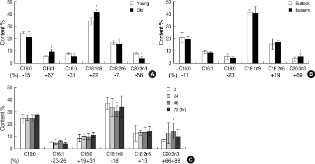
The palmitic acid (C16:0), stearic acid (C18:0), palmitoleic acid (C16:1), oleic acid (C18:1), linoleic acid (C18:2), and (all-cis)-11,14,17-eicosatrienoic acid (ETA, C20:3n-3) were determined as major fatty acid components in the human epidermis (Fig. 1). Among them, linoleic acid and ETA belong to PUFAs. The linoleic acid, one of essential fatty acids, is well known as the precursor of arachidonic acid synthesis. However, the physiological function of ETA has not been well investigated. The levels of SFAs such as palmitic acid and stearic acid, PUFAs such as linoleic acid and ETA were decreased in aged skin by 15%, 31%, 7%, and 56%, compared with those in young skin, respectively. Especially, ETA was most significantly decreased in aged skin, indicating that it may relate with intrinsic aging. In contrast, palmitoleic acid and oleic acid were increased in aged skin by 67% and 22%, respectively, compared with those in young skin (Fig. 1A).
Fig. 1.
The changes of free fatty acid (FFA) composition in the epidermis of human skin. (A) The changes of FFA composition in aged epidermis. Young human (mean age 26.5 yr; age range 21-33 yr, n=4) buttock skin and aged human (mean age 72.7 yr; age range 70-75 yr, n=4) buttock skin were obtained by punch biopsy. Total lipids were extracted with chroloform/methanol/water (1:2:0.8, v/v/v). Lipid extracts were analyzed by typical gas chromatography (GC). *P<0.05, †P<0.01, C16:0-palmitic acid (PA), C16:1-palmitoleic acid (PtA), C18:0-stearic acid (SA), C18:1n9-oleic acid (OA), 18:2n6-linoleic acid (LA), C20:3n3-(All-cis)-11,14,17-eicosatrienoic acid (ETA). (B) The change of FFA composition in photoaged epidermis. Aged human (mean age 72.7 yr; age range 70-75 yr, n=5) buttock/forearm skin were obtained by punch biopsy. Total lipids were extracted with chroloform/methanol/water (1:2:0.8, v/v/v). Lipid extracts were analyzed by typical gas chromatography (GC). †P<0.01, C16:0-palmitic acid (PA), C16:1-palmitoleic acid (PtA), C18:0-stearic acid (SA), C18:1n9-oleic acid (OA), 18:2n6-linoleic acid (LA), C20:3n3-(All-cis)-11,14,17-eicosatrienoic acid (ETA). (C) The change of FFA composition in acutely UV-irradiated buttock epidermis. Young human (mean age 26.5 yr; age range 21-33 yr, n=4) buttock skin was obtained by punch biopsy at the indicated time points after UV irradiation (2 MED). Total lipids were extracted with chroloform/methanol/water (1:2:0.8, v/v/v). Lipid extracts were analyzed by typical gas chromatography (GC). *P<0.05, C16:0-palmitic acid (PA), C16:1-palmitoleic acid (PtA), C18:0-stearic acid (SA), C18:1n9-oleic acid (OA), 18:2n6-linoleic acid (LA), C20:3n3-(All-cis)-11,14,17-eicosatrienoic acid (ETA).
To investigate the alteration of fatty acid composition by photoaging process, the fatty acids composition were compared in the epidermis between photoaged forearm and intrinsically aged buttock tissues of the same elderly individuals (70-75 yr, n=5). The levels of palmitic acid and stearic acid in photoaged forearm epidermis were decreased by 11% and 23%, respectively, compared to those in the buttocks of the same elderly individuals. On the other hand, the amounts of linoleic acid and ETA in photoaged forearm epidermis were increased by 19% and 69%, respectively, compared to those in the buttocks of the same elderly individuals (Fig. 1B).
Next, in order to investigate the acute effect of UV irradiation on free fatty acid composition in the epidermis of human skin, we analyzed the change of free fatty acid composition in acutely UV-irradiated buttock epidermis. Young human (21-33 yr, n=4) buttock skin was obtained by punch biopsy at 24, 48, and 72 hr after UV irradiation (2 MED). The levels of palmitoleic acid and oleic acid showed reduced pattern after UV-irradiation. In contrast, the levels of stearic acid, linoleic acid, and ETA were increased after UV-irradiation from 13% linoleic acid to 88% (ETA) at certain time point (Fig. 1C).
Interestingly, ETA, which is one of the omega-3 PUFA, was significantly increased in photoaged human epidermis in vivo and also in the acutely UV-irradiated human skin in vivo, while it was significantly decreased in intrinsically aged human epidermis. These results suggest that ETA may be closely related to intrinsic aging and photoaging processes. Some of PUFAs such as eicosapentaenoic acid (C20:5n-3) and docosahexaenoic acid (C22:6n-3), representative substances of n-3 PUFAs, are well known to have functional effects in many kinds of diseases. They are previously reported to reduce the risk of coronary heart disease by decreasing plasma triglyceride level and also by their antithrombotic and antiatherosclerotic actions through anti-inflammatory effects (6, 10-12). However, the physiological function of ETA is still not clear.
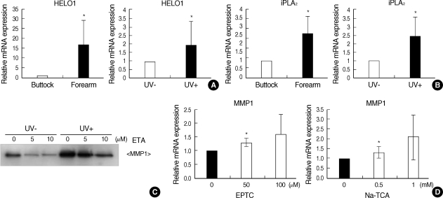
Next, to elucidate the mechanism by which ETA content is increased by photoaging process and acute UV irradiation in the epidermis, we evaluated the gene expressions involved in ETA synthesis from precursors and the release of ETA from phospholipids. The synthesis of long-chain fatty acids is mediated by elongase family members, ELOVL (Elongation of very long chain fatty acid) 1-6. Elongation of SFAs and MUFAs is mediated by ELOVL1, 3, and 6, and ELOVL2, 4, and 5 are responsible for elongation of PUFAs (7). Human elongase 1 (HEOL1/ELOVL5) was involved in the elongation of long-chain PUFAs such as conversion of α-linolenic acid (C18:3n-3) into ω3-ETA (C20:3n-3) (8). The expression of HELO1 was assessed by real-time PCR in photoaged forearm skin and sun-protected buttock skin of the elderly volunteers (n=4). The mRNA level of HELO1 was increased in photoaged forearm skin (Fig. 2A). Furthermore, the mRNA level of HELO1 in normal human keratinocytes was increased 24 hr post-UV (100 mJ/cm2) irradiation (Fig. 2A). These finding indicates that the increased ETA content in the epidermis of photoaged human skin and acute UV-irradiated human skin is associated with enhanced expression of elongase.
Fig. 2.
The expressions of genes involved in ETA production and the effect of ETA on MMP-1 expression. (A) The expression of HELO1 involved in the elongation of fatty acids. (Left) Aged human (mean age 72.7 yr; age range 70-75 yr) buttock/forearm skin were obtained by punch biopsy. Levels of HELO1 were determined by real-time PCR (n=4). *P<0.05. (Right) Normal human keratinocytes were incubated for 24 hr after UV irradiation (100 mJ/cm2). Levels of iPLA2 were determined by real-time PCR (n=3). *P<0.05. (B) The expression of iPLA2 involved in the release of ETA from phospholipids. (Left) Aged human (mean age 72.7 yr; age range 70-75 yr) buttock/forearm skin were obtained by punch biopsy. Levels of iPLA2 were determined by real-time PCR (n=4). *P<0.05.(Right) Normal human keratinocytes were incubated for 24 hr after UV irradiation (100 mJ/cm2). Levels of iPLA2 were determined by real-time PCR (n=3). *P<0.05. (C) The effect of ETA on UV-induced MMP-1 expression. Normal human keratinocytes were incubated with ETA for 24 hr after UV irradiation (100 mJ/cm2). Levels of MMP-1 were determined by Western blots. (n=3). (D) Normal human keratinocytes were incubated with each elongase inhibitor for 24 hr. Levels of MMP-1 were determined by real-time PCR (n=3). *P<0.05.
Phospholipase A2 (PLA2) family members are also involved in the production of free fatty acid from phospholipids. Calcium-dependent PLA2 (cPLA2) is considered as signaling PLA2, regulating stimulus-induced arachidonic acid metabolism, while the release of ω3-PUFA such as DHA is mediated by calcium-independent PLA2 (iPLA2) (9). Levels of iPLA2 were determined by real-time PCR (n=4). The mRNA of iPLA2 was increased significantly in the forearm skin samples (Fig. 2B). In addition, we analyzed the mRNA levels of iPLA2 in normal human keratinocytes, which were incubated for 24 hr after UV (100 mJ/cm2) irradiation. Our data showed that the level of iPLA2 was increased after UV-irradiation (Fig. 2B). An increase of PLA2 in photoaged forearm skin and UV-irradiated human epidermis may lead to an increase of ETA. Hence, up-regulation of iPLA2 may be related to photoaging process. The expression of cPLA2 mRNA was also increased in photoaged forearmskin (data not shown).
Finally, we investigated the relationship between ETA and MMP-1 expression. MMP-1 is known to promote UV-triggered extracellular protein degradation and thereby contributing to photoaging of human skin. Normal human keratinocytes were incubated with ETA (5 and 10 µM) for 24 hr after UV irradiation (100 mJ/cm2). The mRNA level of MMP-1 was increased by UV and ETA inhibited MMP-1 expression after UV-irradiation (Fig. 2C). In contrast, EPTC and NA-TCA, which are elongase inhibitors, increased MMP-1 expression (Fig. 2D). Therefore, our results suggest that the prevention of UV-triggered MMP-1 expression by ETA represents a potential protective strategy in the human skin.
In conclusion, our results suggest that the UV increases the ETA levels, which may have a photoprotective effect in the human skin.
Footnotes
This study was supported by a grant of the Korea Health 21 R&D Project, Ministry of Health & Welfare, the Republic of Korea (A060160) and by a research agreement with AMOREPACIFIC Corporation.
References
- 1.Jenkins G. Molecular mechanisms of skin ageing. Mech Ageing Dev. 2002;123:801–810. doi: 10.1016/s0047-6374(01)00425-0. [DOI] [PubMed] [Google Scholar]
- 2.Fisher GJ, Kang S, Varani J, Bata-Csorgo Z, Wan Y, Datta S, Voorhees JJ. Mechanisms of photoaging and chronological skin aging. Arch Dermatol. 2002;138:1462–1470. doi: 10.1001/archderm.138.11.1462. [DOI] [PubMed] [Google Scholar]
- 3.Trommer H, Wagner J, Graener H, Neubert RH. The examination of skin lipid model systems stressed by ultraviolet irradiation in the presence of transition metal ions. Eur J Pharm Biopharm. 2001;51:207–214. doi: 10.1016/s0939-6411(01)00140-0. [DOI] [PubMed] [Google Scholar]
- 4.Hansen HS, Jensen B. Essential function of linoleic acid esterified in acylglucosylceramide and acylceramide in maintaining the epidermal water permeability barrier. Evidence from feeding studies with oleate, linoleate, arachidonate, columbinate and alpha-linolenate. Biochim Biophys Acta. 1985;834:357–363. doi: 10.1016/0005-2760(85)90009-8. [DOI] [PubMed] [Google Scholar]
- 5.Jump DB. Fatty acid regulation of gene transcription. Crit Rev Clin Lab Sci. 2004;41:41–78. doi: 10.1080/10408360490278341. [DOI] [PubMed] [Google Scholar]
- 6.Siddiqui RA, Harvey KA, Zaloga GP. Modulation of enzymatic activities by n-3 polyunsaturated fatty acids to support cardiovascular health. J Nutr Biochem. 2008;19:417–437. doi: 10.1016/j.jnutbio.2007.07.001. [DOI] [PubMed] [Google Scholar]
- 7.Jakobsson A, Westerberg R, Jacobsson A. Fatty acid elongases in mammals: their regulation and roles in metabolism. Prog Lipid Res. 2006;45:237–249. doi: 10.1016/j.plipres.2006.01.004. [DOI] [PubMed] [Google Scholar]
- 8.Leonard AE, Bobik EG, Dorado J, Kroeger PE, Chuang LT, Thurmond JM, Parker-Barnes JM, Das T, Huang YS, Mukerji P. Cloning of a human cDNA encoding a novel enzyme involved in the elongation of long-chain polyunsaturated fatty acids. Biochem J. 2000;350(Pt 3):765–770. [PMC free article] [PubMed] [Google Scholar]
- 9.Strokin M, Sergeeva M, Reiser G. Docosahexaenoic acid and arachidonic acid release in rat brain astrocytes is mediated by two separate isoforms of phospholipase A2 and is differently regulated by cyclic AMP and Ca2+ Br J Pharmacol. 2003;139:1014–1022. doi: 10.1038/sj.bjp.0705326. [DOI] [PMC free article] [PubMed] [Google Scholar]
- 10.Daviglus ML, Stamler J, Orencia AJ, Dyer AR, Liu K, Greenland P, Walsh MK, Morris D, Shekelle RB. Fish consumption and the 30-year risk of fatal myocardial infarction. N Engl J Med. 1997;336:1046–1053. doi: 10.1056/NEJM199704103361502. [DOI] [PubMed] [Google Scholar]
- 11.Kushi L, Giovannucci E. Dietary fat and cancer. Am J Med. 2002;113(Suppl 9B):63S–70S. doi: 10.1016/s0002-9343(01)00994-9. [DOI] [PubMed] [Google Scholar]
- 12.Horrobin DF, Bennett CN. Depression and bipolar disorder: relationships to impaired fatty acid and phospholipid metabolism and to diabetes, cardiovascular disease, immunological abnormalities, cancer, ageing and osteoporosis. Possible candidate genes. Prostaglandins Leukot Essent Fatty Acids. 1999;60:217–234. doi: 10.1054/plef.1999.0037. [DOI] [PubMed] [Google Scholar]


