Abstract
The goal of this study was to identify objective criteria that would reliably predict imminent death in aged mice. Male and female ICR mice (age, 8 mo) were subcutaneously implanted with an identification chip for remote measurement of body temperature. Mice then were weighed and monitored regularly until spontaneous death occurred or until euthanasia was administered for humane reasons. Clinical signs that signaled implementation of euthanasia included inability to walk, lack of response to manipulation, large or ulcerated tumors, seizures, and palpable hypothermia. In mice that died spontaneously, gradual weight loss was the most frequent and earliest sign of imminent death. Hypothermia developed during the 2 wk prior to death. Slow or labored breathing were observed in about half of the mice before death. A composite score of temperature × weight can be used to provide an objective benchmark to signal increased observation or euthanasia of individual mice. Such assessment may allow the collection of terminal tissue samples without markedly altering longevity data, although application of this criterion may not be appropriate for all studies of longevity. Timely euthanasia of mice based on validated markers of imminent death can allow implementation of endpoints that alleviate terminal distress in aged mice, may not significantly affect longevity data, and can permit timely collection of biologic samples.
Abbreviations: T×W, product of temperature and body weight
Laboratory mice allow the study of age-related physiologic changes in a manner that is not possible in humans. The controlled environment, short life span, and genetic homogeneity of mice make them an ideal species for such research. Mice used in longevity represent a large investment in research time and other resources. Data obtained from these mice would be maximized if objective accurate markers for imminent death could be established to allow timely euthanasia and the subsequent collection of biologic samples. In addition, because mice undergo rapid autolysis after death, determination of the cause of death is often impossible to discern in mice that have been dead for only a few hours. Detection of subtle decreases in health status together with accurate prediction of imminent death could be used as triggers for performing in vivo studies that might reveal valuable information about the pathophysiologic mechanisms that contribute to the physical decline of elderly mice.
Experimental endpoints in biomedical research should be determined based on a combination of scientific, ethical, legal, practical, and humane considerations (for example, reference 19). However, to our knowledge, humane endpoints for mice used in longevity studies have rarely been addressed, despite the expectation that health problems will become more common as the mice age. However, using health-related endpoints to signal euthanasia can be problematic for such studies because their goal is often determination of life span. Premature euthanasia could skew the data and thereby potentially lead to erroneous conclusions, creating a conflict between the collection of necessary data and minimization of animal pain and distress. Furthermore, despite the anticipated increase in health problems as aging progresses, geriatric mice can be found dead in the cage without obvious antemortem signs of overt illness.
Our goal in this study was to identify practical and objective criteria that could be used to predict imminent death in aged mice, particularly those maintained on longevity studies. Data were collected to determine the proportion of aging mice that either did or did not display signs of illness or debilitation before death and how soon these signs could be detected in relation to death. Three factors were monitored as potential signs of imminent death: body weight, body temperature, and respiratory effort. Identifying clinical signs that are predictive of imminent death would not only promote animal wellbeing in longevity studies but also would permit timely euthanasia that facilitates collection of valuable samples and measurements.
Materials and Methods
A total of 110 8-mo-old male and female Hsd:ICR (CD1) mice (retired breeders) were purchased from Harlan (Indianapolis, IN). Fifty male and 20 female mice were singly housed in 11 in. × 7 in. × 5 in. cages, and 40 female mice were housed in groups of 10 in 18 in. × 10 in. × 6 in. cages. Cages were solid-bottom shoebox-style open-top cages that were maintained by using conventional husbandry practices and woodchip bedding (Beta Chip, Northeastern Products, Warrensburg, NY). Cages were changed weekly. Room temperature was maintained at 70 ± 2 °C and relative humidity at 40% to 60%. Food (LabDiet 5001, PMI Nutrition International, St Louis, MO) and tap water were available ad libitum. All mice were free of known infections with common rodent microbial and parasitic agents, as monitored by using monthly testing of sentinel mice housed in the same room. The Laboratory Animal Care and Use Committee at Southern Illinois University School of Medicine approved all animals and experimental procedures used in this study.
Mice were subcutaneously implanted under isoflurane anesthesia with a microchip that allows remote measurement of body temperature by using a wand-type reader (model IPTT300, BioMedic Data Systems, Seaford, DE). The microchip was implanted by using a 12-gauge needle delivery device without an incision or wound closure. Individual chips were not tested for accuracy or otherwise calibrated prior to use but were used according to manufacturer's recommendations.
The husbandry staff monitored all mice for signs of illness or deviation from normal at the daily census and health check and in association with changing cages, as is the standard practice of the Division of Laboratory Animal Medicine. Husbandry personnel are trained to detect signs of illness in mice including, but not limited to, those changes mentioned below as defining the moribund condition. Sick animals were immediately brought to the attention of the veterinarian and the research team, who then evaluated the animal. Mice then underwent either euthanasia or increased frequency of collection of objective data (that is, temperature and body weight), as warranted by the animal's condition.
The research staff evaluated mice at least once every 4 wk, and more frequently if signs of illness or impairment were noted. Clinical signs that were monitored included body weight, temperature, general body condition, pattern of respiration, dehydration, posture, movement, response to manipulation, and condition of hair coat. Measurements typically were made during the afternoon hours. Although the goal of the study was to monitor mice until the time of spontaneous death, some mice underwent euthanasia based on clinical signs that included palpable hypothermia (in our experience based on mice that were implanted with telemetric temperature transmitters during various infectious conditions, palpable hypothermia generally reflects a body temperature of less than 25 °C), inability or unwillingness to walk, lack of response to manipulation, severe dyspnea or cyanosis, and large, bleeding, or ulcerated tumors.
Blood was collected from some aged mice (22 ± 2 mo of age, n = 7 or 8) that underwent euthanasia for various reasons. These sera were used for measurement of serum cytokines and adipokines in the same assay as sera collected from Hsd:ICR (CD1) mice that were held under the same conditions until euthanasia at 2 to 3 mo of age (n = 8). Protein concentrations of IL1α, IL1β, IL2, IL3, IL4, IL5, IL6, IL9, IL10, IL12 p40, IL12 p70, IL13, IL17, granulocyte colony-stimulating factor, granulocyte–macrophage colony-stimulating factor, IFNγ, chemokine (C-C motif) ligand 5 (RANTES), keratinocyte chemoattractant, monocyte chemoattractant protein 1, macrophage inhibitory proteins 1α and 1β, and TNFα were measured in serum by using mouse cytokine multiplex assay (Bio-Plex, BioRad, Hercules, CA). Concentrations of insulin, monocyte chemoattractant protein 1 (CCL2), IL6, TNFα, resistin, leptin, and total plasminogen activator inhibitor 1 were measured in plasma samples using a multiplex adipokine assay (Millipore, Billerica, MA), Luminex technology (100IS System, Luminex, Austin, TX), and commercial software (BioPlex Manager 5.0, BioRad) according to the manufacturers’ instructions.
The overall design of this study consisted of 2-factor mixed-model ANOVA in which the between factor was group membership and the repeated factor was time in weeks. Descriptive measures consisted of means and standard errors. Statistical analysis was conducted with independent t tests, χ2 (Fisher exact test), and ANOVA. The data used for the repeated measurements were derived, for each subject, as a percentage of baseline, where the baseline was the average of the 4-wk time that preceded death by 10 to 13 wk. The ANOVA examined the effect of group, time, and the interaction of group × time, with statistical significance set at the 5% level.
Results
Of the 110 mice that began the study, data from 105 were included for analysis. Data from 5 mice (3 individually housed males and 2 individually housed females) were excluded. Four of these mice either died or underwent euthanasia due to injury or water bottle leaks, whereas one mouse died within 1 wk of arrival of an undetermined cause.
Of the 105 mice whose data are reported here, 81 died spontaneously without obvious clinical signs of severe illness or impending death. The remaining 24 underwent euthanasia due to the development of a moribund condition that was characterized by one or more of the following signs: large or ulcerated tumors (n = 13), ascites (n = 3), inability or unwillingness to walk, palpable hypothermia, and lack of overt response to manipulation.
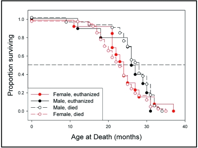
Geriatric mice developed a typical appearance that was characterized by a ruffled hair coat, a rounded appearance (including rounding of the head), pale skin, and lesions at the base of the tail. Mice that underwent euthanasia were killed at a mean age of 25.5 ± 1.9 and 24.2 ± 1.7 mo for males and females, respectively, whereas among mice that died spontaneously, the mean age at death was 27.1 ± 0.8 and 23.1 ± 0.8 mo for males and females, respectively (Figure 1, Table 1). Housing singly or in groups did not significantly influence outcomes in female mice (data not shown), and therefore values from these female mice were combined for analysis. Data collected from mice that died spontaneously or underwent euthanasia were analyzed to identify and compare objective measures that preceded death or a moribund state, respectively.
Figure 1.
Kaplan–Meier survival plot. The lifespans of mice used in this study are depicted.
Table 1.
Temperature and weight (mean ± SEM) in male and female mice that experienced euthanasia or spontaneous death
| Temperature (°C) |
Body weight (g) |
Temperature × Weight |
Respiratory difficulty before death | |||||||||
| Type of death | n | Age at death (mo) | Basal | Final | % | Basal | Final | % | Basal | Final | % | |
| Euthanasia | ||||||||||||
| Male | 11 | 25.5 ± 1.9 | 35.9 ± 0.7 | 30.8 ± 1.2 | 86 ± 3 | 44.4 ± 1.6 | 40.2 ± 1.2 | 91 ± 3 | 1593 ± 66 | 1275 ± 69 | 80 ± 4 | 7 of 11 (64%) |
| Female | 13 | 24.2 ± 1.7 | 36.5 ± 0.3 | 29.0 ± 1.3 | 79 ± 4 | 41.7 ± 2.4 | 33.5 ± 1.6b | 80 ± 4 | 1527 ± 94 | 968 ± 57b | 63 ± 4 | 7 of 13 (54%) |
| Spontaneous | ||||||||||||
| Male | 36 | 27.1 ± 0.8 | 35.8 ± 0.3 | 32.2 ± 0.8 | 89 ± 2 | 45.6 ± 0.9 | 40.4 ± 1.0 | 90 ± 2 | 1644 ± 37 | 1327 ± 53 | 81 ± 3 | 13 of 36 (36%) |
| Female | 45 | 23.1 ± 0.8c | 36.2 ± 0.2 | 32.2 ± 0.7 | 88 ± 2 | 41.9 ± 1.0b | 36.2 ± 1.1b | 86 ± 2 | 1497 ± 36b | 1147 ± 49a | 77 ± 3 | 25 of 41 (61%)c |
Basal values are the average of those taken during weeks 10 through 13 prior to death. Occasional mice had missing values for some measures.
P ≤ 0.05 (t test) compared with value for males that experienced the same type of death.
P ≤ 0.01 (t test) compared with value for males that experienced the same type of death.
P ≤ 0.05 (Fisher exact test) compared with value for males that experienced the same type of death.
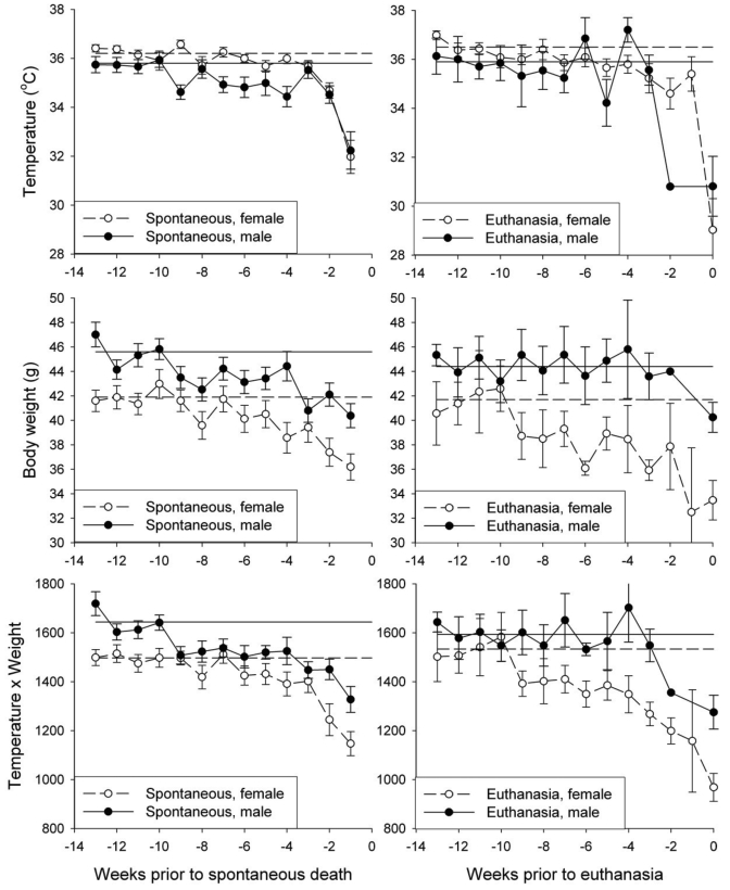
Gradual subtle weight loss was the earliest sign of deterioration in mice that either died spontaneously or underwent euthanasia (Figure 2). Individual mice gradually lost up to 20% of their body weight during the 3-mo period that preceded spontaneous death or euthanasia. Among the mice that died spontaneously, female mice weighed significantly (P ≤ 0.01, t test) less than male mice at both the start and end of the study (Table 1). However, the general pattern of decline was similar in both sexes.
Figure 2.
Temperature, body weight, and the temperature × weight in the final 13 wk of life. Values of temperature and body weight were aligned with respect to the time of collection prior to spontaneous death or euthanasia and are plotted in order of collection prior to death. Horizontal lines represent the average of all values collected during weeks 10 to 13 prior to death for all mice in that group.
Body temperatures were generally stable until the final few weeks of life but then fell by more than 1 °C (Figure 2). The temperatures of male and female mice did not differ significantly (P < 0.05) at either start or the end of the study (Table 1). Slow or labored respiration was observed in 14 of 24 euthanized mice and 39 of 81 mice that died spontaneously (Table 1). Among the mice that died spontaneously, respiratory problems were significantly (P ≤ 0.05, Fisher exact test) more frequent in female mice.
In general, body weights dropped gradually during the 2-mo period prior to death, either spontaneously or by euthanasia, and body temperature dropped precipitously during the preceding week. To permit simple use of both variables, the product of temperature and weight (T×W) was calculated for each mouse during the 3-mo period preceding either spontaneous death or euthanasia (Figure 2). Among mice that underwent euthanasia, the T×W value was significantly influenced by gender (P = 0.0002) and week (P ≤ 0.0001) without interaction (P = 0.19, ANOVA). Among mice that died spontaneously, values were significantly influenced by week (P ≤ 0.0001) but not gender (P = 0.71) with no interaction (P = 0.25, ANOVA).
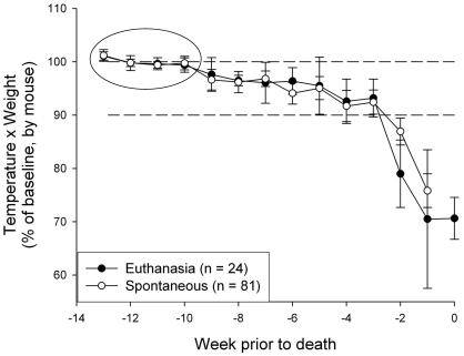
The absence of an interaction of week and gender with regard to T×W permitted combined analysis of both male and female mice as a function of time. This combined analysis was accomplished by transformation of the T×W values for each mouse into a percentage of the mean values collected for that mouse during the initial month of the terminal 3-mo interval (Figure 3A). With this transformation, 3 distinct phases of decline could be discerned. During the first 4 wk, which are assumed to provide the benchmark for normal health in these geriatric mice, values were generally stable. During the next 4 to 6 wk, values fell, on average, to between 100% and 90% of the benchmark mean. During the last 2 to 3 wk before death, values were generally less than 90% of the benchmark value. Patterns of decline were not different between mice that either died spontaneously or underwent euthanasia.
Figure 3.
Body weight and temperature in the last 14 wk of life, combining male and female mice. The panel represents the average of the calculated values of temperature × weight for each mouse. The circle highlights values that were averaged to generate the 100% value for each mouse, represented by the upper horizontal dashed line. In the last 2 wk of life, values fell to below 90% of the 100% benchmark (indicated as the lower dashed line).
As an example of the potential utility of euthanasia in anticipation of imminent death, serum cytokines and adipokines were measured in a random subset of mice that underwent euthanasia in this study (Table 2). These values were compared with those obtained from mice of the same strain that underwent euthanasia at a relatively young age (2 to 3 mo). Notably, values for some analytes were highly variable within the aged cohort. In addition, aged mice showed significant (P < 0.05) differences in some measures as compared with the young mice. For example, based on the adipokine assay, aged mice showed significantly (P < 0.05) higher levels of the inflammatory cytokines monocyte chemoattractant protein 1, total plasminogen activator inhibitor 1, IL6, and TNFα, whereas levels of resistin and leptin were lower (P < 0.05). In the Bio-Plex assay, a number of cytokines (IL3, IL12 p40 and p70, IL17) and RANTES were significantly (P < 0.05) lower in the aged mice as compared with the younger mice.
Table 2.
Adipokines and cytokines (pg/mL; mean ± SEM) in young and aged Hsd:ICR (CD1) mice
| Age at euthanasia |
||||
| 2 to 3 mo (n = 8) | 22 ± 2 mo (n = 7 or 8) | % | Pa | |
| Plasma adipokines | ||||
| Insulin | 439 ± 105 | 253 ± 74 | 58 ± 17 | 0.184 |
| Monocyte chemoattractant protein 1 | 19 ± 7 | 78 ± 24 | 411 ± 126 | 0.034 |
| IL6 | 2.6 ± 0.2 | 178 ± 97 | 59,333 ± 32,333 | 0.091 |
| TNFα | 5.3 ± 1.0 | 18.2 ± 4.8 | 393 ± 91 | 0.017 |
| Total plasminogen activator inhibitor 1 | 6429 ± 1234 | 14,503 ± 4250 | 226 ± 66 | 0.089 |
| Resistin | 2232 ± 174 | 1528 ± 274 | 68 ± 12 | 0.048 |
| Leptin | 789 ± 174 | 284 ± 170 | 36 ± 22 | 0.060 |
| Serum cytokinesb | ||||
| IL3 | 7.0 ± 0.5 | 3.5 ± 1.0 | 50 ± 14 | 0.007 |
| IL12 p40 | 559 ± 54 | 214 ± 59 | 38 ± 11 | 0.001 |
| IL12 p70 | 118 ± 18 | 52 ± 22 | 44 ± 19 | 0.033 |
| IL17 | 275 ± 13 | 120 ± 37 | 44 ± 14 | 0.002 |
| Granulocyte colony-stimulating factor | 63 ± 9 | 302 ± 120 | 479 ± 190 | 0.053 |
| IFNγ | 217 ± 13 | 150 ± 35 | 69 ± 17 | 0.094 |
| Chemokine (C-C motif) ligand 5 | 100 ± 10 | 34 ± 8 | 34 ± 8 | 0.001 |
Grubbs analysis uses transformed Z scores and P < 0.05 to identify outlier values. The Grubbs analysis revealed 1 outlier value for each of the following substances: insulin, leptin, TNFα (adipokine assay), IL3, granulocyte colony-stimulating factor, monocyte chemoattractant protein 1 (cytokine assay), and chemokine (C-C motif) ligand 5 (RANTES). These outlier values were in all cases more than 2.5 SD beyond the mean. These values were excluded from further analysis and were not included in calculation of the means, SEM, and P values shown in this table.
P value based on Student t test.
In the cytokine assay, trends (0.01 > P > 0.05) or significant differences (P < 0.05) were not detected for IL1α, IL1β, IL2, IL4, IL5, IL6, IL9, IL10, IL13, granulocyte–macrophage colony-stimulating factor, keratinocyte chemoattractant, monocyte chemoattractant protein 1, monocyte inhibitory proteins 1α and 1β, and TNFα.
Discussion
We propose that evaluation of aged mice based on reductions in core temperature and body weight can provide a reasonably valid benchmark for performing euthanasia on geriatric mice without markedly influencing outcomes in terms of longevity. Mice that died spontaneously had average life spans of 27.1 ± 0.8 mo (108 ± 3 wk) for males and 23.1 ± 0.8 mo (92 ± 3 wk) for females. Using the benchmark of a reduction in temperature × weight values to 90% of average stable values would theoretically have allowed prediction of death in these mice to within 2 wk. This degree of accuracy would translate into an underestimation of only about 2% in the total survival time. This underestimation may be acceptable for many studies, particularly if euthanasia allows the collection of valuable biologic samples that would be lost if the mouse died spontaneously. These markers can also be used to signal the need for increased observation or monitoring of specific animals in large colonies. Such increased monitoring could perhaps reveal additional markers that would more closely precede death, thereby potentially further refining the approach presented here.
Temperature and weight loss are often recommended as markers for disease severity, a moribund state, imminent death, and the need for euthanasia in experimental models with high likelihood of spontaneous death; these models most often involve infectious disease, sepsis, cancer, and toxicity.1,9,16,18,24,28,29,33,34 Suggested temperature benchmarks for euthanasia of mice range from 28 to 34 °C, depending on the disease model.9,23,30,34,35 Current technology allows rapid measurement of body temperature in mice yet requires minimal handling. Methods include surgically implanted abdominal transmitters, subcutaneous microchips, or surface body temperature.12,16,17,29,32,34 The use of subcutaneous microchips for surface body temperature appears feasible for large colonies of mice that are maintained for the duration of their life spans. However, several important considerations apply to using hypothermia as a marker for imminent death and preemptive euthanasia in mice. First, the body temperatures of mice can be influenced by numerous factors, including the time of day, ambient temperature, the presence and type of bedding, and the number of cage mates.7,8,20 For example, one study found that the hypothermia induced by injection of toxic doses of nickel was influenced markedly by acute exposure to low ambient temperatures.10 Second, different strains of mice can show widely different temperature responses to the same experimental challenge (for example, reference 29). Third, some measurements techniques (for example, rectal temperatures and scanned chips) provide a temperature only at a discrete time point, as opposed to a continuous pattern of change. Because the hypothermia may develop acutely in advance of imminent death, frequent sampling may be necessary in some experimental situations. Furthermore, suitable temperature benchmarks should be identified and validated for each specific experimental model.
Weight and weight loss are often considered as benchmarks of wellbeing in animal models of acute and chronic disease. However, use of these measures can be complex, and they do not always indicate the severity of illness or the likelihood of imminent death.16 Several examples illustrate this complexity. First, in a study of bacterial endotoxemia in mice, weight loss was not found to be an accurate predictor of death.14 Second, in a study of endotoxemic shock induced by cecal ligation and puncture in mice, weight gain was a predictor of imminent death, whereas weight loss was not.16 Third, in a study of the survival of rats with CNS tumors, weight loss for 7 or 8 consecutive days signaled imminent death, although relative weight loss and reduced food intake were not predictive.22 For tumor models in particular, body condition scoring or evaluation of emaciation, rather than absolute body weight, is reported to provide a more accurate assessment of animal health and wellbeing.21,31,33 Finally, with regard to longevity, a recent report26 found that the combined measurement of body weight and T-cell subsets prior to 2 y of age can allow prediction of the lifespan quartile of an individual mouse with 35% accuracy. However, this degree of precision may be inadequate for many studies.
Benchmark values for weight and weight loss can be difficult to glean from the literature, because some studies present such data as experimental findings, rather than as criteria for assessment of wellbeing or implementation of euthanasia.2,5,11,27 For example, a study of experimental rabies reported that infected animals lost 15% of their body weight,2 and a study of avian influenza virus reported that mice with genetic deletion of receptors for interferon α/β experienced more weight loss and died more rapidly than did genetically normal control mice.27 Some authorities recommend using weight loss to assess wellbeing but do not offer guidelines or benchmarks for amount of weight loss that would be acceptable.15,18
Our study was conducted in an effort to add to the body of knowledge available to improve both animal welfare and scientific discovery. We suggest that monthly or weekly determination of the product of body weight and body temperature provides a benchmark that can signal the need for close (for example, daily) monitoring of individual mice for weight loss, hypothermia, and respiratory difficulty as penultimate markers of imminent death. According to our data, a sustained reduction to between 100% and 90% of stable basal values signals the need for either euthanasia or closer monitoring of individual mice. We suggest maintaining a benchmark value that is recalculated to incorporate values obtained during weeks 10 to 13 prior to the current date, as illustrated in our study. Using this marker appears to allow prediction of death to within 1 or 2 wk, even among mice that die without obvious clinical signs. Among the mice we studied here, 1 wk represents on average about 1% of the median lifespan. Another alternative is to maintain actual, rather than percentage, benchmark values for temperature, body weight, and T×W that are calculated based on values collected from all male or female mice during a given index interval (for example, 3 mo prior to the current date). This approach, which essentially is illustrated in Figure 2 without showing the 90% benchmark value for male and female mice, allows quick determinations of a deteriorating condition in any mouse based on measurements made in real time, without the need to refer to records for individual mice. Values from any individual mouse that reaches the group benchmark could carefully be reviewed for determination of whether that animal should either undergo euthanasia or be signaled out for closer individual assessment. Closer evaluation of high-risk mice with attention to these and other variables (for example, capillary refill time, hemaocrit) potentially could allow even more accurate prediction. Other benchmarks may also be identified and could be used after validation.
By taking advantage of markers such as those we suggest here, pathophysiologic changes in aged mice can be studied in the context of clinical deterioration that predicts or precipitates end-of-life decline. To exemplify such potential, we measured serum cytokines, chemokines, and adipokines in aged mice that underwent euthanasia based on clinical signs. Our data suggest that the mice that died by euthanasia would have died spontaneously within 1 or 2 wk. Had spontaneous death occurred, their terminal concentrations of cytokines, chemokines, and adipokines could not have been measured. The elevation in cytokines and chemokines in serum of mice that underwent euthanasia is consistent with age-related chronic inflammation that develops in healthy and frail elderly individuals, including mice.4,13,25 Cytokines that are generated during inflammation can influence the brain, directly or indirectly, to induce common symptoms of illness, including loss of appetite, sleepiness, withdrawal from normal social activities, fever, and fatigue.6 Prolonged activation of an inflammatory response and associated changes in the production of pro- and antiinflammatory cytokines may contribute to the development of fatigue during aging and age-related chronic disease.4,6,13 For example, aging is accompanied by 2- to 4-fold increases in circulating concentrations of inflammatory mediators, including IL6.3,13 Although the magnitude of these age-related increases is far below what would arise in response to acute infection, these elevated levels nonetheless suggest increased inflammatory ‘tone’ in elderly mice. The high variability we observed in the aged mice could be related to variation in the specific causes of deteriorating health among the mice. Most importantly for purposes of this study, despite high variability among aged mice and discrepancies between some measures in the adipokine and cytokine assays, collecting serum and obtaining such measurements would have been impossible had these mice died spontaneously.
Another clear advantage of an ability to predict imminent death in mice is related to the rapid autolysis that mice undergo after death. This deterioration can render accurate determination of the cause of death essentially impossible even in mice that have been dead for only a few hours. Furthermore, in addition to the advantages of allowing accurate diagnosis of the cause of death and of performing postmortem studies or analyses using tissue collected from dying mice, the combined detection of subtle decreases in health status and the accurate prediction of imminent death, as reported in our current study, could also provide a temporal signal for performing in vivo assessments relevant to the pathophysiologic mechanisms that contribute to end-of-life decline in elderly mice.
Determination of humane endpoints for laboratory animals is increasingly important to institutional animal care and use committees and regulatory agencies. Technologic advances now increase the feasibility of gathering data relevant to the identification of markers of illness in rodents. In light of our data, we suggest that investigators who conduct similar studies of aging and longevity using other strains of mice validate our approach in their ongoing studies and report their findings to the scientific community.
Acknowledgments
This work was supported in part by NIH grant K26-RR17543 and the Southern Illinois University School of Medicine. We thank Christine Bosgraaf and Lisa Cox for providing excellent technical assistance.
References
- 1.Aldred AJ, Cha MC, Meckling-Gill KA. 2002. Determination of a humane endpoint in the L1210 model of murine leukemia. Contemp Top Lab Anim Sci 41:24–27 [PubMed] [Google Scholar]
- 2.Blum SA, Braunschweiger M, Kramer B, Rubmann P, Duchow K, Cubetaler K. 1998. How to prove complete virus inactivation in rabies vaccines: a comparison of an in vivo to an in vitro method. ALTEX 15:46–49 [PubMed] [Google Scholar]
- 3.Bruunsgaard H. 2002. Effects of tumor necrosis factor α and interleukin 6 in elderly populations. Eur Cytokine Netw 13:389–391 [PubMed] [Google Scholar]
- 4.Bruunsgaard H, Pedersen BK. 2003. Age-related inflammatory cytokines and disease. Immunol Allergy Clin North Am 23:15–39 [DOI] [PubMed] [Google Scholar]
- 5.Cubetaler K, Morton D, Hendriksen CF. 1998. Humane endpoints as a replacement for the estimation of lethality rates in the potency testing of rabies vaccines. ALTEX 15:40–42 [PubMed] [Google Scholar]
- 6.Dantzer R, Kelley KW. 2007. Twenty years of research on cytokine-induced sickness behavior. Brain Behav Immun 21:153–160 [DOI] [PMC free article] [PubMed] [Google Scholar]
- 7.Gordon CJ. 2004. Effect of cage bedding on temperature regulation and metabolism of group-housed female mice. Comp Med 54:63–68 [PubMed] [Google Scholar]
- 8.Gordon CJ, Becker P, Ali JS. 1998. Behavioral thermoregulatory responses of single- and group-housed mice. Physiol Behav 65:255–262 [DOI] [PubMed] [Google Scholar]
- 9.Gordon CJ, Fogelson L, Highfill JW. 1990. Hypothermia and hypometabolism: sensitive indices of whole-body toxicity following exposure to metallic salts in the mouse. J Toxicol Environ Health 29:185–200 [DOI] [PubMed] [Google Scholar]
- 10.Gordon CJ, Fogelson L, Stead AG. 1989. Temperature regulation following nickel intoxication in the mouse: effect of ambient temperature. Comp Biochem Physiol C 92:73–76 [DOI] [PubMed] [Google Scholar]
- 11.Hartinger J, Folz T, Cussler K. 2001. Clinical endpoints during rabies vaccine control tests. ALTEX 18:37–40 [PubMed] [Google Scholar]
- 12.Kort WJ, Hekking-Weijima JM, TenKate MT, Somm V, VanStrik R. 1998. A microchip implant system as a method to determine body temperature of terminally ill rats and mice. Lab Anim 32:260–269 [DOI] [PubMed] [Google Scholar]
- 13.Krabbe KS, Pedersen M, Bruunsgaard H. 2004. Inflammatory mediators in the elderly. Exp Gerontol 39:687–699 [DOI] [PubMed] [Google Scholar]
- 14.Krarup A, Chattopadhyay P, Bhattacharjee AK, Burge JR, Ruble GR. 1999. Evaluation of surrogate markers of impending death in the galactosamine-sensitized murine model of bacterial endotoxemia. Lab Anim Sci 49:545–550 [PubMed] [Google Scholar]
- 15.Morton DB. 2000. A systematic approach for establishing humane endpoints. ILAR J 41:80–86 [DOI] [PubMed] [Google Scholar]
- 16.Nemzek JA, Xiao HY, Minard AE, Bolgos GL, Remick DG. 2004. Humane endpoints in shock research. Shock 21:17–25 [DOI] [PubMed] [Google Scholar]
- 17.Newsom DM, Bolgos GL, Colby L, Nemzek JA. 2004. Comparison of body surface temperature measurement and conventional methods for measuring temperature in the mouse. Contemp Top Lab Anim Sci 43:13–18 [PubMed] [Google Scholar]
- 18.Olfert ED, Goodson DL. 2000. Humane endpoints for infectious disease animal models. ILAR J 41:99–104 [DOI] [PubMed] [Google Scholar]
- 19.Organization for Economic Cooperation and Development [Internet] 2000. Guidance document on the recognition, assessment, and use of clinical signs as humane endpoints for experimental animals used in safety evaluation. Environmental health and safety publication series on testing and assessment no. 19 [Cited Oct 2009]. Available at http://www.oecd.org/dataoecd/31/33/44229192.pdf [Google Scholar]
- 20.Overton JM, Williams TD. 2004. Behavioral and physiologic responses to caloric restriction in mice. Physiol Behav 81:749–754 [DOI] [PubMed] [Google Scholar]
- 21.Paster EV, Villines KA, Dickman DL. 2009. Endpoints for mouse abdominal tumor models: refinement of current criteria. Comp Med 59:234–241 [PMC free article] [PubMed] [Google Scholar]
- 22.Redgate ES, Deutsch M, Boggs SS. 1991. Time of death of CNS tumor-bearing rats can be reliably predicted by body weight-loss patterns. Lab Anim Sci 41:269–273 [PubMed] [Google Scholar]
- 23.Soothill JS, Morton DB, Ahmad A. 1992. The HID50 (hypothermia-inducing dose 50): an alternative to the LD50 for measurement of bacterial virulence. Int J Exp Pathol 73:95–98 [PMC free article] [PubMed] [Google Scholar]
- 24.Stiles BG, Campbell YG, Castle RM, Grove SA. 1999. Correlation of temperature and toxicity in murine studies of staphylococcal enterotoxins and toxic shock syndrome toxin 1. Infect Immun 67:1521–1525 [DOI] [PMC free article] [PubMed] [Google Scholar]
- 25.Swain SL, Nikolich-Zugich J. 2009. Key research opportunities in immune system aging. J Gerontol A Biol Sci Med Sci 64:183–186 [DOI] [PMC free article] [PubMed] [Google Scholar]
- 26.Swindell WR, Harper JM, Miller RA. 2008. How long will my mouse live: machine learning approaches for prediction of mouse life span. J Gerontol A Biol Sci Med Sci 63:895–906 [DOI] [PMC free article] [PubMed] [Google Scholar]
- 27.Szretter KJ, Gangappa S, Belser JA, Zeng H, Chen H, Matsuoka Y, Sambhara S, Swayne DE, Tumpay TM, Katz JM. 2009. Early control of H5N1 influenza virus replication by the type I interferon response in mice. J Virol 83:5825–5834 [DOI] [PMC free article] [PubMed] [Google Scholar]
- 28.Toth LA. 2000. Defining the moribund condition as an experimental endpoint for animal research. ILAR J 41:72–79 [DOI] [PubMed] [Google Scholar]
- 29.Toth LA, Hughes LF. 2006. Sleep and temperature responses of inbred mice with Candida albicans-induced pyelonephritis. Comp Med 56:252–261 [PubMed] [Google Scholar]
- 30.Toth LA, Rehg JE, Webster RG. 1995. Strain differences in sleep and other pathophysiological sequelae of influenza virus infection in naive and immunized mice. J Neuroimmunol 58:89–99 [DOI] [PubMed] [Google Scholar]
- 31.Ullman-Cullere MH, Foltz CJ. 1999. Body condition scoring: a rapid and accurate method for assessing health status in mice. Lab Anim Sci 49:319–323 [PubMed] [Google Scholar]
- 32.Vlach KD, Boles JW, Stiles BG. 2000. Telemetric evaluation of body temperature and physical activity as predictors of mortality in a murine model of staphylococcal enterotoxic shock. Comp Med 50:160–166 [PubMed] [Google Scholar]
- 33.Wallace J. 2000. Humane endpoints and cancer research. ILAR J 41:87–93 [DOI] [PubMed] [Google Scholar]
- 34.Warn PA, Brampton MW, Sharp A, Morrissey G, Steel N, Denning DW, Priest T. 2003. Infrared body temperature measurement of mice as an early predictor of death in experimental fungal infections. Lab Anim 37:126–131 [DOI] [PubMed] [Google Scholar]
- 35.Wong ML, Bongiorno PB, Rettori V, McCann SM, Licinio J. 1997. Interleukin (IL) 1β, IL1 receptor antagonist, IL10, and IL13 gene expression in the central nervous system and anterior pituitary during systemic inflammation: pathophysiological implications. Proc Natl Acad Sci USA 94:227–232 [DOI] [PMC free article] [PubMed] [Google Scholar]


