History
A 12 year old girl reported to our department with the chief complaint of a swelling below the tongue producing difficulty in speaking and in closing her mouth for past one year.
Clinical Features
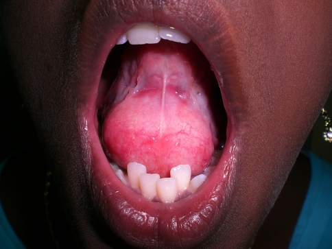
Examination revealed the presence of a solitary midline swelling in the sublingual region measuring 4 × 3.5 cm, which was non-tender, fluctuant, soft, non-mobile with the overlying mucosa showing no secondary changes. There were no inflammatory signs or lymphadenopathy associated with the swelling (Fig. 1). Transillumination was negative.
Fig. 1.
Clinical presentation of a large sub-lingual, midline swelling
Pre-Operative Investigations
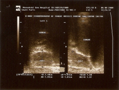
Ultrasonogram: Ultrasonographic study suggested that the mass was a unilocular cystic lesion (Fig. 2), adherent to and moving with the frenum and the tongue.
Aspiration cytology: Aspiration cytology revealed a cheesy material, which contained numerous non nucleated epithelial cells.
Fig. 2.
Ultra-sonogram demonstrating a uni-locular cystic lesion
Treatment
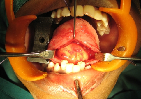
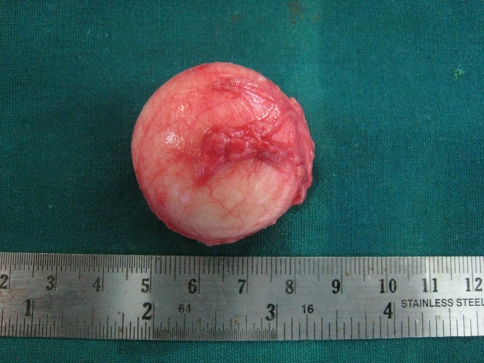
The aspiration cytology helped us arrive at a working diagnosis of an epidermiod or dermoid cyst. The patient was then prepared for surgery under general anesthesia. An intraoral midline incision extending from the base of the tongue to the floor of the mouth was used to access the lesion (Fig. 3). Special attention was paid to Whartons ducts which were dissected and protected bilaterally. The cyst was completely exposed and on evaluation no caudal herniation through the mylohyoid muscle was seen. A combination of sharp and blunt dissection was used to free the cyst and it was delivered per oral in total (Fig. 4). The wound was closed in layers with a non-vacuum drain kept in situ for 24 h.
Fig. 3.
Intra-oral midline incision from the base of the tongue to the floor of the mouth providing excellent exposure of the lesion
Fig. 4.
Specimen measuring approximately 4 cm in diameter delivered in total
Histopathology
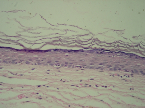
Examination with H&E staining showed a cystic lesion with stratified squamous epithelial lining and fibro-vascular connective tissue capsule covering the cystic lumen (Fig. 5).
Fig. 5.
Photomicrograph showing ortho-keratinised stratified squamous epithelium with flat epithelial-connective tissue interface lining the cystic cavity (H&E staining under 40 × magnification)
Discussion
Epidermoid and dermoid cysts of the oral cavity represent less than 0.01% of all oral cavity cysts [1]. The cyst is described as epidermoid when the lining presents only epithelium, dermoid when skin adnexa are found and as teratoid cyst when other tissues like muscle, cartilage, bone are present within the cyst [2]. Histologically, this distinction of the cysts in the floor of the mouth was presented by Meyer in 1955 [3]. Dermoid cysts of the floor of the mouth are dis-embryogenetic lesions derived from entrapment and subsequent growth of epithelial cells during the midline fusion between the first and second branchial arches in the third and fourth embryonic weeks [4]. Acquired forms are derived from either iatrogenic or traumatic inclusion of epithelium and skin appendages. Dermoid cysts are generally diagnosed in the second and third decades of life, however can present in all ages [4]. Anatomically, three different types of dermoid cysts can be distinguished; median genio-glossal (sublingual), median geniohyoid (submental), and lateral, according to the anatomic relationship between the cyst and the muscles of the floor of the mouth [5]. The floor of the mouth is the second most common site for dermoid cysts in the head and neck region after the lateral eyebrow [6].
Meyer divided the cysts of the floor of the mouth into 3 groups histologically, epidermoid, dermoid, and teratoid. Although dermoid cysts represent a separate entity, the term “dermoid” is generally used to indicate all 3 categories: (a) Epidermoid cyst—lined with simple squamous epithelium with a fibrous wall and no adnexal structures, (b) True dermoid cyst—an epithelial-lined cavity with keratinization and skin appendages (sebaceous and sweat glands and hair follicles in the cyst wall), also known as a compound cyst and (c) Teratoid cyst—lined with a range of epithelia, from simple squamous epithelium to ciliated respiratory type, containing derivatives of ectoderm, mesoderm, and endoderm, also known as a complex cyst [6]. All 3 types contain a cheesy keratinous material.
Treatment is by enucleation via an intraoral or extraoral approach. An intraoral approach is recommended by most authors for sublingual cysts of small or moderate dimensions above the mylohyoid muscle, whereas an extraoral approach is preferred for larger sublingual cysts [5].
Recurrence is very rare with complete excision of the lesion however a 5% rate of malignant transformation of oral dermoid cysts of the teratoid type has been reported in literature [7].
References
- 1.Rajayogeswaran V, Eveson JW. Epidermoid cyst of the buccal mucosa. Oral Surg Oral Med Oral Pathol. 1989;67:181–184. doi: 10.1016/0030-4220(89)90326-5. [DOI] [PubMed] [Google Scholar]
- 2.Calderon S, Kaplan I. Concomitant sublingual and submental epidermoid cysts: a case report. J Oral Maxillofac Surg. 1993;51:790–792. doi: 10.1016/S0278-2391(10)80425-2. [DOI] [PubMed] [Google Scholar]
- 3.Rapidis AD. Dermoid cyst of the floor of the mouth. Report of a case. Br J Oral Surg. 1981;19:43–51. doi: 10.1016/0007-117X(81)90020-2. [DOI] [PubMed] [Google Scholar]
- 4.Kim IK, Kwak HJ, Choi J, Han JY, Park SW. Coexisting sublingual and submental dermoid cysts in an infant. Oral Surg Oral Med Oral Pathol Oral Radiol Endod. 2006;102:778–781. doi: 10.1016/j.tripleo.2005.09.016. [DOI] [PubMed] [Google Scholar]
- 5.Di Francesco A, Chiapasco M, Biglioli F, Ancona D. Intraoral approach to large dermoid cysts of the floor of the mouth: a technical note. Int J Oral Maxillofac Surg. 1995;24:233–235. doi: 10.1016/S0901-5027(06)80135-9. [DOI] [PubMed] [Google Scholar]
- 6.Teszler CB, El-Naaj IA, Emodi O, Luntz M, Peled M. Dermoid cysts of the lateral floor of the mouth: a comprehensive anatomo-surgical classification of cysts of the oral floor. J Oral Maxillofac Surg. 2007;65:327–332. doi: 10.1016/j.joms.2005.06.022. [DOI] [PubMed] [Google Scholar]
- 7.Kandogan T, Koç M, Vardar E, Selek E, Sezgin O. Sublingual epidermoid cyst: a case report. J Med Case Reports. 2007;17(1):87. doi: 10.1186/1752-1947-1-87. [DOI] [PMC free article] [PubMed] [Google Scholar]


