Abstract
PURPOSE
To measure the tear meniscus dynamics in aqueous tear deficiency dry eye patients using optical coherence tomography (OCT).
DESIGN
Clinical research study of a laboratory technique.
METHODS
Twenty-five aqueous tear deficiency dry eye patients and thirty healthy subjects were recruited. Upper and lower tear menisci of one randomly selected eye of each participant were imaged during normal and delayed blinking using OCT. Measured parameters included upper tear meniscus height and volume, lower tear meniscus height and volume, the blink outcome defined as the meniscus volume change during blink action, and open eye outcome defined as the meniscus volume change during open eye period.
RESULTS
During normal blinking, both tear meniscus height and volume before blink in dry eye patients were significantly smaller than those in healthy subjects, except for the upper tear meniscus volume. During normal blinking, the blink outcome and open eye outcome of lower tear meniscus were significantly smaller in dry eye patients compared to healthy subjects. During delayed blinking, the upper and lower tear menisci heights and volumes significantly increased in both groups. However, dry eye patients had smaller increases than healthy subjects. During delayed blinking, the open eye outcomes of upper and lower tear menisci were smaller in dry eye patients than healthy subjects.
CONCLUSIONS
Dry eye patients appeared to have reduced tear meniscus dynamics during normal blinking and smaller increases of meniscus volume during delayed blinking. Analysis of tear meniscus dynamics may provide more insight in the altered tear system in dry eye patients.
INTRODUCTION
The tear system is dynamic and blinking plays the important role of distributing the tears on the ocular surface so that physiologic equilibrium is maintained.1–3 Blinking redistributes the tears to the ocular surface from the tear menisci around both upper and lower eyelids, and it facilitates tear drainage.3–5 Blinking may also influence tear evaporation.6 Dynamic changes of the tear menisci are the result of the interaction between blinking and tears, which has been studied in normal subjects.7 In dry eye patients, lack of the tears may result in abnormal tear distribution on the ocular surface and irregular interaction during blinking. Distortion of the tear dynamics and the tear distribution on ocular surface with each blink may contribute to ocular discomfort and dryness, which are the prevalent complaints by dry eye patients with aqueous tear deficiency (ATD).8–11 The tear system has been evaluated in many studies by measuring tear breakup time, wetting of Schirmer strips, and determining lower tear meniscus dimensions in aqueous tear deficiency dry eye patients.1,11–13 However, tear meniscus dynamics during the blink cycle in dry eye patients remains untested, mainly due to the difficulty of real-time imaging in capturing the alterations of the tear system. Knowledge of these dynamics in dry eye patients may lead to better understanding of the etiology of the tear deficiency and its resulting damage to the ocular surface. In this study, we used real-time optical coherence tomography (OCT) to measure tear meniscus dynamics in aqueous tear deficiency dry eye patients.
SUBJECTS AND METHODS
Twenty-five clinically diagnosed aqueous tear deficiency dry eye patients (15 women and 10 men, mean ± standard deviation age: 49.6 ± 17.4 years) and thirty healthy subjects (17 women and 13 men, age: 31.0 ± 8.4 years) were enrolled. Dry eye patients who complained ocular discomfort were evaluated and diagnosed at Bascom Palmer Eye Institute, University of Miami Miller School of Medicine and Department of Ophthalmology, University of Rochester. They had no ocular diseases except for dry eye, and no history of ophthalmic surgery and trauma. The patients were recruited in aqueous tear deficiency dry eye group if they had Schirmer I test score less than 5 mm in 5 minutes, tear breakup time less than 10 seconds, no Meibomian glands or eyelid diseases through slit-lamp examination, and one or more of the dry eye symptoms in the questionnaire occurring at least often.14,15 The healthy subjects, serving as the control group, were recruited as they had no history of ocular discomfort, ocular diseases, contact lens wear and had a Schirmer I test score more than 10 mm.
The specification of the custom-built OCT instrument and the experimental procedure were described in previous studies.7,16,17 The repeatability of tear meniscus measurement with the OCT has been proved.18 In present study, central air conditioning and two humidifiers maintained the temperature at 15 to 25% and the relative humidity at 30 to 50%. Ambient room light was used while imaging each subject. The subjects were evaluated by OCT after 10:00 am and were required not to use eye drops 24 hours prior to the visit. One randomly selected eye of each subject was tested. The custom-built OCT instrument with real-time scanning at 8 frames per second made 12 mm scans at the vertical meridian across the corneal apex. The subjects were asked to look at an external target while blinking normally (normal blinking). The upper and lower tear menisci were monitored simultaneously using OCT. Then, two consecutive normal blink actions with one blink interval (open eye period) were recorded. After that, the subjects were asked to delay each blink as long as possible (delayed blinking), and the video including two consecutive delayed blink actions with one blink interval were obtained.
The frames acquired immediately before and after each blink action were processed with custom software as detailed in our previous studies.7,19 In brief, both upper and lower tear meniscus variables were obtained, including upper and lower tear meniscus heights and cross-sectional areas. Tear meniscus volumes in upper and lower tear menisci were calculated as described in previous studies. The volume was calculated as the product of the meniscus cross-sectional area, the constant eyelid length of 25 mm, and a factor of 0.75.19,20 Since we recorded two consecutive blink actions, the meniscus variables obtained before the two blink actions were averaged. The meniscus variables obtained after the two blink actions were averaged as the same way. Since the two consecutive blink actions formed one open eye period, the meniscus variables at the beginning of open eye period were obtained after the first blink action. Similarly, the meniscus variables at the end of open eye period were obtained before the second blink action.7,19 The blink outcome was defined as the difference in meniscus volume before and after blink action. The open eye outcome was calculated as the difference in meniscus volume at the beginning and the end of the open eye period.19
All the data were analyzed electronically. Paired t-test was used to identify the differences in tear meniscus variables between normal blinking and delayed blinking. Independent t-test was performed to compare the meniscus variables between dry eye group and control group.
RESULTS
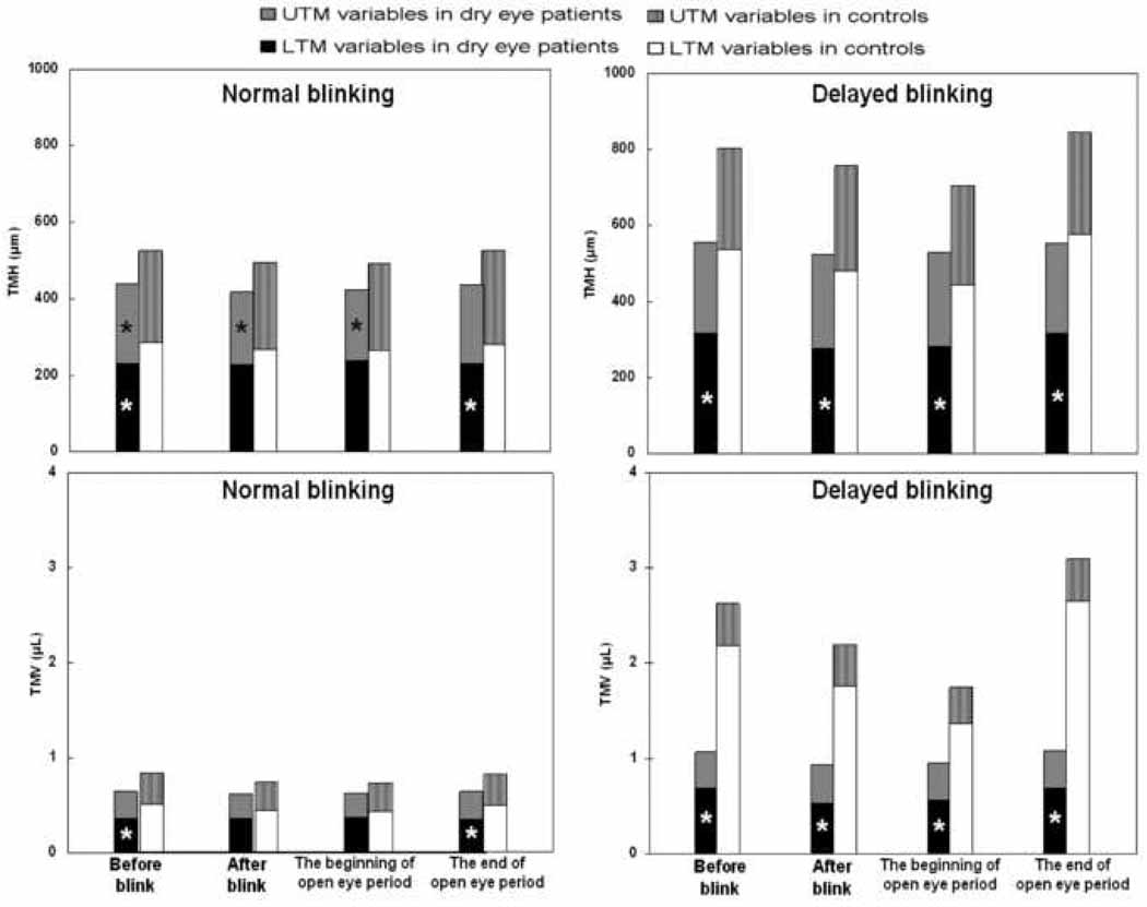
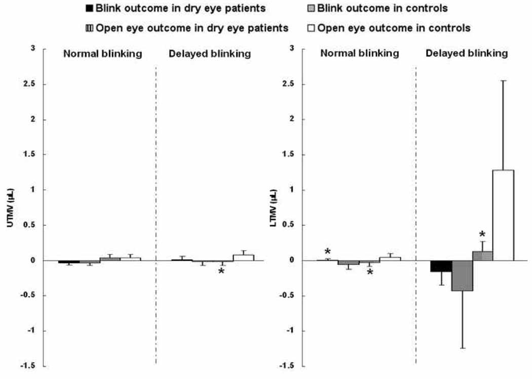
With normal blinking in dry eye patients, the upper tear meniscus height measured before and after blink, and at the beginning of open eye period was significantly smaller than those in controls (P < .05, Table 1, Fig. 3). However, there were no significant differences in upper tear meniscus volume (P > .05). During normal blinking, lower tear meniscus height and volume measured before blink and at the end of open eye period were smaller in dry eye patients compared to controls (P < .05, Table 1, Fig. 3). The dry eye patients also had smaller blink outcome and open eye outcome in lower tear meniscus volume (P < .05, Table 2, Fig. 4).
Table 1.
Tear meniscus height and volume during normal and delayed blinking in dry eye patients and control subjects.
| UTMH (µm) | UTMV (µL) | LTMH (µm) | LTMV (µL) | |||||
|---|---|---|---|---|---|---|---|---|
| Dry eye | Controls | Dry eye | Controls | Dry eye | Controls | Dry eye | Controls | |
| Normal blinking | ||||||||
| Blink action | ||||||||
| Before blink | ||||||||
| Mean | 207* | 241 | 0.28 | 0.34 | 233* | 287 | 0.36* | 0.50 |
| ± SD | 74 | 66 | 0.20 | 0.16 | 90 | 100 | 0.25 | 0.33 |
| After blink | ||||||||
| Mean | 190* | 230 | 0.25 | 0.31 | 227 | 268 | 0.36 | 0.44 |
| ± SD | 87 | 57 | 0.20 | 0.14 | 93 | 97 | 0.28 | 0.32 |
| Open eye period | ||||||||
| The beginning of open eye period | ||||||||
| Mean | 187* | 230 | 0.25 | 0.30 | 237 | 265 | 0.38 | 0.43 |
| ± SD | 89 | 66 | 0.20 | 0.14 | 100 | 97 | 0.33 | 0.35 |
| The end of open eye period | ||||||||
| Mean | 205 | 242 | 0.29 | 0.34 | 232* | 283 | 0.35* | 0.49 |
| ± SD | 88 | 79 | 0.21 | 0.19 | 92 | 103 | 0.23 | 0.35 |
| Delayed blinking | ||||||||
| Blink action | ||||||||
| Before blink | ||||||||
| Mean | 241 | 269 | 0.38 | 0.44 | 317* | 538 | 0.69* | 2.19 |
| ± SD | 73 | 73 | 0.27 | 0.20 | 169 | 464 | 0.90 | 3.73 |
| After blink | ||||||||
| Mean | 244 | 278 | 0.39 | 0.44 | 278* | 479 | 0.54* | 1.76 |
| ± SD | 87 | 81 | 0.32 | 0.23 | 122 | 390 | 0.48 | 3.42 |
| Open eye period | ||||||||
| The beginning of open eye period | ||||||||
| Mean | 246 | 257 | 0.40 | 0.37 | 283* | 445 | 0.56* | 1.37 |
| ± SD | 93 | 79 | 0.34 | 0.20 | 118 | 338 | 0.49 | 2.32 |
| The end of open eye period | ||||||||
| Mean | 237 | 275 | 0.39 | 0.43 | 318* | 577 | 0.69* | 2.66 |
| ± SD | 80 | 76 | 0.32 | 0.20 | 150 | 558 | 0.78 | 4.73 |
UTMH, upper tear meniscus height; UTMV, upper tear meniscus volume; LTMH, lower tear meniscus height; LTMV, lower tear meniscus volume.
The asterisks (*) indicate the tear meniscus variables in dry eye patients were significantly smaller than those in controls (P < .05).
Figure 3. Tear meniscus height and volume during normal and delayed blinking in dry eye patients and control subjects.
Both upper and lower tear menisci heights (Top right) and volumes (Bottom right) during delayed blinking were significantly larger than those during normal blinking (Top left, Bottom left) in both dry eye patients and controls (P < .05). The asterisks (*) indicate the tear meniscus variables in dry eye patients were significantly smaller than those in controls (P < .05). UTM, upper tear meniscus; LTM, lower tear meniscus; TMH: tear meniscus height; TMV: tear meniscus volume.
Table 2.
Blink outcome and open eye outcome of tear meniscus volume during normal and delayed blinking in dry eye patients and control subjects.
| UTMV (µL) | LTMV (µL) | |||
|---|---|---|---|---|
| Dry eye | Controls | Dry eye | Controls | |
| Normal blinking | ||||
| Blink outcome | ||||
| Mean | −0.03 | −0.03 | 0.004* | −0.06 |
| ± SD | 0.07 | 0.11 | 0.06 | 0.15 |
| Open eye outcome | ||||
| Mean | 0.04 | 0.04 | −0.03* | 0.05 |
| ± SD | 0.13 | 0.13 | 0.13 | 0.12 |
| Delayed blinking | ||||
| Blink outcome | ||||
| Mean | 0.02 | −0.01 | −0.16 | −0.43 |
| ± SD | 0.11 | 0.14 | 0.46 | 1.97 |
| Open eye outcome | ||||
| Mean | −0.01* | 0.07 | 0.13* | 1.29 |
| ± SD | 0.14 | 0.17 | 0.36 | 3.08 |
UTMV, upper tear meniscus volume; LTMV, lower tear meniscus volume.
The asterisks (*) indicate the outcomes in dry eye patients were significantly smaller than those in controls (P < .05).
Figure 4. Blink outcome and open eye outcome of tear meniscus volume during normal and delayed blinking in dry eye patients and control subjects.
During normal blinking, dry eye patients had smaller blink outcome and open eye outcome of lower tear meniscus volume during normal blinking (Middle right). Furthermore, open eye outcomes of both tear menisci in dry eye patients were smaller during delayed blinking compared to controls (Middle left, Far right). The asterisks (*) indicate the outcomes in dry eye patients were significantly smaller than those in controls (P < .05). Vertical bars denote 95% confidence intervals. UTMV: upper tear meniscus volume; LTMV: lower tear meniscus volume.
During delayed blinking, all variables of upper and lower tear menisci in both dry eye and control groups were significantly increased compared to normal blinking (P < .05, Table 1, Fig. 3). There were no significant differences in upper tear meniscus height and volume between groups (P > .05). In contrast, the lower tear meniscus height and volume in dry eye patients were significantly smaller than those in controls (P < .05, Table 1, Fig. 3). Furthermore, the open eye outcomes of both upper and lower tear menisci were smaller in dry eye patients compared to controls (P < .05, Table 2, Fig. 4). However, there were no significant differences in blink outcomes between groups (P > .05).
DISCUSSION
Using a custom-built, real-time OCT instrument with a wide scan width, Wang and associates16,18 were the first to image both upper and lower tear menisci simultaneously to investigate the tear dynamics during the blink cycle. In normal subjects, the tear system is highly dynamic and alterations in the menisci occurred during blinking.7 Using a similar device, Shen et al.21 studied the tear menisci in dry eye patients and demonstrated the diagnostic value of dry eye using OCT instrument. According to the published data in the measurement of tear menisci in normal and dry eye patients using a similar device, a sample size of 10 subjects in each group would be enough to detect the true difference of the tear menisci with detection power of 0.9, according to a software program (Gpower, Ver. 3.0) developed by Erdfelder et al.22 In the present study, double the sample size would ensure the enough power to detect the true difference of the tear meniscus changes by blinking. The present study extends our understanding of tear meniscus dynamics in dry eye patients. As expected, smaller dimensions of the tear menisci were found in dry eye patients, which is in agreement with previous studies.13,23–25 The measured values of healthy subjects were in agreement with that of other studies as well.23–31 Most importantly, the method has revealed the lower outcomes of tears in dry eye patients, which means that the tear meniscus dynamics are reduced compared to normal subjects. Potentially, OCT imaging of tear dynamics holds promise for the development of a new, less invasive, more rapid, and more reliable diagnosis of dry eye in routine clinical exams.
Changes in the regulated tear system, such as reductions in blink outcome and open eye outcome, are the basis of the reduced tear meniscus dynamics in the dry eye patients. With sufficient volume in healthy subjects, blinking spreads the tears onto the ocular surface, and then upper and lower tear menisci possibly exchange content when they meet. The drainage system removes tears by drawing the tears into the canaliculi by a negative pressure.3 During the open eye period, freshly secreted tears are collected in the upper tear meniscus and transferred into the lower tear meniscus via the connection at the junctions between upper and lower lids.3,7 In dry eye patients, lower tear volume and reduced tear secretion were well documented.11,13,23,24 This resulted in the reduced open eye outcome during normal blinking in present study, since not much tears were added into the tear system compared to controls. Extremely small blink outcome of lower tear meniscus may indicate the possible shutting down of the drainage system. This helps to maintain the presence of tears on the ocular surface. Thus, the system becomes less dynamic in response to the aqueous deficient situation.
Based on these observations, we hypothesize that there exists an auto-regulatory mechanism in the tear system that adjusts ocular tear volume through changes in tear secretion and tear drainage. With sufficient tear volume, the drainage opens and excessive tears are removed. With deficient tear volume, shutting down of the drainage helps to preserve the tears. Although the actual mechanism of the regulating system is unclear, some hints exist, which suggests the presence of auto-regulatory mechanism of tear system. Francois et al.32 found that decreased tear outflow results from a decrease of tear secretion, and suggested that tear drainage might adjust to the tear production via sensitive receptors in the lacrimal sac. Tsubota and Yamada33 developed a device covering the eye to evaluate the humidity in healthy and dry eye patients. They suggested that tear production, drainage, and evaporation were interlinked and maintained a low level of tears in dry eye patients. Though the dry eye patients adjust the drainage and try to preserve tears to distribute tear film on ocular surface by blinking,1–3 the decreased drainage may lead inappropriately to decreased production.32 This may result in a vicious cycle that produces even fewer tears. Thus the tear system becomes less dynamic as tear renewal slows down and the tear quality decreases, which is the main cause of dry eye symptoms. This is also supported by the reduced values of fluorescein clearance test in dry eye patients.34,35
The tear menisci act as reservoirs, and the tears newly secreted by the lacrimal gland first flow to the upper tear meniscus and then to the lower tear meniscus via the lid junctions if no blinking takes place.3,7 Thus the tear menisci are connected at the lid junctions during open eye period. In the present study, the lower tear meniscus volume was larger than upper tear meniscus volume, and the upper tear meniscus volume did not change significantly. Gravity and the upper eyelid structure reduce the amount of tears that can be held.7 However, during normal blinking, while the open eye outcome of lower tear meniscus volume was extremely small in dry eye patients, the open eye outcome of upper tear meniscus volume in dry eye patients was similar to that in controls. It suggested the upper tear meniscus held most of the newly secreted tears, and the lower tear meniscus volume remain nearly unchanged. This indicates that the tear menisci may be disconnected at the lid junction when the tear volume is extremely deficient.
Delayed blinking usually causes reflex tearing, which occurred with significant increases of the tear menisci as we expected in both groups. With increased tear volume in dry eye patients, both blink outcome and open eye outcome recovered to some degree. This indicates that the higher tear secretion was accompanied by higher drainage, which provides support for our hypothesized autoregulatory mechanism of tear system. Since the blink outcomes were similar between the two groups, the capability of drainage system appeared similar between dry eye and control subjects. With increased tear secretion, the tear menisci seemed reconnected at the lid junctions during open eye period, so that the open eye outcome of lower tear meniscus volume in dry eye patients significantly increased compared to normal blinking. However, the meniscus volume in dry eye patients was still lower than that in controls. Additionally, the open eye outcome of the upper tear meniscus volume in dry eye patients decreased, whereas it increased in the controls. This may have been due to the smaller tear production in dry eye patients compared to controls during delayed blinking. Farris et al.36 used to report that dry eye patients were deficient in both basal and reflex tears. Secreted tears were transferred to the lower lid as the tear menisci were reconnected, but the secretion rate was likely lower than the tear flow rate. That may explain why the open eye outcome of upper tear meniscus volume decreased to a minus value while the open eye outcome of lower tear meniscus volume increased in present study.
There are some limitations in the present study. (1) The tear volume was estimated using a constant value of the lid length as we did in a previous study.20 This could have resulted in some measurement error. However, this may not impact the calculation of blink and open eye outcomes or the small measurement error may not contaminate our data for conclusion. Significant differences between the groups were evident even with the measurement error. (2) Blinking rate is a factor of tear dynamics,4,5 but it is difficult to control. In addition, controlling blink rate may alter the tear system of the subjects and induce errors in the calculation of the open eye outcome. (3) The tear film was not directly visualized during blinking. Tear film thickness, which is related to the tear meniscus and blinking, was not measured. Ultra-high resolution OCT may be an ideal tool and future studies may focus on the tear film dynamics and its interaction with tear meniscus in dry eye patients. (4) The two study groups were not age-matched, which may induce errors. This will be considered in future studies. However, no study reported that the tear meniscus variables were influenced by aging. Other possible errors in the OCT methods have been discussed in detail elsewhere.16
In summary, aqueous tear deficiency dry eye patients had reduced tear meniscus dynamics during normal blink cycle. During delayed blinking, they also had a smaller increase of the tear volume than healthy subjects. The reduction of tear meniscus dynamics may be the result of lower tear secretion and the possible reduction or shutting down of the tear drainage system. Analysis of tear meniscus dynamics may provide more insight in the altered tear system in dry eye patients.
Figure 1. A diagram of the instruction for measuring tear meniscus variables during blinking.
Two consecutive blink actions were recorded by OCT during both normal and delayed blinking. The frames acquired immediately before (checkpoint A, C) and after (checkpoint B, D) each blink action were processed to yield tear meniscus variables. The mean values of checkpoint A and C were defined as the tear meniscus variables before blink. Similarly, the mean values of checkpoint B and D were defined as the tear meniscus variables after blink. The tear meniscus variables at the beginning and the end of the open eye period were obtained from checkpoint B (after first blink) and checkpoint C (before second blink), respectively.
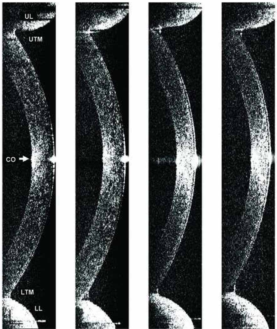
Figure 2. Upper and lower tear menisci in a dry eye patient and a control subject imaged immediately before normal and delayed blinks.
The images obtained from a dry eye patient (Far left, Middle left) and a control subject (Middle right, Far right) were demonstrated. Compared to normal blinking (Far left, Middle right), both upper and lower tear menisci were swollen with delayed blinking (Middle left, Far right). The tear menisci in the dry eye patient (Far left, Middle left) appeared smaller than those in the control subject (Middle right, Far right). CO, cornea; UTM: upper tear meniscus; LTM: lower tear meniscus; UL, upper eyelid; LL lower eyelid. Bars = 500 µm.
ACKNOWLEDGMENTS/DISCLOSURE
Funding / Support: This study was supported by research grants from NIH/NEI (R03 EY016420), Bausch & Lomb, Allergan, NEI Center Grant P30 EY014801, Research to Prevent Blindness (RPB) and Wallace H. Coulter Center.
Financial Disclosure: The authors have no proprietary interest in any materials or methods described within this article.
Contributions of Authors: Design and conduct of the study (J.W, Y.Y); Collection, management, analysis, and interpretation of the data (J.W, Y.Y, Q.C, A.T, M.A, M.S); Preparation, review and approval of the manuscript (J.W, Y.Y, M.S).
Statement about Conformity with Author Information: This study was approved by the Institutional Review Boards of University of Rochester and University of Miami. Informed consent was obtained from each participant, who was treated in accordance with the tenets of the Declaration of Helsinki.
Other Acknowledgments: We thank Britt Bromberg of Xenofile Editing for providing editing services for this manuscript.
Biography

Yimin Yuan, MD, graduated from Wenzhou Medical College in 2004, and received master’s degree in ophthalmology and optometry in 2007. Afterwards he completed the residency and became an ophthalmologist at the affiliated hospital. From November 2008, he spent one year at Bascom Palmer Eye Institute (Miami), doing research regarding the clinical application of optical coherence tomography. Currently, he is a PhD student at Wenzhou Medical College, and continues researching in dry eye and contact lens.
Footnotes
Publisher's Disclaimer: This is a PDF file of an unedited manuscript that has been accepted for publication. As a service to our customers we are providing this early version of the manuscript. The manuscript will undergo copyediting, typesetting, and review of the resulting proof before it is published in its final citable form. Please note that during the production process errors may be discovered which could affect the content, and all legal disclaimers that apply to the journal pertain.
REFERENCES
- 1.Lemp MA. Advances in understanding and managing dry eye disease. Am J Ophthalmol. 2008;146:350–356. doi: 10.1016/j.ajo.2008.05.016. [DOI] [PubMed] [Google Scholar]
- 2.Doane MG. Interactions of eyelids and tears in corneal wetting and the dynamics of the normal human eyeblink. Am J Ophthalmol. 1980;89:507–516. doi: 10.1016/0002-9394(80)90058-6. [DOI] [PubMed] [Google Scholar]
- 3.Doane MG. Blinking and the mechanics of the lacrimal drainage system. Ophthalmology. 1981;88:844–851. doi: 10.1016/s0161-6420(81)34940-9. [DOI] [PubMed] [Google Scholar]
- 4.Sahlin S, Laurell CG, Chen E, Philipson B. Lacrimal drainage capacity, age and blink rate. Orbit. 1998;17:155–159. doi: 10.1076/orbi.17.3.155.2757. [DOI] [PubMed] [Google Scholar]
- 5.Sahlin S, Chen E. Gravity, blink rate, and lacrimal drainage capacity. Am J Ophthalmol. 1997;124:758–764. doi: 10.1016/s0002-9394(14)71692-7. [DOI] [PubMed] [Google Scholar]
- 6.Tsubota K, Nakamori K. Effects of ocular surface area and blink rate on tear dynamics. Arch Ophthalmol. 1995;113:155–158. doi: 10.1001/archopht.1995.01100020037025. [DOI] [PubMed] [Google Scholar]
- 7.Palakuru JR, Wang J, Aquavella JV. Effect of blinking on tear dynamics. Invest Ophthalmol Vis Sci. 2007;48:3032–3037. doi: 10.1167/iovs.06-1507. [DOI] [PubMed] [Google Scholar]
- 8.Schaumberg DA, Sullivan DA, Buring JE, Dana MR. Prevalence of dry eye syndrome among US women. Am J Ophthalmol. 2003;136:318–326. doi: 10.1016/s0002-9394(03)00218-6. [DOI] [PubMed] [Google Scholar]
- 9.Schein OD, Munoz B, Tielsch JM, Bandeen-Roche K, West S. Prevalence of dry eye among the elderly. Am J Ophthalmol. 1997;124:723–728. doi: 10.1016/s0002-9394(14)71688-5. [DOI] [PubMed] [Google Scholar]
- 10.Doughty MJ, Fonn D, Richter D, Simpson T, Caffery B, Gordon K. A patient questionnaire approach to estimating the prevalence of dry eye symptoms in patients presenting to optometric practices across Canada. Optom Vis Sci. 1997;74:624–631. doi: 10.1097/00006324-199708000-00023. [DOI] [PubMed] [Google Scholar]
- 11.Lemp MA. Report of the National Eye Institute/Industry workshop on Clinical Trials in Dry Eyes. CLAO J. 1995;21:221–232. [PubMed] [Google Scholar]
- 12.Khanal S, Tomlinson A, McFadyen A, Diaper C, Ramaesh K. Dry eye diagnosis. Invest Ophthalmol Vis Sci. 2008;49:1407–1414. doi: 10.1167/iovs.07-0635. [DOI] [PubMed] [Google Scholar]
- 13.Oguz H, Yokoi N, Kinoshita S. The height and radius of the tear meniscus and methods for examining these parameters. Cornea. 2000;19:497–500. doi: 10.1097/00003226-200007000-00019. [DOI] [PubMed] [Google Scholar]
- 14.Lin PY, Tsai SY, Cheng CY, Liu JH, Chou P, Hsu WM. Prevalence of dry eye among an elderly Chinese population in Taiwan: the Shihpai Eye Study. Ophthalmology. 2003;110:1096–1101. doi: 10.1016/S0161-6420(03)00262-8. [DOI] [PubMed] [Google Scholar]
- 15.Schein OD, Tielsch JM, Munoz B, Bandeen-Roche K, West S. Relation between signs and symptoms of dry eye in the elderly. A population-based perspective. Ophthalmology. 1997;104:1395–1401. doi: 10.1016/s0161-6420(97)30125-0. [DOI] [PubMed] [Google Scholar]
- 16.Wang J, Aquavella J, Palakuru J, Chung S, Feng C. Relationships between central tear film thickness and tear menisci of the upper and lower eyelids. Invest Ophthalmol Vis Sci. 2006;47:4349–4355. doi: 10.1167/iovs.05-1654. [DOI] [PubMed] [Google Scholar]
- 17.Wang J, Thomas J, Cox I, Rollins A. Noncontact measurements of central corneal epithelial and flap thickness after laser in situ keratomileusis. Invest Ophthalmol Vis Sci. 2004;45:1812–1816. doi: 10.1167/iovs.03-1088. [DOI] [PubMed] [Google Scholar]
- 18.Wang J, Aquavella J, Palakuru J, Chung S. Repeated measurements of dynamic tear distribution on the ocular surface after instillation of artificial tears. Invest Ophthalmol Vis Sci. 2006;47:3325–3329. doi: 10.1167/iovs.06-0055. [DOI] [PMC free article] [PubMed] [Google Scholar]
- 19.Palakuru JR, Wang J, Aquavella JV. Effect of blinking on tear volume after instillation of midviscosity artificial tears. Am J Ophthalmol. 2008;146:920–924. doi: 10.1016/j.ajo.2008.06.020. [DOI] [PMC free article] [PubMed] [Google Scholar]
- 20.Wang J, Simmons P, Aquavella J, et al. Dynamic distribution of artificial tears on the ocular surface. Arch Ophthalmol. 2008;126:619–625. doi: 10.1001/archopht.126.5.619. [DOI] [PubMed] [Google Scholar]
- 21.Shen M, Li J, Wang J, et al. Upper and lower tear menisci in the diagnosis of dry eye. Invest Ophthalmol Vis Sci. 2009;50:2722–2726. doi: 10.1167/iovs.08-2704. [DOI] [PubMed] [Google Scholar]
- 22.Erdfelder E, Faul F, Buchner A. GPower: a general power analysis program. Behav Res Methods Instrum Comput. 1996;28:1–11. [Google Scholar]
- 23.Uchida A, Uchino M, Goto E, et al. Noninvasive interference tear meniscometry in dry eye patients with Sjogren syndrome. Am J Ophthalmol. 2007;144:232–237. doi: 10.1016/j.ajo.2007.04.006. [DOI] [PubMed] [Google Scholar]
- 24.Mainstone JC, Bruce AS, Golding TR. Tear meniscus measurement in the diagnosis of dry eye. Curr Eye Res. 1996;15:653–661. doi: 10.3109/02713689609008906. [DOI] [PubMed] [Google Scholar]
- 25.Savini G, Barboni P, Zanini M. Tear meniscus evaluation by optical coherence tomography. Ophthalmic Surg Lasers Imaging. 2006;37:112–118. [PubMed] [Google Scholar]
- 26.Johnson ME, Murphy PJ. The agreement and repeatability of tear meniscus height measurement methods. Optom Vis Sci. 2005;82:1030–1037. doi: 10.1097/01.opx.0000192352.78935.e0. [DOI] [PubMed] [Google Scholar]
- 27.Johnson ME, Murphy PJ. Temporal changes in the tear menisci following a blink. Exp Eye Res. 2006;83:517–525. doi: 10.1016/j.exer.2006.02.002. [DOI] [PubMed] [Google Scholar]
- 28.Doughty MJ, Laiquzzaman M, Button NF. Video-assessment of tear meniscus height in elderly Caucasians and its relationship to the exposed ocular surface. Curr Eye Res. 2001;22:420–426. doi: 10.1076/ceyr.22.6.420.5487. [DOI] [PubMed] [Google Scholar]
- 29.Golding TR, Bruce AS, Mainestone JC. Relationship between tear-meniscus parameters and tear-film breakup. Cornea. 1997;16:649–661. [PubMed] [Google Scholar]
- 30.Lamberts DW, Foster CS, Perry HD. Schirmer test after topical anesthesia and the tear meniscus height in normal eyes. Arch Ophthalmol. 1979;97:1082–1085. doi: 10.1001/archopht.1979.01020010536004. [DOI] [PubMed] [Google Scholar]
- 31.Miller WL, Doughty MJ, Narayanan S, et al. A comparison of tear volume (by tear meniscus height and phenol red thread test) and tear fluid osmolality measures in non-lens wearers and in contact lens wearers. Eye Contact Lens. 2004;30:132–137. doi: 10.1097/01.icl.0000138714.96401.2b. [DOI] [PubMed] [Google Scholar]
- 32.Francois J, Neetens A. Tear flow in man. Am J Ophthalmol. 1973;76:351–358. doi: 10.1016/0002-9394(73)90490-x. [DOI] [PubMed] [Google Scholar]
- 33.Tsubota K, Yamada M. Tear evaporation from the ocular surface. Invest Ophthalmol Vis Sci. 1992;33:2942–2950. [PubMed] [Google Scholar]
- 34.Macri A, Rolando M, Pflugfelder S. A standardized visual scale for evaluation of tear fluorescein clearance. Ophthalmology. 2000;107:1338–1343. doi: 10.1016/s0161-6420(00)00101-9. [DOI] [PubMed] [Google Scholar]
- 35.Pflugfelder SC, Tseng SC, Sanabria O, et al. Evaluation of subjective assessments and objective diagnostic tests for diagnosing tear-film disorders known to cause ocular irritation. Cornea. 1998;17:38–56. doi: 10.1097/00003226-199801000-00007. [DOI] [PubMed] [Google Scholar]
- 36.Farris RL, Stuchell RN, Mandel ID. Basal and reflex human tear analysis. I. Physical measurements: osmolarity, basal volumes, and reflex flow rate. Ophthalmology. 1981;88:852–857. doi: 10.1016/s0161-6420(81)34939-2. [DOI] [PubMed] [Google Scholar]


