Abstract
Introduction:
The aim of this study was to investigate the effect of quantification method on estimates of voluntary quadriceps muscle activation.
Methods:
Twenty-two people with no history of serious lower extremity injuries underwent voluntary quadriceps activation testing at 60° of knee flexion. Estimates of quadriceps activation were derived with: 1) a formula based on the interpolated twitch technique, 2) the central activation ratio, and 3) a modified central activation ratio. Predictive equations were developed that describe the relationships between the three methods.
Results:
Significant differences (P < 0.001) were observed between the estimates of voluntary quadriceps muscle activation obtained using the three methods (ITT percent activation = 93.0 ± 6.4%, CAR = 95.9 ± 3.8%, modified CAR = 98.5 ± 4.1%). Excellent correlation (r = 0.995) was observed between ITT based percent activation and the CAR method. The associations between these methods and the modified CAR approach were weaker.
Discussion:
Quantification method affects activation estimates. The equations developed will assist scientists in accurately comparing results of studies that use different methods of quantifying activation.
Keywords: Burst Superimposition, Central Activation Ratio, Interpolated Twitch Technique, Isometric, Knee Strength
INTRODUCTION
Voluntary activation failure is defined as the inability of the central nervous system to maximally drive a muscle during voluntary contraction.1,2 There are two primary contributing factors to this activation failure: 1) inability to fully recruit the motor units in a muscle/muscle group, and/or 2) suboptimal firing of the motor units that are recruited. Voluntary activation of the quadriceps muscle group has been studied extensively in both healthy people and those with knee joint pathology.3-9 Incomplete activation of the quadriceps muscle can significantly affect knee extensor strength and has been theorized as a mechanism contributing to the rapid quadriceps muscle atrophy typically observed after knee joint trauma and intra-articular knee surgery.7,9,10,11 Accordingly, quadriceps muscle activation tests are commonly used by scientists who study the effects of knee joint pathology and the outcomes of intervention strategies in people with knee pathology.
Evidence indicates that voluntary activation failure of the quadriceps muscle group is common in people after knee trauma or knee surgery and in those with knee osteoarthritis.4,5,9,12,13 Reported activation magnitudes in studies investigating these populations have varied considerably. For example, Chmielewski et al12 reported a mean activation failure of 8% in people with anterior cruciate ligament (ACL) injuries, whereas Urbach et al14 reported a mean deficit of 25% in a similar population. The inconsistency in reported magnitudes of activation failure is not unique to the ACL injury population; it is observed in most test populations including those with no known knee pathology.4,7,11 Although multiple factors likely contribute to this variability in activation estimates, large amounts of the variability may be attributable to the use of different quantification methods when estimating muscle activation levels.
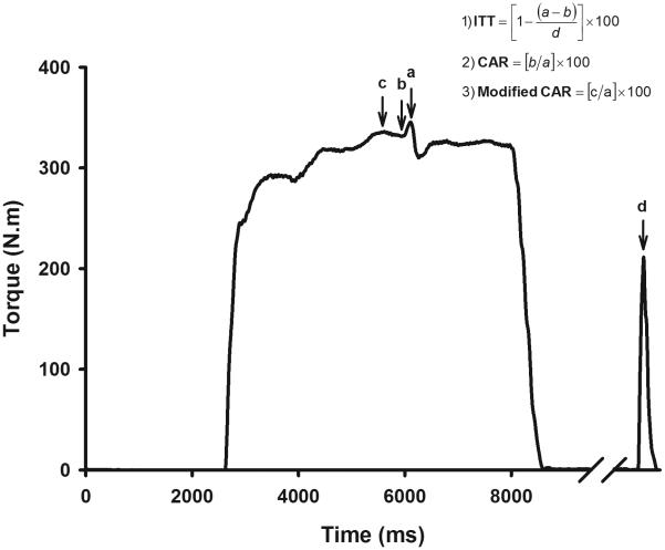
Voluntary muscle activation is most frequently assessed by introducing a maximal or supramaximal electrical stimulus during a maximal voluntary isometric contraction (MVIC).15-17 When incomplete activation is present, the superimposed electrical stimulus augments the force/torque generated by the muscle. Conversely, when there is complete or nearly complete voluntary activation, there is little to no increase in force/torque when the electrical stimulus is introduced. Several methods of quantifying voluntary activation have been described in the literature.17 The two most commonly used superimposition based quantification methods include calculating percent activation from data collected using the interpolated twitch technique (ITT) and the central activation ratio (CAR) method.1,3,7,9 The ITT involves superimposition of an electrical stimulus during an MVIC and the application of an identical stimulus while the subject sits at rest. Percent voluntary activation is quantified by expressing the stimulus evoked force/torque during contraction as a percentage of the stimulus evoked force/torque during rest (Figure 1, formula 1). In the CAR method, the electrical stimulus is delivered during the MVIC alone. Voluntary activation is estimated by comparing the voluntary torque at the time of stimulus delivery to the peak force/torque measured during superimposition of electrical pulses (Figure 1, formula 2). Error is introduced in both of these methods if the electrical stimulus is not delivered at or near voluntary peak torque.17,18 A modified CAR method was developed in an attempt to minimize the error associated with stimulus timing precision.8 In the modified CAR method, the voluntary peak force/torque observed at any point prior to stimulation is used to estimate voluntary activation failure instead of the voluntary force/torque measured at the time of stimulus delivery (Figure 1, formula 3).
Figure 1.
Schematic representation of the quantification techniques used to estimate voluntary quadriceps muscle activation. Incomplete activation of the quadriceps muscle is visualized by the increment in torque associated with the superimposed electrical pulses. When estimating percent voluntary activation based on the ITT, the user expresses stimulus evoked torque during contraction as a percentage of stimulus evoked torque at rest. When using the CAR method, the user estimates voluntary activation by comparing the voluntary torque at the time of stimulus delivery with the peak torque evoked with electrical burst superimposition. When using the modified CAR method, the user estimates voluntary activation by comparing the voluntary peak torque at any time prior to the stimulus delivery with the peak torque evoked with electrical burst superimposition. (a = peak torque evoked due to the superimposition of the electrical pulses, b = voluntary torque at the time of stimulus delivery, c = voluntary peak torque any time prior to the stimulus delivery, & d = stimulus evoked torque at rest).
Although scientists have long recognized that differences in quantification methods can lead to different voluntary activation estimates, there is a lack of evidence to describe the effects of quantification technique on voluntary muscle activation estimates. Moreover, we are unaware of a research report that includes predictive equations to describe the relationships between estimates resulting from the use of the quantification methods described above. Such equations would facilitate accurate comparisons of the results of studies in which different quantification methods have been used in similar samples of subjects. This would be a meaningful development, as it is unlikely that a consensus will be reached on using a single approach. Therefore, the purpose of this study was to evaluate the effect using different quantification methods (ITT based percent activation, CAR, and modified CAR) on estimates of voluntary quadriceps muscle activation and to develop equations that describe the associations between the resulting estimates. We hypothesized that the estimates of voluntary quadriceps muscle activation derived from the ITT, CAR, and modified CAR methods would be significantly different, but significantly associated.
METHODS
The study sample included twenty-two volunteers (11 males, 11 females) with no history of serious lower extremity injuries. All subjects were regular participants in fitness or sports activities (Tegner Activity Score19 ≥ 4). This study was approved by the University of Iowa Human Subjects Research Institutional Review Board, and all subjects provided written informed consent to participation using a form approved by this review board.
Testing Procedures
Subjects were asked to refrain from any strenuous physical activity for 24 hours prior to participation. The test session began by having subjects perform a 5-minute “warm-up” on a cycle ergometer, which was followed by self-directed stretching of the quadriceps, hamstrings, and gastrocnemius muscles. The skin of the anterior thigh was then cleaned with alcohol swabs, and two self-adhesive surface electrodes (2.75 in × 5 in, Dura-Stick II, Chattanooga Group, Hixson, TN, USA) were applied over the proximal and distal surface of the quadriceps muscles. These electrodes were connected with wires to a high voltage constant current stimulator (DS7AH, Digitimer Ltd., Hertfordshire, England) that introduced electrical stimuli during testing. Subjects were then positioned on and tightly secured to a HUMAC NORM Testing and Rehabilitation System (Computer Sports Medicine, Inc., Stoughton, MA, USA) with their test knee fixed at 60° of flexion and their hips fixed at approximately 90° of flexion. Each subject's dominant leg was tested. The dominant leg was defined by the leg the subject preferred to use to kick a ball for distance.
Subjects performed three submaximal practice trials at 50% to 85% maximum effort and one maximal practice trial to familiarize them with isometric contractions and potentiate the quadriceps muscles. Three electrical pulses at sub-maximal intensity were subsequently delivered to familiarize the subjects with electrical stimuli. Subject-specific maximal current intensities were used during testing. Maximal current intensity was determined by sequentially stimulating the quadriceps muscle with pulse trains (10 pulse, 100 Hz, 200 μs pulse duration, 400 V) in current steps of 100 mA until the torque associated with the electrically evoked muscle contractions reached a plateau and then decreased. Current was then reduced by 50 mA, and a final stimulus was provided. Approximately 10 seconds transpired between stimuli. The current that produced the greatest evoked knee extensor torque at rest was selected for use in testing. After a two minute rest period, subjects performed three knee extensor MVIC test trials with three minutes of rest between each trial. Loud verbal encouragement and visual feedback of the real-time torque signal were provided in order to facilitate maximal effort. Burst superimposition at the predetermined subject-specific stimulus intensity occurred three seconds after the onset of each MVIC. Software written in LabVIEW (v. 7.0, National Instruments Corporation, Austin, TX, USA) was used to trigger the stimulator and record data.
Data Management and Analysis
Torque signals were sampled at 1000 Hz. The signals from the dynamometer were passed through a 3rd order Butterworth low pass filter with a cut-off frequency of 4 Hz and then converted to torque values (N·m) using calibrated conversion factors that were validated onsite prior to testing. The average of the torque values obtained in the three test trials was used for estimating quadriceps muscle activation based on mean values. The single trial that produced the highest peak torque during testing was used for estimating voluntary quadriceps muscle activation based on peak values. Voluntary quadriceps muscle activation was estimated using the ITT based percent activation, the CAR, and the modified CAR methods (Figure 1).
Statistical analyses were performed using SPSS for Windows (SPSS Inc., Chicago, IL, USA). Descriptive statistics were calculated for subject demographics, and the voluntary activation values were obtained using each method. A one-way analysis of variance (ANOVA) was used to evaluate if there were statistical differences in the male and female demographics. Repeated measures ANOVA with one within-subjects factor (method) and one between-subjects factor (sex) was used to evaluate voluntary activation differences between quantification methods and between the sexes. Post-hoc analysis with Bonferonni correction was performed to identify differences within main effects for quantification method. The corrected level of significance was α = 0.0166. There is no consensus in the literature on whether to use the mean force/torque values from a series of test trials or the peak force/torque value observed during testing when assessing strength and voluntary activation. Therefore, we examined whether significant differences in voluntary activation failure estimates resulted from using mean torque values derived using the results of the three test trials versus the peak force/torque values observed during testing. This was accomplished using repeated measures ANOVAs. Linear regression was used to assess and describe the associations between the three quantification methods.
RESULTS
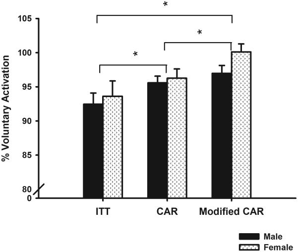
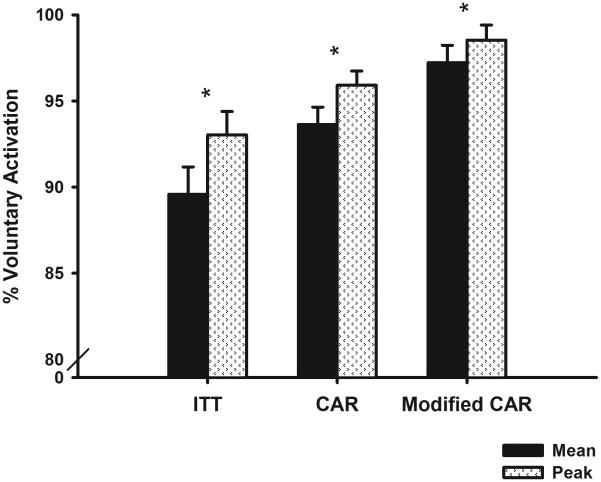
The male and female participants were similar in demographics with the exception of their height and weight (Table 1). The mean current intensities used were 359.1 mA for males and 286.4 mA for females. The mean electrically evoked knee extensor torque at rest was 57.6% of the subjects' maximum voluntary torque. The estimates of voluntary quadriceps muscle activation obtained using the three methods were significantly different (P < 0.001, ηp2 = 0.571; Figure 2). Post hoc analyses revealed that the voluntary activation estimates obtained from all the three methods were significantly different (P < 0.001 for ITT based percent activation vs. CAR, P = 0.002 for CAR vs. modified CAR, and P < 0.001 for ITT based percent activation vs. modified CAR). Voluntary activation did not differ significantly by sex (P = 0.392, ηp2 = 0.037). There was no significant interaction for quantification method by sex (P = 0.242, ηp2 = 0.067). The use of mean versus peak values resulted in significantly different estimates of voluntary activation (P < 0.001, ηp2 = 0.558). This was a consistent finding regardless of the approach used to estimate voluntary activation (ITT based percent activation: P < 0.001, ηp2 = 0.529; CAR: P < 0.001, ηp2 = 0.546; and modified CAR: P = 0.005, ηp2 = 0.319; Figure 3). Significant correlations were observed in the voluntary activation estimates obtained using the three methods (P < 0.001 for each comparison; ITT based percent activation vs. CAR: r = 0.998, ITT based percent activation vs. modified CAR: r = 0.688, and CAR vs. modified CAR: r = 0.672; Figure 4). As is typical with voluntary activation estimates, our activation estimates were negatively skewed. Transforming the negatively skewed data using reflected square root transformation to approximate a normal distribution did not alter the results significantly (ITT based percent activation vs. CAR: r = 0.998, ITT based percent activation vs. modified CAR: r = 0.679, and CAR vs. modified CAR: r = 0.664).
Table 1.
Demographics of the male and female subjects
| Variable | Sex | n | Mean | SD | P |
|---|---|---|---|---|---|
| Age | Female | 11 | 24.36 | 2.84 | .740 |
| Male | 11 | 24.00 | 2.19 | ||
| Total | 22 | 24.18 | 2.48 | ||
| Height (in) | Female | 11 | 65.14 | 2.39 | <.001* |
| Male | 11 | 71.00 | 2.65 | ||
| Total | 22 | 68.07 | 3.88 | ||
| Weight (lbs) | Female | 11 | 140.27 | 10.93 | <.001* |
| Male | 11 | 176.91 | 18.52 | ||
| Total | 22 | 158.59 | 23.91 | ||
| BMI | Female | 11 | 23.20 | 2.17 | .157 |
| Male | 11 | 24.57 | 2.20 | ||
| Total | 22 | 23.88 | 2.25 | ||
|
Tegner Activity
Score |
Female | 11 | 6.00 | 1.00 | .229 |
| Male | 11 | 6.45 | 0.69 | ||
| Total | 22 | 6.23 | 0.87 |
Figure 2.
Mean estimates of peak voluntary quadriceps muscle activation obtained using the three quantification techniques. The estimates obtained using the three methods were significantly different (*) from one another (P < 0.001). This finding was consistent for both males and females. Voluntary quadriceps muscle activation estimates did not differ significantly by sex. Error bars represent standard error of the mean.
Figure 3.
Estimates of mean (average of the 3 trials) and peak voluntary quadriceps muscle activation (voluntary activation value from the trial that had peak voluntary torque) obtained using the three quantification techniques. There was a significant difference (*) in the estimates of voluntary activation obtained using the mean and peak values of the three trials (P < 0.001). Error bars represent standard error of the mean.
Figure 4.
Scatterplots demonstrating the relationships between the three commonly used voluntary activation quantification techniques. There were significant correlations between all the three methods (P < 0.001). Note that the correlation between the CAR method and the ITT based percent activation method was very strong, however, the relationships between these methods and the modified CAR method was not very strong. R2 values represent the amount of variability explained by the model. The equations in the figure describe the relationships between the different quantification methods.
DISCUSSION
The purpose of this study was to evaluate the effect of quantification method selection on estimates of voluntary quadriceps muscle activation and to define the relationships between the three most commonly used methods available in the literature. The results indicate that the three quantification methods examined in this investigation lead to statistically significant differences in voluntary activation estimates. The absolute difference in estimated voluntary activation directly attributable to quantification method averaged 5.5% in this sample of healthy individuals. Larger differences in activation estimates are expected in patient populations that typically exhibit significant activation failure (e.g., ACL injury, osteoarthritis).
The voluntary quadriceps muscle activation estimates obtained in this study are consistent with values reported by other investigators who have studied “healthy” young people using like techniques.3,8,14,20 The estimates derived based on the ITT method were significantly lower than those derived using the CAR and the modified CAR methods. This finding is consistent with the results of prior studies that have included both the ITT based percent activation and CAR methods.3,21 Calculating percent activation based on the ITT method produces lower activation estimates than the CAR methods, because the electrically evoked torque at rest is typically lower than maximal voluntary isometric torque. This is not surprising, as there is currently no feasible method of introducing electrical stimuli in humans that reproducibly evokes maximal quadriceps muscle torque.22,23 In our study, evoked torque at rest averaged 57.6% of the subjects' maximal voluntary isometric torque. The general inability to electrically evoke maximum muscle force/torque leads to measurement error (typically an overestimation of activation) in each of the quantification methods, as all three approaches are based on the premise that the stimulus introduced during voluntary contraction maximally activates the muscle of interest. Based on the calculation formulas, the CAR and modified CAR approaches are likely to overestimate activation to a greater degree than calculating percent activation based on the ITT approach, because the formula used to derive percent activation from the ITT includes electrically evoked force/torque in both the numerator and denominator of the quantification equation. Consideration of this methodological limitation has lead some scientists to advocate using the ITT or curvilinear predictive models when estimating muscle activation based on stimulus superimpositon.3,23 Another advantage of the ITT method is that assessment of electrically evoked torque at rest provides meaningful insight on peripheral adaptations in muscle (e.g., trophic changes) that are often associated with injury, surgery, or other physiologic processes in addition to its application in quantifying voluntary activation levels.
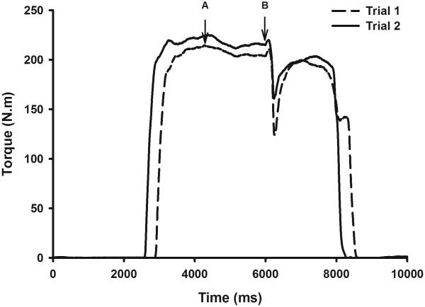
The electrical stimuli introduced in voluntary activation tests are typically triggered either manually at the point the examiner perceives to be peak force/torque while watching the subject's real time force/torque curves or automatically at a set time-point following the onset of volitional contraction.8,10 However, it is rare for the stimulus to be delivered at peak torque with either of these approaches, because MVICs are characteristically somewhat unsteady. Consequently, some measurement error is typically present in voluntary activation estimates regardless of the equations used.24 This difficulty in delivering the stimulus at or near peak torque is recognized as a limitation of voluntary activation testing.8,17 As identified in the introduction section of the paper, the modified CAR approach was introduced as an effort to minimize measurement error associated with this difficulty in delivering stimuli at peak force/torque.8 This modified approach is based on the theory that voluntary peak force/torque values measured prior to stimulus delivery better represent “true” voluntary peak force/torque than lower force/torque values measured at the time of stimulus delivery. As expected, the modified CAR method consistently produced higher activation estimates than were obtained using the ITT based percent activation method and the CAR method. Concern arises, however, from the fact that the modified CAR approach provided non-physiologic voluntary activation values (> 100%) in several subjects. These non-physiologic values were obtained when the voluntary peak torque measured prior to stimulus delivery exceeded the values measured during burst superimposition. Although it would be logical to assume that these subjects had complete activation, higher torque values were observed at the time of stimulus delivery in many of the subsequent trials (see Figure 5 for an example). This finding suggests that activation was not complete in at least some of the trials with activation estimates of ≥ 100%. Therefore, although the theoretical basis for the modified CAR approach is reasonable, the results of this study suggest that this approach is an imperfect solution to the stimulus timing precision problem. Improved stimulus timing can be achieved using a torque-based triggering technique in which the stimuli are delivered when torque values drop by a threshold value (e.g., 1 N·m). Evidence indicates that such torque-based triggering can reduce stimulus timing errors by about 75% when compared with the conventional triggering approaches described in this report.18 In addition to the limitations described above, it should be noted that stimulation superimposition based activation tests are generally unable to reliably detect subtle differences in voluntary activation. Therefore, it is best to reserve these tests for use in situations when the expected effects sizes are moderate to large.
Figure 5.
Representative example that demonstrates error in estimating voluntary activation using the modified CAR approach. The torque curve from trial 1 indicates that the subject had complete activation when estimated using the modified CAR approach. The subject produced slightly higher torque at the time of stimulus delivery (Arrow B) in trial 2 when compared to the voluntary peak torque of trial 1 (Arrow A). The torque curve from trial 2, however, shows a small stimulus evoked torque during contraction, which indicates incomplete activation in the preceding trial.
There is currently no gold standard to use in comparing the validity and reliability of voluntary activation estimates. Hence, the user must choose a technique based on its theoretical framework and other practical considerations such as the historical practice within a group of researchers. Scientists have recently described using transcranial magnetic stimulation and T2-weighted magnetic resonance imaging to assess voluntary activation.25,26 These newer activation estimation methods are promising, but they have their own inherent limitations.
To our knowledge this is the first study to provide equations that describe the relationships between the ITT based percent activation, CAR, and modified CAR methods of quantifying voluntary quadriceps muscle activation. Although there were significant differences in the activation estimates obtained with the three techniques, the results of the three approaches were significantly associated. The association between results obtained with the ITT based percent activation and the CAR methods was especially strong (r = 0.995). The associations with the modified CAR method were weaker. Although our data are consistent with those in the literature for similar samples of subjects,3,8,14,20 we acknowledge that the use of healthy subjects with high levels of activation is a potential limitation of the study. To assess the practicality of these equations, we tested the hypothesis that using the equation developed in this study to transform voluntary activation data from previous studies that included similar samples of subjects but used different methods of calculating quadriceps activation would minimize the variability in reported voluntary activation values. Three population categories were chosen for the purpose of this analysis: older people, knee osteoarthritis, and ACL injury. In each population, values from studies that used the CAR method were transformed using the predictive equation describing the relationship between the CAR method and ITT based percent activation estimates. The transformed results were subsequently compared with the estimates from studies that used the ITT method. We were unable to assess the practicality of the modified CAR equations due to the fact that we did not find studies in which the modified CAR approach had been used in the target populations. The voluntary quadriceps muscle activation estimates reported in studies using the CAR method were: 94% in the older subjects,27,28 85% in osteoarthritis patients,11 and 90% in ACL injured subjects.9 Inputting these values into the linear equation [ITT based percent activation = 1.661(CAR) - 66.260] provided voluntary activation estimates of 89.9% (older people), 74.9% (osteoarthritis), and 83.2% (ACL). These values closely match estimates of voluntary activation reported by researchers who have used the ITT when studying quadriceps activation (older people: 90.9%, osteoarthritis: 76.2%, and ACL: 83.9%).4,20,29 The results of this transformation test suggest that the predictive equations provided in this study will enable clinicians and scientists to more accurately compare the results of studies in which the ITT, CAR, or modified CAR methods have been used. However, we acknowledge that direct verification of the equations in patient populations is the only way to confirm the generalizability of the equations. It should be noted that we specifically selected studies that used testing positions and stimulus parameters that approximated those in our study for this transformation test. It is unclear if the equations would remain robust with data collected under significantly different test conditions. Therefore, we recommend that users consider study methodology when applying the predictive equations.
There is no consensus on whether to use mean or peak force/torque values when estimating activation. The results of this study indicate that these data management approaches produce significantly different estimates of activation. This finding was consistent regardless of which quantification formula was selected. This suggests that the subjects were generally unable to provide maximal effort in all the trials, which is consistent with other reports.8,30 Hence, the selection of mean versus peak force/torque values is another factor that should be considered when designing and comparing studies.
No significant differences in voluntary quadriceps muscle activation were observed between the male and female subjects. This is a meaningful finding, as voluntary activation differences between the sexes has been put forth as a potential explanation for the finding of neurophysiological differences by sex in previous electromyography-based research.31 The results of this study indicate that there is no difference in the ability of “healthy” men and women to volitionally activate their quadriceps muscles during MVICs.
In conclusion, the results of this study provide evidence that one's choice of quantification technique (ITT based percent activation, CAR, modified CAR) has a significant effect on the resulting estimate of quadriceps muscle activation. Consequently, quantification technique should be considered when designing studies including voluntary activation tests or comparing the results of studies in which voluntary muscle activation has been estimated using different approaches. Although the activation estimates obtained with the ITT based percent activation and CAR methods are significantly different in terms of magnitude, the estimates are strongly associated. The associations between these methods and the modified CAR method are weaker. The predictive equations developed in this study provide a solution to the variability in activation estimates associated with the use of different quantification methods. These equations can be used to more accurately compare and contrast the results of studies in which quadriceps activation has been assessed with different quantification techniques in similar samples of subjects.
Funding Acknowledgement
Supported in part by NIH Grant K12 HD055931.
Abbreviations
- MVIC
Maximum Voluntary Isometric Contraction
- ITT
Interpolated Twitch Technique
- CAR
Central Activation Ratio
- ACL
Anterior Cruciate Ligament
Footnotes
This study was presented at the 2008 Combined Sections Meeting of the American Physical Therapy Association.
References
- 1.Kent-Braun JA, Le Blanc R. Quantitation of central activation failure during maximal voluntary contractions in humans. Muscle Nerve. 1996;19:861–869. doi: 10.1002/(SICI)1097-4598(199607)19:7<861::AID-MUS8>3.0.CO;2-7. [DOI] [PubMed] [Google Scholar]
- 2.Todd G, Gorman RB, Gandevia SC. Measurement and reproducibility of strength and voluntary activation of lower-limb muscles. Muscle Nerve. 2004;29:834–842. doi: 10.1002/mus.20027. [DOI] [PubMed] [Google Scholar]
- 3.Behm D, Power K, Drinkwater E. Comparison of interpolation and central activation ratios as measures of muscle inactivation. Muscle Nerve. 2001;24:925–934. doi: 10.1002/mus.1090. [DOI] [PubMed] [Google Scholar]
- 4.Berth A, Urbach D, Neumann W, Awiszus F. Strength and voluntary activation of quadriceps femoris muscle in total knee arthroplasty with midvastus and subvastus approaches. J Arthroplasty. 2007;22:83–88. doi: 10.1016/j.arth.2006.02.161. [DOI] [PubMed] [Google Scholar]
- 5.Fitzgerald GK, Piva SR, Irrgang JJ, Bouzubar F, Starz TW. Quadriceps activation failure as a moderator of the relationship between quadriceps strength and physical function in individuals with knee osteoarthritis. Arthritis Rheum. 2004;51:40–48. doi: 10.1002/art.20084. [DOI] [PubMed] [Google Scholar]
- 6.Grindstaff TL, Jackson KR, Garrison JC, Diduch DR, Ingersoll CD. Decreased quadriceps activation measured hours prior to a noncontact anterior cruciate ligament tear. J Orthop Sports Phys Ther. 2008;38:508–516. doi: 10.2519/jospt.2008.2761. [DOI] [PubMed] [Google Scholar]
- 7.Hurley MV, Scott DL, Rees J, Newham DJ. Sensorimotor changes and functional performance in patients with knee osteoarthritis. Ann Rheum Dis. 1997;56:641–648. doi: 10.1136/ard.56.11.641. [DOI] [PMC free article] [PubMed] [Google Scholar]
- 8.Miller M, Holmback AM, Downham D, Lexell J. Voluntary activation and central activation failure in the knee extensors in young women and men. Scand J Med Sci Sports. 2006;16:274–281. doi: 10.1111/j.1600-0838.2005.00479.x. [DOI] [PubMed] [Google Scholar]
- 9.Williams GN, Buchanan TS, Barrance PJ, Axe MJ, Snyder-Mackler L. Quadriceps weakness, atrophy, and activation failure in predicted noncopers after anterior cruciate ligament injury. Am J Sports Med. 2005;33:402–407. doi: 10.1177/0363546504268042. [DOI] [PubMed] [Google Scholar]
- 10.Petterson SC, Barrance P, Buchanan T, Binder-Macleod S, Snyder-Mackler L. Mechanisms underlying quadriceps weakness in knee osteoarthritis. Med Sci Sports Exerc. 2008;40:422–427. doi: 10.1249/MSS.0b013e31815ef285. [DOI] [PMC free article] [PubMed] [Google Scholar]
- 11.Stevens JE, Mizner RL, Snyder-Mackler L. Quadriceps strength and volitional activation before and after total knee arthroplasty for osteoarthritis. J Orthop Res. 2003;21:775–779. doi: 10.1016/S0736-0266(03)00052-4. [DOI] [PubMed] [Google Scholar]
- 12.Chmielewski TL, Stackhouse S, Axe MJ, Snyder-Mackler L. A prospective analysis of incidence and severity of quadriceps inhibition in a consecutive sample of 100 patients with complete acute anterior cruciate ligament rupture. J Orthop Res. 2004;22:925–930. doi: 10.1016/j.orthres.2004.01.007. [DOI] [PubMed] [Google Scholar]
- 13.Suter E, Herzog W, Bray RC. Quadriceps inhibition following arthroscopy in patients with anterior knee pain. Clin Biomech (Bristol, Avon) 1998;13:314–319. doi: 10.1016/s0268-0033(98)00098-9. [DOI] [PubMed] [Google Scholar]
- 14.Urbach D, Nebelung W, Becker R, Awiszus F. Effects of reconstruction of the anterior cruciate ligament on voluntary activation of quadriceps femoris a prospective twitch interpolation study. J Bone Joint Surg Br. 2001;83:1104–1110. doi: 10.1302/0301-620x.83b8.11618. [DOI] [PubMed] [Google Scholar]
- 15.Herbert RD, Gandevia SC. Twitch interpolation in human muscles: mechanisms and implications for measurement of voluntary activation. J Neurophysiol. 1999;82:2271–2283. doi: 10.1152/jn.1999.82.5.2271. [DOI] [PubMed] [Google Scholar]
- 16.Merton PA. Voluntary strength and fatigue. J Physiol. 1954;123:553–564. doi: 10.1113/jphysiol.1954.sp005070. [DOI] [PMC free article] [PubMed] [Google Scholar]
- 17.Shield A, Zhou S. Assessing voluntary muscle activation with the twitch interpolation technique. Sports Med. 2004;34:253–267. doi: 10.2165/00007256-200434040-00005. [DOI] [PubMed] [Google Scholar]
- 18.Krishnan C, Allen EJ, Williams GN. Torque-based triggering improves stimulus timing precision in activation tests. Muscle Nerve. 2009;40:130–133. doi: 10.1002/mus.21279. [DOI] [PMC free article] [PubMed] [Google Scholar]
- 19.Tegner Y, Lysholm J. Rating systems in the evaluation of knee ligament injuries. Clin Orthop Relat Res. 1985:43–49. [PubMed] [Google Scholar]
- 20.Urbach D, Nebelung W, Weiler HT, Awiszus F. Bilateral deficit of voluntary quadriceps muscle activation after unilateral ACL tear. Med Sci Sports Exerc. 1999;31:1691–1696. doi: 10.1097/00005768-199912000-00001. [DOI] [PubMed] [Google Scholar]
- 21.Bampouras TM, Reeves ND, Baltzopoulos V, Maganaris CN. Muscle activation assessment: effects of method, stimulus number, and joint angle. Muscle Nerve. 2006;34:740–746. doi: 10.1002/mus.20610. [DOI] [PubMed] [Google Scholar]
- 22.Stackhouse SK, Dean JC, Lee SC, Binder-MacLeod SA. Measurement of central activation failure of the quadriceps femoris in healthy adults. Muscle Nerve. 2000;23:1706–1712. doi: 10.1002/1097-4598(200011)23:11<1706::aid-mus6>3.0.co;2-b. [DOI] [PubMed] [Google Scholar]
- 23.Stackhouse SK, Stevens JE, Johnson CD, Snyder-Mackler L, Binder-Macleod SA. Predictability of maximum voluntary isometric knee extension force from submaximal contractions in older adults. Muscle Nerve. 2003;27:40–45. doi: 10.1002/mus.10278. [DOI] [PubMed] [Google Scholar]
- 24.Oskouei MA, Van Mazijk BC, Schuiling MH, Herzog W. Variability in the interpolated twitch torque for maximal and submaximal voluntary contractions. J Appl Physiol. 2003;95:1648–1655. doi: 10.1152/japplphysiol.01189.2002. [DOI] [PubMed] [Google Scholar]
- 25.Kendall TL, Black CD, Elder CP, Gorgey A, Dudley GA. Determining the extent of neural activation during maximal effort. Med Sci Sports Exerc. 2006;38:1470–1475. doi: 10.1249/01.mss.0000228953.52473.ce. [DOI] [PubMed] [Google Scholar]
- 26.Todd G, Taylor JL, Gandevia SC. Measurement of voluntary activation of fresh and fatigued human muscles using transcranial magnetic stimulation. J Physiol. 2003;551:661–671. doi: 10.1113/jphysiol.2003.044099. [DOI] [PMC free article] [PubMed] [Google Scholar]
- 27.Stevens JE, Stackhouse SK, Binder-Macleod SA, Snyder-Mackler L. Are voluntary muscle activation deficits in older adults meaningful? Muscle Nerve. 2003;27:99–101. doi: 10.1002/mus.10279. [DOI] [PubMed] [Google Scholar]
- 28.Stackhouse SK, Stevens JE, Lee SC, Pearce KM, Snyder-Mackler L, Binder-Macleod SA. Maximum voluntary activation in nonfatigued and fatigued muscle of young and elderly individuals. Phys Ther. 2001;81:1102–1109. [PubMed] [Google Scholar]
- 29.Berth A, Urbach D, Awiszus F. Improvement of voluntary quadriceps muscle activation after total knee arthroplasty. Arch Phys Med Rehabil. 2002;83:1432–1436. doi: 10.1053/apmr.2002.34829. [DOI] [PubMed] [Google Scholar]
- 30.Hales JP, Gandevia SC. Assessment of maximal voluntary contraction with twitch interpolation: an instrument to measure twitch responses. J Neurosci Methods. 1988;25:97–102. doi: 10.1016/0165-0270(88)90145-8. [DOI] [PubMed] [Google Scholar]
- 31.Krishnan C, Huston K, Amendola A, Williams GN. Quadriceps and hamstrings muscle control in athletic males and females. J Orthop Res. 2008;26:800–808. doi: 10.1002/jor.20592. [DOI] [PubMed] [Google Scholar]


