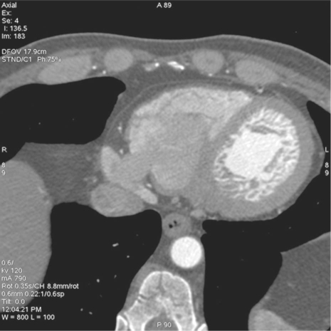
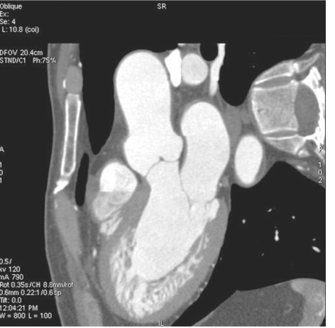
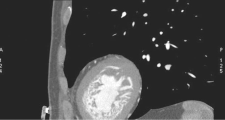
A 52-year-old man went to his primary care physician with mild dyspnea. He was initially evaluated with echocardiography, which revealed a left ventricular ejection fraction of 0.42. The patient was subsequently referred to a cardiologist, who ordered a multidetector computed tomogram (MDCT) to establish the cause of the heart failure. The MDCT images (Figs. 1, 2, and 3) showed the presence of deep intertrabecular recesses in the noncompacted left ventricular myocardium during the end-diastolic phase. The recesses filled with blood, and prominent trabeculations extended into the ventricular cavity. The 3-chamber view (Fig. 2), in particular, clearly showed that the ratio between noncompacted and compacted myocardium exceeded 2.3. The coronary arteries and remaining cardiac chambers were normal. Noncompaction was not noted upon echocardiography.
Fig. 1 Axial multidetector computed tomogram shows prominent intertrabecular recesses filled with blood. Note also the prominent trabeculations in the left ventricular noncompacted myocardium.
Fig. 2 Three-chamber multidetector computed tomogram shows that the ratio of the maximal thickness of the noncompacted abnormal myocardium to the compacted normal myocardium exceeds 2.3.
Fig. 3 Short-axis multidetector computed tomogram depicts multiple ventricular trabeculations and deep intertrabecular recesses that communicate freely with the left ventricular cavity.
Comment
Noncompaction is a rare congenital form of cardiomyopathy that results from the arrest of the normal compaction process during embryogenesis.1 As a result, the ventricular wall is markedly trabeculated with deep recesses that communicate with the ventricular cavity. Patients may present with congestive heart failure, arrhythmias, or emboli. Echocardiography is considered to be the diagnostic test of choice; however, limited knowledge about this rare disorder often results in misdiagnosis. Echocardiographic diagnostic criteria include a noncompacted-to-compacted myocardial thickness ratio of >2 at end-systole.2 A recent publication has proposed that a ratio of >2.3 at end-diastole can be used to diagnose noncompaction upon magnetic resonance imaging.3 The magnetic resonance imaging diagnostic criteria were extrapolated in order to make the diagnosis of noncompaction of the left ventricular myocardium with use of MDCT in our patient. Further research will most likely show that MDCT is accurate for the diagnosis of myocardial noncompaction while it excludes other causes of cardiomyopathy, including ischemic heart disease.
Footnotes
Address for reprints: Matthew Budoff, MD, Division of Cardiology, Los Angeles Biomedical Research Institute, 1000 W. Carson St., Torrance, CA 90502
E-mail: mbudoff@labiomed.org
References
- 1.Weiford BC, Subbarao VD, Mulhern KM. Noncompaction of the ventricular myocardium. Circulation 2004;109(24): 2965–71. [DOI] [PubMed]
- 2.Chrissoheris MP, Ali R, Vivas Y, Marieb M, Protopapas Z. Isolated noncompaction of the ventricular myocardium: contemporary diagnosis and management. Clin Cardiol 2007;30 (4):156–60. [DOI] [PMC free article] [PubMed]
- 3.Petersen SE, Selvanayagam JB, Wiesmann F, Robson MD, Francis JM, Anderson RH, et al. Left ventricular non-compaction: insights from cardiovascular magnetic resonance imaging. J Am Coll Cardiol 2005;46(1):101–5. [DOI] [PubMed]


