Abstract
Objective
To determine whether ankle orthoses that provide medial and lateral support, and have been found to decrease gait variability in older persons with peripheral neuropathy, decrease (improve) frontal plane ankle proprioceptive thresholds or increase unipedal stance time in that same population.
Design
Observational study in which unipedal stance time was determined with a stopwatch, and frontal plane ankle (inversion and eversion) proprioceptive thresholds were quantified during bipedal stance with and without the ankle orthoses, in 11 older diabetic subjects with peripheral neuropathy (8 men; age 72 ± 7.1 years) using a foot cradle system which presented a series of 100 rotational stimuli.
Results
The subjects demonstrated no change in combined frontal plane (inversion + eversion) proprioceptive thresholds or unipedal stance time with versus without the orthoses (1.06 ± 0.56 versus 1.13 ± 0.39 degrees, respectively; p = 0.955 and 6.1 ± 6.5 versus 6.2 ± 5.4 seconds, respectively; p = 0.922).
Conclusion
Ankle orthoses which provide medial-lateral support do not appear to change ankle inversion/eversion proprioceptive thresholds or unipedal stance time in older persons with diabetic peripheral neuropathy. Previously identified improvements in gait variability using orthoses in this population are therefore likely related to an orthotically-induced stiffening of the ankle rather than a change in ankle afferent function.
Keywords: Ankle, Orthosis, Stance, Gait, Peripheral Neuropathy, Proprioception
Older persons with peripheral neuropathy (PN) due to diabetes and other causes have a markedly increased rate of falling and fall-related injury as compared to similarly-aged populations without neuropathy.1,2 Accordingly, patients with PN have difficulty maintaining unipedal stance.3,4 This inability to reliably stand on one foot has been strongly correlated with impairments in frontal plane (inversion/eversion) ankle motor function5 and proprioceptive thresholds.6 Moreover, the markedly increased injury potential of lateral falls7 provides further impetus for the study of ankle neuromuscular function in the frontal plane.
Other work, predominantly in young or athletic populations, suggests that ankle orthoses can improve postural stability and/or proprioceptive thresholds, 8–14 although such results are not universal.15–17 Ankle orthoses have also been found to decrease (improve) temporospatial measures of gait variability in older subjects with PN walking on an irregular surface, suggesting improved postural control during this challenging gait task. 18 Similarly, high collar shoes, as compared to shoes with low collars or bare feet, have been found to improve standing balance in older women.19 However, these studies aside, little research has been devoted to the effects of ankle orthoses in older populations or patients with peripheral neurologic disease, and the need for study of these populations has been emphasized.20 Therefore, we elected to study the effect of ankle orthoses on frontal plane ankle proprioceptive thresholds and unipedal stance time (UST). More specifically, we hypothesized that older persons with PN wearing ankle orthoses, which provide medial-lateral support at the ankle, would demonstrate: 1) decreased (improved) frontal plane ankle proprioceptive thresholds and; 2) increased UST, as compared to when not wearing orthoses.
MATERIALS AND METHODS
Overview
Eleven older subjects with PN participated in the study. Ankle inversion and eversion proprioceptive thresholds were quantified with specialized hardware and software, and then UST was determined clinically. Odd-numbered subjects underwent these evaluations with the ankle orthoses initially, and then without them, whereas even-numbered subjects were evaluated first without the orthoses. The orthoses (Active Ankle T2; Active Ankle Systems, Inc., Louisville, KY) are semi-circular shaped shells lined with foam that lie on either side of the malleoli and extend proximally over the lower leg. They are held tightly to the lower leg and ankle by hook and loop straps that wrap circumferentially around the leg. The shells are connected inferiorly by a sling. The biomechanical effect of the orthoses is such that extremes of ankle inversion and eversion are resetricted but ankle plantar and dorseflexion occur freely. So as to allow the subjects to accommodate to the orthoses, subjects wore them and walked in a hallway ad lib for 5 minutes prior to testing In addition, at least 5 minutes of rest was allowed between proprioceptive threshold testing and UST testing.
Subjects
Of the 11 subjects, 8 were men (age and BMI of all subjects were 72 ± 7.1 yrs and 30.9 ± 5.0 kg/m2, respectively). The subjects were recruited from the University of Michigan Electrodiagnostic Laboratory and the University of Michigan Orthotics and Prosthetics Center. All procedures were approved by the Institutional Review Board and all subjects provided written informed consent.
Inclusion criteria for all subjects were: 1) age between 50 and 85 yrs; 2) ability to speak and understand English; 3) ankle dorsiflexion strength ≥ 4 by manual muscle testing; and 4) ability to ambulate at least household distances without an assistive device. Subjects were excluded if they had a history of abnormal vision despite correction; weight greater than 136 kg (300 lbs); evidence on physical examination of central neurologic dysfunction or musculoskeletal abnormality such as severe scoliosis or amputation.
All of the subjects had a known history of diabetes mellitus treated by diet, oral hypoglycemic and/or insulin therapy. Subjects all reported symptoms consistent with PN; i.e., symmetrically altered sensation in the distal lower extremities that improved proximally. Most subjects reported “negative” symptoms such as a sensation of “deadness” in the distal limbs, and a few reported positive symptoms at night as well. All subjects had signs consistent with PN as determined by the examination necessary for determination of the Michigan Diabetes Neuropathy Score (MDNS).21 The MDNS is a 0 to 46 point scale, with higher score reflecting more severe diabetic PN, which correlates well with more extensive neuropathy staging scales. The scale includes muscle stretch reflexes at the biceps, triceps, patella and Achilles; ability to perceive a pin, a 10 gram monofilament and 128 Hz tuning fork at the great toes; and strength of hand dorsal interossei, great toe extensors and ankle dorsiflexors determined bilaterally. All PN subjects had MDNS scores of 9 or greater which, in previous work, was found to be an appropriate cut-off for older persons.22 Lastly, all subjects had electrodiagnostic evidence of a diffuse PN as evidenced by abnormal sural and peroneal motor responses. These abnormal responses were obtained bilaterally, as has been suggested in the definition of PN for the purposes of clinical research.23 Abnormal sural responses were defined as being absent, or with amplitudes of less than 6 μV and/or latencies of greater than 4.2 msec stimulating 14 cm from the recording site posterior to the lateral malleolus. Peroneal motor responses were obtained by recording over the extensor digitorum brevis muscle and stimulating 9 cm proximally over the deep peroneal nerve and then distal to the fibular head. Abnormal peroneal motor responses were defined as absent or with amplitudes less than 2 mV, and/or latencies greater than 6.2 msec, and/or conduction velocities less than 41.0 m/sec.
Experimental Apparatus
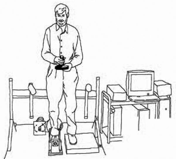
A servo motor-driven foot cradle system, which we have used in previous work,24 was created to evaluate ankle proprioceptive thresholds in the frontal plane (Figure 1). An Aerotech servo motor (Model Number: 1000DC, Aerotech, Inc., Pittsburg, PA; continuous torque capability: 80 oz-in) was connected to the foot cradle via an aircraft cable and pulley system so as to produce rotation of the cradle system. To allow for finer control of the cradle, an additional 6000 line pulse encoder was installed on the rotational axis of the cradle. The depth of the cradle was matched to the height of the subtalar axis of ankle rotation so that the axes of the two were collinear, and a raised dummy plate was placed under the contralateral foot bringing the feet to the same level. The system was controlled by a custom LabVIEW® (National Instrument) program on an IBM-compatible PC platform. The LabVIEW program interfaced with a Mektronix MC-03 (Mektronics, Inc., Australia) motor controller board and National Instrument high-resolution multi-function I/O board (AT-MIO-16XE-50). A PC-game joystick was installed so the subject could provide feedback on the direction of any perceived ankle rotation to the computer. To ensure subject safety, infrared light beams were attached to each handrail to serve as emergency cut-off switches should the rails be touched. An additional emergency cutoff switch was available to the operator as well. Finally, limit switches and mechanical stops were provided that limited the range of ankle rotation to a maximum of 15 degrees of inversion or eversion beyond neutral ankle position.
Figure 1.
Experimental apparatus used for quantifying frontal plane proprioceptive thresholds at the ankle.
Subject Protocols
Determining Proprioceptive Thresholds
The subject was asked to place his/her right foot inside the cradle which was adjusted so the sub-talar joint axis was aligned as closely as possible with the rotational axis of the cradle, and with the heel and second metatarsal joint of the foot aligned with the longitudinal axis of the cradle. The left foot was placed on a fixed plate at the same height as the surface of the cradle. The subject was instructed to place half their body weight on each foot and stand without assistance, looking forward while holding a joystick in front of them at the level of the umbilicus. (Figure 1) When the LabVIEW program started to run, it provided an audible warning cue, after which the control program generated a single ankle rotation (inversion or eversion) at 5 degrees per second, or no rotation (dummy trial). The subject then pressed the joystick handle in the direction of the perceived foot rotation. Prior to data collection subjects were allowed sufficient practice to confirm their understanding of the direction to push the joystick in response to the inversion and eversion stimuli. Further stimuli of randomized magnitude were then presented in a similar fashion using a staircase approach.24 The program evaluated the proprioceptive threshold of the subject in real-time so as to adjust the ankle rotation displacements by 0.1 degree increments between 0.1 and 1.0 degrees, and then by 0.5 degree increments between 1.0 and 3.0 degrees, to fit the subject’s proprioceptive ability. A total of 100 trials were presented to each subject, with 40 being inversion and 40 eversion. The subject was instructed not to guess the direction of rotation, and to evaluate this 20 dummy no-motion trials were randomly placed into the 100 data trials. During these dummy trials, the motor was active so that auditory clues were not available to the subject to assist in discriminating between real and dummy trials.
Determining UST
Subjects were instructed to maintain unipedal stance for as long as possible, and not to touch their lifted foot to the floor, or the stance limb, unless it was necessary to restore balance. The starting point for unipedal stance was lift-off of the non stance limb and the endpoint was touching the non stance limb to the floor or the stance limb, or shifting/sliding of the stance limb. A stopwatch was used to determine the elapsed time up to a maximum of 30 seconds. Subjects stood with arms crossed in front of the chest during unipedal stance testing. Given the absence of a laterality effect on UST,25,26 the subjects performed 3 trials using the right foot followed by 3 trials using the left foot with rest as required given between each trial. An experienced physical therapist stood near the subject, acting as a spotter in the event of an uncontrolled loss of balance.
Statistical Analyses
Determining proprioceptive thresholds
Ankle proprioception was represented by an outcome measure, TH75, which was defined as the smallest rotational displacement of the ankle that a subject could identify with a 75% probability of success.27 Given the displacement protocol described above, the resolution of TH75 was 0.1 degree for thresholds up to 1.0 degree, and 0.5 degree for thresholds > 1.0 degree. To find TH75 the data, which were assumed to have the form of the Gaussian cumulative distribution function, were filtered using weighted three-point averaging. A ceiling algorithm was then applied to transform the data into a monotonic function. Because many results showed step-wise increases in the probability graphs, with jumps from 0% to 100% of success rate for successive stimuli amplitudes, a continuous regression function (probit) was used to induce greater error. The intersection at 50% success rate (TH50) was identified using linear interpolation between the two points just above and below 50%. If two consecutive points shared identical probability then the next nearest point was used for the analysis. This point was assumed to be the mean of the Gaussian probability distribution function and the slope at the point was assumed to be the standard deviation of that probability distribution function. The TH75 was calculated from this information using a custom MATLAB® (The Math Works, Cambridge, MA) program developed specifically for that purpose. Microsoft Excel and SPSS (SPSS Inc., Chicago, IL) were used for statistical processing.
Determining UST times
UST was calculated as the maximum value, or the longest, of the total (6) unipedal balance trials.
Determining condition differences in proprioceptive thresholds and UST
Within subject means for proprioceptive thresholds and UST with, and without, the orthoses were compared using paired two-sided t tests. A p-value of 0.05 or less was considered significant. Given the relatively small number of subjects, normality could not be confirmed and so the data were also analyzed using the Wilcoxon sign rank test.
RESULTS
Comparison of Proprioceptive Thresholds with and Without Orthoses
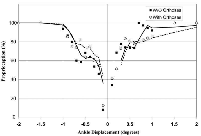
The subjects demonstrated no difference in ankle proprioceptive thresholds with (1.06 ± 0.56 degrees) as compared to without (1.13 ± 0.39 degrees) the ankle orthoses (p = 0.955). A similar p value was obtained using the Wilcoxon sign rank test (0.578). This is further appreciated by observing plots of ankle inversion and eversion displacements against percentage of accurate responses for a given displacement. As can be seen in Figure 2, the plots do not suggest any difference between the curve with, versus without, the ankle orthosis. Of interest, the subjects responded to just 7% of the dummy trials, indicating that they usually did not guess when they were uncertain as to direction of rotation.
Figure 2.
Accuracy of subject responses (as a percentile in the y axis) to ankle rotational displacements (in degrees in the x axis, with inversion + and eversion −) with (circles) and without (squares) ankle orthoses. The close approximation of the response accuracy with and without the orthoses suggests that they had no effect on perception of motion. Note: The percentage refers to the mean group accuracy at a particular ankle displacement.
Comparison of UST With and Without Orthoses
The subjects demonstrated no significant difference in UST with (6.1 ± 6.5 sec) and without (6.2 ± 5.4 sec) the ankle orthoses (p = 0.922). A similar p value was obtained using the Wilcoxon sign rank test (0.765). Observing these same data in a more categorical fashion, four of the 11 subjects increased UST slightly (maximum of 4.5 sec) and seven decreased UST slightly (maximum of 3.6 sec) while wearing the orthosis. (Table 1)
Table 1.
Comparisons in UST time and proprioceptive thresholds
| Without Orthosis | With Orthosis | With: without orthosis | p value | |
|---|---|---|---|---|
| Unipedal Balance Time (seconds) | ||||
| Right Foot | 5.2 ± 5.1 | 5.7 ± 6.6 | 1.10 | 0.625 |
| Left Foot | 5.2 ± 5.4 | 5.2 ± 4.9 | 0.99 | 0.901 |
| Right or Left | 6.2 ± 5.4 | 6.1 ± 6.5 | 0.99 | 0.922 |
| Proprioceptive Threshold (TH75 in degrees) | ||||
| Eversion | 0.66 ± 0.25 | 0.66 ± 0.34 | 1.00 | 0.872 |
| Inversion | 0.56 ± 0.37 | 0.47 ± 0.31 | 1.19 | 0.680 |
| Combined (Eversion + Inversion) | 1.13 ± 0.39 | 1.06 ± 0.56 | 0.94 | 0.955 |
DISCUSSION
The main finding of this work is that ankle orthoses which provide medial-lateral support improved neither ankle inversion/eversion proprioceptive thresholds nor UST in older persons with PN, a population with known impairments in both outcomes. This finding is likely real given that a concern with the design of this study was the inability to adequately blind subjects and examiners. This lack of blinding would be expected to provide bias in favor of the intervention; however, no positive effect was found. This absence of an effect is in contrast to other reports which describe improvements in joint position sense and/or postural control in response to ankle orthoses of various types.8–14
There are several potential explanations for this contrast in results. The most obvious explanation is that we studied older persons with neuropathy, whereas other work studied young, predominantly healthy populations. These latter populations, even those with a history of ankle sprain, are likely to have superior residual afferent somatosensory function allowing them to take advantage of ankle orthoses in ways that our PN subjects could not. Providing a counterpoint, however, Menz and colleagues demonstrated that a light touch stimulus to the lower leg decreased anterior-posterior postural sway in bipedal stance in older PN subjects similar to ours28 suggesting that our subjects also likely had sufficient somatosensory function to allow some improvement. Another key difference between our study and those that report improvements in proprioception or joint position sense with orthoses9–11,14 is that our study evaluated subjects while weight-bearing. This is likely relevant given that during gait the cyclical activity of the ankle musculature influences muscle spindle responses instrumental in determining proprioceptive thresholds.29 Moreover, determining proprioceptive thresholds while weight-bearing allows the inclusion of plantar cutaneous input. Other work inferred that ankle proprioceptive thresholds in the frontal plane improved because of postural changes with use of orthotic devices, but did not actually measure these thresholds.8,12,13
We also did not find that the orthoses had an effect on UST, again in contrast with other work which found postural improvements in association with orthotic devices.8,11–14 A potential explanation is that our postural task, one legged balance, is more challenging than bipedal standing,30 which was used as the outcome in one of the studies reporting that ankle orthoses positively influenced balance.13 However, three other studies (two in young subjects with “functionally unstable” ankles and one in healthy young subjects) evaluated unipedal balance and found improvements in response to ankle orthoses.8,11,14 These studies evaluated the steadiness or dynamic control of unipedal balance, rather than the ability to maintain it, indicating that the subjects likely had superior balance and distal neuromuscular function as compared to our subjects. Therefore, although ankle orthoses may improve balance “quality” in younger persons, they were unable to improve unipedal balance quantity in older subjects with PN. This might be explained by the fact that older persons, as compared to younger, employ more of a “hip strategy” for maintaining postural control during one-legged balance.31 This is likely even more true for older persons with PN.32
In previous work we found the orthosis used in this study to decrease step time and step width variability while older neuropathic subjects walked on an irregular surface.18 The present work suggests that this improvement in gait regularity under challenging circumstances was not due to an accentuation of afferent function, at least during stance. Therefore, it seems likely that the orthoses exerted their effect by means of a modification in motor function. In support of this, modeling of a neuropathic ankle encountering a perturbation suggests that stiffening the ankle reduces post-perturbation step width alteration.33 If this occurs sequentially on an irregular surface during gait then a reduction in gait variability would be anticipated. Alternatively, the orthoses may exert their effect by maintaining a stable ankle foot alignment during the swing phase of gait, although the importance of swing limb alignment is uncertain given other work finding that PN patients in the community are much more likely to fall on an irregular, rather than flat, surface.34
Determining proprioceptive thresholds is challenging, and the techniques presented had disadvantages. For example, the normal sway that occurs during quiet standing could, when in or out of phase with the direction of ankle rotation, reduce or augment the ankle rotation stimulus intensity. Testing performed in an open chain fashion, with the foot and ankle non-weight bearing, avoids this potential source of error but does so at the cost of loss of construct validity due to reduced muscle spindle activation and the absence of plantar pressure sensation, both of which are normally present during the stance phase of gait. The testing required sustained attention which may be affected by diabetes mellitus.35 However, other studies suggest that there is no relationship between neuropsychologic testing and PN36 and that the influence of diabetes mellitus on cognitive function is uncertain.37 Therefore it seems unlikely that cognitive dysfunction significantly influenced the results, particularly given work by Uciolli et al. which found postural stability in subjects with diabetic neuropathy to be quantitatively related to peripheral, but not central, nerve conduction study parameters.38 Although the techniques used were quantitative, it is possible that an insufficient number of subjects was recruited. However this appears less likely given a post-hoc power analysis which found that 10 subjects would be required to identify an orthotically-related proprioceptive threshold improvement of 0.5 degrees, given the standard deviation from this work, with a power of 0.80.
In summary, the data provided no evidence that ankle orthoses which provide medial-lateral support positively influence ankle inversion or eversion proprioceptive thresholds or UST in older persons with diabetic PN, a group at high risk for falls. The previously identified improvement (reduction) in gait variability in this same population when using these orthoses while walking on an irregular surface is therefore more likely related to the stiffening of the ankle than a change in ankle proprioceptive function.
Acknowledgments
All three authors were supported by PHS grant 1P30 AG 08808. Additionally, James K. Richardson was supported by PHS grants K23 AG 00989-01 and RO1 AG 026569-02
Footnotes
Disclosures: Financial disclosure statements have been obtained, and no conflicts of interest have been reported by the authors or by any individuals in control of the content of this article.
References
- 1.Cavanagh PR, Derr JA, Ulbrecht JS, Maser RE, Orchard TJ. Problems with gait and posture in neuropathic patients with insulin-dependent diabetes mellitus. Diabetes Med. 1992;9:469–474. doi: 10.1111/j.1464-5491.1992.tb01819.x. [DOI] [PubMed] [Google Scholar]
- 2.Richardson JK, Ching C, Hurvitz EA. The relationship between electromyographically documented peripheral neuropathy and falls. J Am Geriatr Soc. 1992;40(10):1008–1012. doi: 10.1111/j.1532-5415.1992.tb04477.x. [DOI] [PubMed] [Google Scholar]
- 3.Richardson JK, Hurvitz EA. Peripheral neuropathy: a true risk factor for falls. J Gerontol: Med Sci. 1995;50A(4):M211–215. doi: 10.1093/gerona/50a.4.m211. [DOI] [PubMed] [Google Scholar]
- 4.Richardson JK, Ashton-Miller JA, Lee SG, Jacob K. Moderate peripheral neuropathy impairs weight transfer and unipedal balance in the elderly. Arch Phys Med Rehabil. 1996;77(11):1152–1156. doi: 10.1016/s0003-9993(96)90139-2. [DOI] [PubMed] [Google Scholar]
- 5.Gutierrez LM, Helber MB, Dealva D, Ashton-Miller JA, Richardson JK. Mild diabetic neuropathy affects ankle motor function. Clin Biomech. 2001;16(6):522–28. doi: 10.1016/s0268-0033(01)00034-1. [DOI] [PubMed] [Google Scholar]
- 6.Son J, Ashton-Miller JA, Richardson JK. Frontal plane ankle proprioceptive thresholds and unipedal balance. Muscle Nerve. 2009;39(2):150–7. doi: 10.1002/mus.21194. [DOI] [PMC free article] [PubMed] [Google Scholar]
- 7.Greenspan SL, Meyers ER, Maitland LA, Resnick NJ, Hayes WC. Fall severity and bone mineral density as risk factors for hip fracture in ambulatory elderly. JAMA. 1994;271(2):128–33. [PubMed] [Google Scholar]
- 8.Baier M, Hopf T. Ankle orthoses effect on single-limb standing balance in athletes with functional ankle instability. Arch Phys Med Rehabil. 1998;79:939–44. doi: 10.1016/s0003-9993(98)90091-0. [DOI] [PubMed] [Google Scholar]
- 9.Feuerbach JW, Graviner MD, Koh TJ, Weiker GG. Effect of an ankle orthosis and ankle ligament anesthesia on ankle joint proprioception. Am J Sport Med. 1994;22(22):223–29. doi: 10.1177/036354659402200212. [DOI] [PubMed] [Google Scholar]
- 10.Hartsell HD. The effects of external bracing on joint position sense awareness for the chronically unstable ankle. J Sport Rehabil. 2000;9:279–89. [Google Scholar]
- 11.Jerosch J, Hoffstetter JJ, Bork H, Bischof M. The influence of orthoses on the propriocepton of the ankle joint. Knee Surg Sports Traumato Arthrosc. 1995;3:39–46. doi: 10.1007/BF01553524. [DOI] [PubMed] [Google Scholar]
- 12.Scheuffelen C, Rapp W, Gollhofer A, Lohrer H. Orthotic devices in functional treatment of ankle sprain: stabilizing effects during real movements. Int J Sports Med. 1993;14(3):1409. doi: 10.1055/s-2007-1021158. [DOI] [PubMed] [Google Scholar]
- 13.Vuillerme N, Demetz S. Do ankle foot orthoses modify postural control during bipedal quiet standing following a localized fatigue of the ankle muscles. Int J Sports Med. 2007;28:243–6. doi: 10.1055/s-2006-924292. [DOI] [PubMed] [Google Scholar]
- 14.You SH, Granata KP, Bunker LK. Effects of circumferential ankle pressure on ankle proprioception, stiffness, and postural stability: a preliminary investigation. J Orthop Sports Phys Ther. 2004;34(8):449–60. doi: 10.2519/jospt.2004.34.8.449. [DOI] [PubMed] [Google Scholar]
- 15.Bennell KL, Goldie PA. The different effects of external ankle support on postural control. J Orthop Sports Phys Ther. 1994;20(6):287–95. doi: 10.2519/jospt.1994.20.6.287. [DOI] [PubMed] [Google Scholar]
- 16.Kernozek T, Durall CJ, Friske A, Mussallem M. Ankle bracing, plantar-flexion angle, and ankle muscle latencies during inversion stress in healthy participants. J Athl Train. 2008;43(1):37–43. doi: 10.4085/1062-6050-43.1.37. [DOI] [PMC free article] [PubMed] [Google Scholar]
- 17.Papadopoulos ES, Nikolopoulos C, Badekas A, Vagenas G, Papadakis SA, Athanasopoulos S. The effect of different skin-ankle brace application pressures on quiet single-limb balance and electromyographic activation onset of lower limb muscles. BMC Musculoskel Dis. 2007;8:89. doi: 10.1186/1471-2474-8-89. [DOI] [PMC free article] [PubMed] [Google Scholar]
- 18.Richardson JK, Thies S, DeMott T, Ashton-Miller JA. Interventions improve gait regularity in patients with peripheral neuropathy while walking on an irregular surface under low light. J Am Geriatr Soc. 2004;52(4):510–15. doi: 10.1111/j.1532-5415.2004.52155.x. [DOI] [PubMed] [Google Scholar]
- 19.Lord SR, Bashford GM, Howland A, Munroe BJ. Effects of shoe collar height and sole hardness on balance in older women. J Am Geriatr Soc. 1999;47(6):681–4. doi: 10.1111/j.1532-5415.1999.tb01589.x. [DOI] [PubMed] [Google Scholar]
- 20.Hijmans JM, Geertzen JHB, Dijkstra PU, Postema K. A systematic review of the effects of shoes and other ankle or foot appliances on balance in older people and people with peripheral nervous system disorders. Gait Posture. 2007;25:316–23. doi: 10.1016/j.gaitpost.2006.03.010. [DOI] [PubMed] [Google Scholar]
- 21.Feldman EL, Stevens MJ, Thomas PK, Brown MB, Canal N, Greene DA. A practical two-step quantitative clinical and electrophysiological assessment for the diagnosis and staging of diabetic neuropathy. Diabetes Care. 1994;17(11):1281–1289. doi: 10.2337/diacare.17.11.1281. [DOI] [PubMed] [Google Scholar]
- 22.Richardson JK. The clinical identification of peripheral neuropathy among older persons. Arch Phys Med Rehabil. 2002;83(11):1553–8. doi: 10.1053/apmr.2002.35656. [DOI] [PubMed] [Google Scholar]
- 23.England JD, Gronseth GS, Franklin G, Miller RG, Asbury AK, Carter GT, et al. Distal symmetrical polyneuropathy: definition for clinical research. Muscle Nerve. 2005;31:113–23. doi: 10.1002/mus.20233. [DOI] [PubMed] [Google Scholar]
- 24.Gilsing MG, Van den Bosch CG, Lee SG, Ashton-Miller JA, Alexander NB, Schultz AB, Ericson WA. Association of age with the threshold for detecting ankle inversion and eversion in upright stance. Age Ageing. 1995;10(3):147–56. 24, 58–66. doi: 10.1093/ageing/24.1.58. [DOI] [PubMed] [Google Scholar]
- 25.Bohannon RW, Larkin PA, Cook AC, Gear J, Singer J. Decrease in timed balance test scores with aging. Phys Ther. 1984;64(7):1067–70. doi: 10.1093/ptj/64.7.1067. [DOI] [PubMed] [Google Scholar]
- 26.Goldie PA, Evans OM, Bach TM. Steadiness in one-legged stance: development of a reliable force-platform testing procedure. Arch Phys Med Rehabil. 1992;73:348–354. doi: 10.1016/0003-9993(92)90008-k. [DOI] [PubMed] [Google Scholar]
- 27.Van den Bosch C, Gilsing MG, Lee SG, Richardson JK, Ashton-Miller JA. Peripheral neuropathy effect on ankle inversion and eversion detection thresholds. Arch Phys Med Rehabil. 1995;76:850–856. doi: 10.1016/s0003-9993(95)80551-6. [DOI] [PubMed] [Google Scholar]
- 28.Menz HB, Lord SR, Fitzpatrick RC. A tactile stimulus applied to the leg improves postural stability in young, old and neuropathic subjects. Neurosci Lett. 2006;406:23–6. doi: 10.1016/j.neulet.2006.07.014. [DOI] [PubMed] [Google Scholar]
- 29.Ashton-Miller JA, Wojtys EM, Huston LJ, Fry-Welch D. Can proprioception really be improved by exercises? Knee Surg Sports Traumato Arthrosc. 2001;9:128–36. doi: 10.1007/s001670100208. [DOI] [PubMed] [Google Scholar]
- 30.Wang CY, Hsieh CL, Olson SL, Wang CH, Sheu CF, Liang CC. Psychometric properties of the Berg Balance Scale in a community-dwelling elderly resident population in Taiwan. J Formosan Med Assoc. 2006;105(12):992–1000. doi: 10.1016/S0929-6646(09)60283-7. [DOI] [PubMed] [Google Scholar]
- 31.Amiridis IG, Hatzitaki V, Arabatzi F. Age-induced modifications of static postural control in humans. Neurosci Lett. 2003;350:137–40. doi: 10.1016/s0304-3940(03)00878-4. [DOI] [PubMed] [Google Scholar]
- 32.Horak FB, Nashner LM, Diener HC. Postural strategies associated with somatosensory and vestibular loss. Exp Brain Res. 1990;82:167–77. doi: 10.1007/BF00230848. [DOI] [PubMed] [Google Scholar]
- 33.Thies S. Doctoral Dissertation. Department of Biomedical Engineering, University of Michigan; Sep, 2004. Human gait on an irregular surface: Effects of age and peripheral neuropathy on step variability. [Google Scholar]
- 34.DeMott T, Richardson JK, Thies SB, Ashton-Miller JA. Falls and gait characteristics among older persons with peripheral neuropathy. Am J Phys Med Rehabil. 2007;86:1250–32. doi: 10.1097/PHM.0b013e31802ee1d1. [DOI] [PubMed] [Google Scholar]
- 35.Manschot SM, Brands AM, van der Grond J, Kessels RP, Algra A, Kappelle LJ, Biessels GJ. Brain magnetic resonance imaging correlates of impaired cognition in patients with type 2 diabetes. Diabetes. 2006;55(4):1106–13. doi: 10.2337/diabetes.55.04.06.db05-1323. [DOI] [PubMed] [Google Scholar]
- 36.Lawson JS, Erdahl DLW, Monga TN, Bird CE, Donald MW, Surridge DHC, Letemendia FJJ. Neuropsychological function in diabetic patients with neuropathy. Br J Psychiatr. 1984;145:263–68. doi: 10.1192/bjp.145.3.263. [DOI] [PubMed] [Google Scholar]
- 37.Coker LH, Shumaker SA. Type 2 diabetes mellitus and cognition: an understudied issue in women’s health. J Psychosom Res. 2003;54(2):129–39. doi: 10.1016/s0022-3999(02)00523-8. [DOI] [PubMed] [Google Scholar]
- 38.Uccioli L, Gicomini PG, Pasqualetti P, DiGirolamo S, Ferrigno P, Monticone G, Bruno E, Boccasena P, Magrini A, Parisi L, Menzinger G, Rossini PM. Contribution of central neuropathy to postural instability in IDDM patients with peripheral neuropathy. Diabetes Care. 1997;20:929–934. doi: 10.2337/diacare.20.6.929. [DOI] [PubMed] [Google Scholar]


