Abstract
Dengue virus is a mosquito-borne human viral pathogen that has recently become a major public health concern particularly in tropical and subtropical countries, predominantly in urban and periurban areas. Plasma from five patients infected by the virus was selected since they have in different degrees prolonged thrombin times: +2.1, +3.4, +5.7, +7.1 and +18.5 s, like a transitory acquired dysfibrinogenemia. The serotype could be determined in only two patients, being DEN-1 and DEN-3. The fibrinogen concentration was normal ranging from 2.5 to 3.2 g/l. In general, the fibrin degradation products of the patients were high, reaching values of 6000 ng/ml. The polymerization process was quite similar to that of the control, except in two cases where the final turbidity was almost half the control value. In one of these patients, the fibrinogen was purified and mixed with normal fibrinogen (v : v); the patients’ fibrinogen impaired normal fibrin polymerization. Studies of the fibrinolytic process revealed that clots from dengue patients started to lyze before they have reached the maximum turbidity, although this was not reflected in the time needed for complete clot dissolution, which was similar to that of the control for all the patients. Fibrinolysis of clots made by mixing normal and patient purified fibrinogen (2.5 : 1) was impaired. Clot images obtained by scanning electron microscopy showed that the patients’ fibrin network had some degree of degradation and the fibers were thicker than those of the control (P < 0.05). This preliminary study seems to indicate that the dengue virus infection modifies the balance of coagulation-fibrinolysis toward hyperfibrinolysis and could modify the normal fibrinogen molecule.
Keywords: D-dimer, dengue virus infection, fibrin formation, fibrinolysis, scanning electron microscopy
Introduction
Dengue fever is the most prevalent mosquito-borne viral disease in people and is most common in tropical Asia, Latin America and the Caribbean. It is caused by four dengue virus serotypes (DEN-1, DEN-2, DEN-3, and DEN-4) of the genus Flavivirus, and transmitted by Aedes aegypti mosquitoes. Infection provides life-long immunity against the infecting viral serotype, but not against the other serotypes [1].
Infection with dengue viruses is associated with symptoms with a wide range of clinical severity, from mild febrile illness, such as dengue fever to severe dengue hemorrhagic fever (DHF) and dengue shock syndrome (DSS) [2–4]. The typical biphasic fever, headache, body pain, and rash characterize clinical manifestations of dengue fever. In some cases, however, patients develop the life-threatening syndrome DHF/DSS, which is characterized by abnormalities of hemostasis and vascular permeability [4]. The risk of severe disease is several times higher in sequential than in primary dengue virus infections [5]. The mechanisms involved in the pathogenesis of DHF/DSS remain unclear [6,7]. The clinical features of DHF/DSS include plasma leakage, bleeding tendency, and liver involvement [8]. Thrombocytopenia, which is common in dengue fever and is a constant finding in DHF/DSS, may be related to the bleeding tendency [3,4,8,9].
The pathogenesis of bleeding in DHF is poorly understood. Thrombocytopenia may enhance the risk, but the primary cause of bleeding is unknown. Limited data suggest that activation of coagulation and fibrinolysis play a role in the pathogenesis of DHF [10,11]. Defects in coagulation and fibrinolysis are believed to be related to disseminated intravascular coagulation, a feature seen only in the severest form of dengue infection, which is generally thought not to be central to the pathogenesis of DHF and DSS [11,12]. However, the defective vascular function resulting in increased permeability seen in DHF of any severity can also lead to changes in other properties of the endothelium. The normal endothelium presents an anticoagulant phenotype that inhibits thrombus formation and therefore contributes to the maintenance of vascular patency. On stimulation by cytokines and microorganisms, the endothelium can lose its nonthrombogenic protective properties by expressing tissue factor, plasminogen activator inhibitor 1, and von Willebrand factor [13]. These responses act with platelets, plasma coagulation, and fibrinolytic and inhibitory factors in a highly integrated way to maintain normal hemostasis. However, if responses are excessive, intravascular thrombosis, bleeding, or both can follow. In-vitro studies have suggested that dengue virus and antibodies from patients infected with the virus can directly influence the fibrinolytic pathway [14–17].
Five dengue patients from the Municipal Blood Bank of Caracas were referred to us since the thrombin time of their blood plasma was found to be prolonged, and behaved as a ‘transient acquired dysfibrinogenemia’. We have performed some biochemical and fibrin structural studies on these plasma samples and purified fibrinogen from the patient with the most prolonged thrombin time.
Material and methods
All chemicals used were of analytical grade. Agarose type II (MEEO) and bovine thrombin were purchased from Sigma Chemical Co. (St. Louis, Missouri, USA). Human thrombin, r-tPA and IMUCLONE D-Dimer ELISA were from American Diagnostica Inc. (Stamford, Connecticut, USA). Human antialbumin was from Dako Corporation (Carpinteria, California, USA).
Patients
Plasma from five patients with dengue virus infection was sent to our laboratory from the Municipal Blood Bank as they had prolonged thrombin times and behaved as plasma from patients with a transient acquired dysfibrinogenemia. We measured the thrombin time from thawed frozen plasma and determined plasma fibrinogen, D-dimer and albumin concentrations. The patients were labelled in ascending order according to the prolonged thrombin time. The laboratory of Virology at IVIC determined the virus serotype directly from plasma samples, using the RT-PCR procedure described by Lanciotti et al. [18]. Case four was DEN-1 and case two was DEN-3, whereas for the rest of the cases the serotype could not be determined.
Thrombin time, fibrinogen, albumin and D-dimer determinations
A pooled plasma was performed from 20 apparently healthy donors. Thrombin time was adjusted between 18–22 s. Fibrinogen concentration was measured using the gravimetric method of Ingram [19]. The albumin was quantified by immunoelectrophoresis, using the method described by Laurell [20]. The fibrin degradation products were measured by ELISA, using the manufacturer’s protocol.
Evaluation of plasma fibrinogen integrity by sodium dodecyl sulfate/polyacrylamide gel electrophoresis
The fibrinogen integrity was evaluated by fibrin monomer formation, using the following protocol: 150 µl of plasma was mixed with an equal volume of 0.05 mol/l EDTA-Na pH 7.4 and 5 U/ml of bovine thrombin (final concentration); the reaction was left for 2 h at 37°C. Then, the clots were removed and extensively washed. The fibrin monomer chains were evaluated in a 6% gel, SDS-PAGE Tricine-system [21].
Fibrinogen purification
As case five had a more prolonged thrombin time and the worse fibrin polymerization process, we chose this patient in order to see if its fibrinogen molecules were altered, impairing the normal fibrin polymerization and fibrinolysis process. Fibrinogen was purified from plasma using the method of Kazal et al. [22]. The coagulability of the purified protein was 100%, both for controls and patients.
Polymerization and internal fibrinolysis
From plasma
Fibrin polymerization and internal fibrin lysis were studied from plasma samples as described elsewhere [23]. Briefly, 100 µl of plasma was diluted 1 : 10 with Tris-buffered saline (TBS) (50 mmol/l Tris, 0.15 mol/l NaCl, pH 7.4) and clotted by adding 0.6 units/ml bovine thrombin and 20 mmol/l CaCl2 (final concentrations). The lag time, slope and maximum turbidity were calculated.
For fibrin lysis experiments, 0.05 µg/ml of r-tPA was added to the 1 : 10 diluted plasma before mixing with the thrombin-calcium solution (same concentrations as for polymerization). The following parameters were calculated: total lysis time (TLT): the time needed for complete clot dissolution, in seconds; the rate of fibrin lysis (V): the decrease of the OD per unit of time (seconds) in the linear part of the descending limb of the curve, just after the plateau; t1/2: the time needed to reduce the maximum turbidity of the clot to the half-maximal value, in seconds; and ιf/ιp: the ratio between the maximum turbidity value obtained from fibrinolysis and the maximum turbidity value obtained from polymerization, expressed in percentage. As we are not measuring the concentrations of any fibrinolytic enzyme neither of any fibrinolytic inhibitors (i.e PAI-1, TAFI), we have defined this parameter in order to have an approximation of the balance between these components. In our system, the quantity of extra t-PA added to control corresponded to the amount needed to trigger fibrinolysis close to the maximum turbidity value obtained in polymerization experiments.
Each sample was analyzed in triplicate. The polymerization and fibrin lysis parameters were calculated individually from each curve and then averaged. The changes in absorbance were followed at 350 nm using a Genesys 2 spectrophotometer (Spectronic Instruments, Rochester, New York, USA).
From purified fibrinogen
In order to investigate if the fibrinogen molecules purified from the patient who had the most prolonged thrombin time were different from normal, control and patient purified fibrinogen was mixed 2.5 : 1 g/g, respectively. The clotting conditions were the same for unmixed and mixed samples: 0.2 mg/ml of fibrinogen was clotted with 0.2 units/ml of thrombin in the presence of 1 mmol/l CaCl2, and for fibrinolysis the same clotting conditions as for polymerization were used but 1 µg/ml of plasminogen and 0.05 µg/ml of t-PA were mixed with the fibrinogen solution before adding calcium and thrombin.
Scanning electron microscopy
One hundred microlitres of plasma was clotted with human thrombin-CaCl2 solution (1 unit/ml and 20 mmol/l, respectively), transferred immediately inside pre-etched plastic tubes and left for 2 h in a humid chamber at room temperature. Then, the clots were washed with TBS and processed for scanning electron microscopy, essentially as described elsewhere [24]. Triplicates of each experiment were done. Commonly 10 digital images, at both 2000× and 10 000×, were collected using a Philips XL20 scanning electron microscope (FEI, Hillsboro, Oregon, USA). The 2000× images were used for analyzing the general clot structure and the 10 000× images were used for fine structure details, and fibrin fiber thickness measurements, using the public domain National Institutes of Health ImageJ program, 1.36b version. Three hundred fibers were measured for each patient, except for case one (280 fibers).
Statistical analysis
The data were analyzed first comparing the variances by applying an F-test. Then if the variances were similar, the t-test for similar variance was applied, otherwise, the Welch approximation was used. A probability (P) less than 0.05 was considered statistically significant. Regression analyses between thrombin time and D-dimer were performed.
Results
Thrombin time, fibrinogen, albumin and D-dimer determination
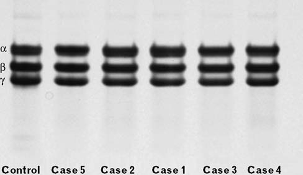
The patients with dengue virus infection had prolonged thrombin times of different degree (Table 1). The patients’ fibrinogen concentration was within the normal range. In general, the patients had elevated fibrin degradation products (D-dimer), especially cases 2, 3 and 5. Only in cases 1 and 4 were the D-dimer concentrations normal. No correlation was found between the D-dimer concentration and thrombin time. The albumin concentration was quite similar to that of control values only in cases 4 and 5; from case 1 to 3, the values were higher than those of the control, but can be considered within the normal range. The α, β and γ chains from the patients’ samples were intact, indicating that there was no fibrinogenolysis (Fig. 1).
Table 1.
Summary of some biochemical, functional and structural characteristics patients infected by the dengue virus
| Parameters | Control | Case 1 | Case 2 | Case 3 | Case 4 | Case 5 |
|---|---|---|---|---|---|---|
| Fg (g/l) | 3.2 | 3.2 | 2.6 | 2.5 | 2.6 | 2.5 |
| TT (s) | 19.9 | 22 | 23.3 | 25.6 | 27 | 38.4 |
| D-dimer (ng/ml) | 625 | 375 | 1300 | 6000 | 750 | 1600 |
| Albumin (%) | 100 | 166 | 166 | 132 | 116 | 108 |
| Polymerization | ||||||
| Lag Time (s) | 0 | 30 | 50 ± 9 | 25 ± 17 | 70 ± 9 | 35 ± 9 |
| Slope (OD unit/s) × 10−3 | 1.9 ± 0.1 | 2.6 ± 0.1 | 2.2 ± 0.2 | 1.9 ± 0.3 | 1.9 ± 0.0 | 1.4 ± 0.0 |
| Maximum ι (OD unit) | 0.230 ± 0.006 | 0.179 ± 0.002 | 0.198 ± 0.009 | 0.203 ± 0.009 | 0.128 ± 0.004 | 0.131 ± 0.005 |
| Fibrinolysis | ||||||
| TLT (s) | 548 ± 11 | 550 ± 9 | 550 ± 17 | 580 ± 23 | 580 ± 23 | 548 ± 11 |
| V (OD unit/s) 10−5 | 100 ± 5 | 70 ± 3 | 90 ± 2 | 70 ± 2 | 50 ± 4 | 50 ± 0.3 |
| t½ (s) | 462 ± 5 | 463 ± 9 | 450 ± 20 | 438 ± 23 | 495 ± 20 | 450 ± 11 |
| (ιf/ιp) × 100 | 98 ± 5 | 68 ± 2 | 83 ± 4 | 71 ± 3 | 87 ± 2 | 67 ± 8 |
| Fiber diameter (nm) | 97 ± 31 | 112 ± 44* | 117 ± 40* | ND | 113 ± 35* | 104 ± 37* |
Fibrinogen concentration was determined by the gravimetric method, albumin was quantified by immunoelectrophoresis and fibrin polymerization and lysis process was measured by turbidity at 350 nm. The mean average of fibrin fibers thickness was calculated from digitized scanning electron microscopy pictures using ImageJ 1.36b software. ND, not done; ι, turbidity; ιf/ιp, ratio between maximum turbidity of fibrinolysis and polymerisation; t½, time to reach 50% of clot degradation; TLT, total lysis time; V, lysis rate.
Statistically significant.
Fig. 1.
Electrophoresis of the fibrinogen on a 6% gel, SDS/PAGE Tricine-system, under reducing conditions. The gel was loaded with 14 µg of non cross-linked fibrin from each sample, prepared as described in the Materials and Methods section. In the control gel lane, the fibrin α, β, and γ chains are indicated on the left-side.
Fibrin polymerization and internal fibrinolysis
From plasma
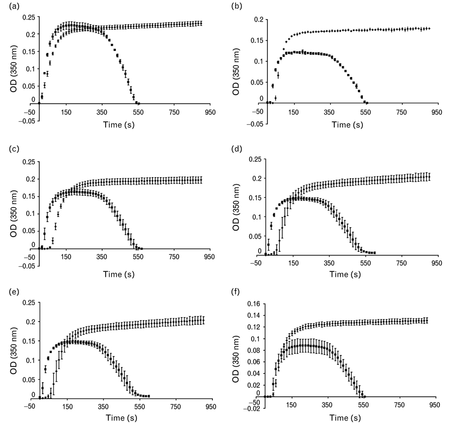
On the basis of the relationship between the maximum turbidity and the plasma fibrinogen concentration from data collected from normal plasma fibrin polymerization of apparently healthy individuals (not shown) and the present control, we consider that only cases 4 and 5 had lower maximum turbidity compared with the expected values (Table 1). The pattern of internal fibrinolysis of the patients was similar to that of the control (Table 1); only the ιf/ιp, the ratio between the maximum turbidity value obtained from fibrinolysis and the maximum turbidity value obtained from polymerization, expressed as a percentage (defined in the Materials and Methods section) was diminished, especially in cases 1, 3 and 5. The polymerization and fibrinolysis curves plotted together for each sample show this parameter as a function of time (Fig. 2). Only the control clots start to dissolve after the maximum turbidity was reached, whereas the patient samples start dissolution earlier. Another feature is that, in general, the lag time of the patient fibrinolysis curves was shortened compared with that of polymerization, except for case 5, where no change was observed.
Fig. 2.
Plasma fibrin polymerization and lysis followed by turbidity at 350 nm. The fibrin polymerization and fibrin lysis curve of each patient are recorded in the same graph. For clot dissolution, t-PA was added before thrombin, with the same clotting conditions as for polymerization. (a) Control, (b–f) Case 1 through 5, respectively.
From purified fibrinogen
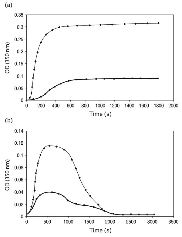
Mixing the normal purified fibrinogen with case 5 purified fibrinogen impaired severely both the polymerization and fibrinolytic process. The lag time was four times longer, the rate of fibrin formation 10 times slower and the final turbidity 3.5 times lower (Fig. 3a). Also, the control fibrinolytic process worsened substantially in the presence of approximately 30% of case 5 fibrinogen molecules. The rate of lysis decreased approximately three-fold (as mentioned in the Materials and Methods section, the rate of lysis was calculated from the linear part of the descending limb of the curve, just after the plateau) (Fig. 3b).
Fig. 3.
Effect of purified fibrinogen from a dengue patient (case 5) on normal polymerization and fibrinolysis. (a) Polymerization process: Upper curve (♦) control fibrinogen; lower curve (■) after mixing 7 µmol/l normal fibrinogen with 3 µmol/l of dengue patient 5 fibrinogen. (b) Internal Fibrinolysis: Upper curve (♦) control fibrinogen; lower curve (■) after mixing 7 µmol/l normal fibrinogen with 3 µmol/l of dengue patient 5 fibrinogen.
Scanning electron microscopy
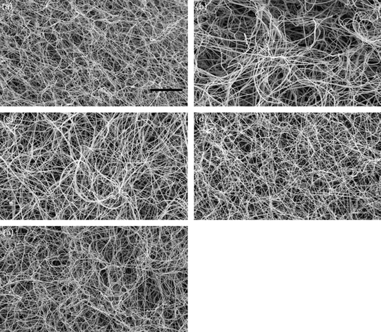
Electron microscopy revealed distinctive differences in clots from some patients (Fig. 4). The fibrin networks from cases 1 and 2 had larger pores and thicker fibers compared with control clots and those of the rest of the patients; the frequency distribution of the fibrin fiber thicknesses corroborated these observations (Table 2). The patients’ fibrin fiber thickness distributions were statistically different from those of the control, with a higher mean of fiber diameter (Table 1, P < 0.01).
Fig. 4.
Images of clot architecture visualized by scanning electron microscopy. The clot was formed from plasma and extensively washed for scanning electron microscopy preparation. Magnification bar is 10 µm. (a) Control, (b–e) Case 1, 2, 4 and 5, respectively.
Table 2.
Frequency distribution of fibrin fibers according to their diameter
| Range (nm) | Control | Case 1 | Case 2 | Case 4 | Case 5 |
|---|---|---|---|---|---|
| 25–40 | 0.04 | 0.01 | 0.01 | 0.02 | 0.04 |
| 40–55 | 0.07 | 0.06 | 0.06 | 0.08 | 0.06 |
| 55–70 | 0.09 | 0.07 | 0.07 | 0.05 | 0.08 |
| 70–85 | 0.11 | 0.09 | 0.08 | 0.06 | 0.10 |
| 85–100 | 0.17 | 0.13 | 0.12 | 0.09 | 0.17 |
| 100–115 | 0.21 | 0.15 | 0.14 | 0.16 | 0.16 |
| 115–130 | 0.16 | 0.17 | 0.16 | 0.23 | 0.18 |
| 130–145 | 0.08 | 0.14 | 0.13 | 0.14 | 0.10 |
| 45–160 | 0.05 | 0.09 | 0.09 | 0.08 | 0.04 |
| 160–175 | 0.01 | 0.08 | 0.07 | 0.06 | 0.04 |
| 175–190 | 0.003 | 0.05 | 0.04 | 0.03 | 0.003 |
| 190–205 | 0 | 0.01 | 0.01 | 0.007 | 0.02 |
| 205–220 | 0 | 0.01 | 0.01 | 0 | 0.003 |
| 220–235 | 0 | 0.004 | 0.003 | 0 | 0 |
| 235–250 | 0 | 0.004 | 0.003 | 0 | 0 |
| 250–265 | 0 | 0 | 0 | 0 | 0 |
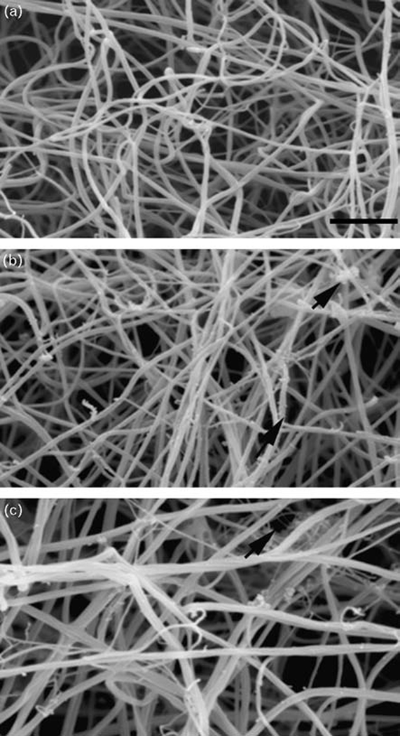
Furthermore, the patients’ clot scanning electron microscopy images showed some distinctive features: many free fiber ends and, in some areas, chunks of fibrin associated with fibrin fibers (Fig. 5, panel b, top-right arrow), an increase in fiber lateral association, fibers with spaced ‘buds’ along them (Fig. 5, panel b, bottom-right arrow), and very thin fibers intertwined with thicker fibers (Fig. 5, panel c, top-right arrow).
Fig. 5.
Evidence of fibrin fiber lysis during clot formation in patients’ samples by scanning electron microscopy. Magnification bar corresponds to 2 µm. (a) Control, (b) Case 4, and (c) Case 5. The black arrows indicate the special fibrinolytic characteristics described in the text.
These are all characteristics of the early stages of fibrinolysis, indicating that a very low quantity of plasmin had formed during the 2-h incubation. The incipient beginning of fibrin reorganization before clot dissolution is characterized by free fiber ends and chunks of fibers arising from lateral transection of fibers, an increase in fibrin thickness from lateral association of cut fibers, a rough surface of fibers that corresponds to bound-plasmin(ogen), tPA and fibrin degradation products, and loosely associated thin fibers from the dissociation that occurs as protofibrils are cleaved. These results were in good accord with the ιf/ιp ratio, calculated from internal fibrinolysis experiments, as lysis began before the clot was fully formed (Table 1).
Discussion
For the first time, clots from individuals infected by the dengue virus were studied by scanning electron microscopy and some studies were performed with the purified fibrinogen. One of the limitations of the present work is that we have studied only five patients, and we are aware that for future studies it would be necessary to increase the number of patients for more conclusive results, although the techniques used in the present work are difficult to perform on a large scale. These patients were sent to us due to the fact that they behaved like a transitory acquired dysfibrinogenemia and there were no other causes that could explain the prolonged thrombin time. We thought that this lengthening might be related to the presence of high D-dimer levels in most of the patients, as they could interact with fibrin oligomers, truncating the progression of oligomer growth [25,26]. However, we have not found a correlation between the degree of thrombin time prolongation and the D-dimer concentration. Other authors have reported that the prolonged thrombin time, increased D-dimer, and decreased fibrinogen concentration in different degrees appear to be related to the severity of the disease in dengue fever and DHF (for a review see Mairuhu et al. [13]). It seems that in DHF the activated partial thromboplastin and thrombin time are more frequently abnormal than the prothrombin time [27,28]. Recently, a study performed with 42 children, 20 with dengue fever and 22 with DHF, found that the concentration of plasma vWF:Ag seemed to be the best indicator of the progression to DHF [29]; we have not measured vWF in the present patients.
Some of the differences observed among the patient clot properties may arise because we do not know at what day of virus infection the plasma samples were obtained, that is, febrile, acute or convalescent stage. In spite the fact that case 1 had normal D-dimer levels, the ιf/ιp ratio was among the lowest (66%). The scanning electron microscopy images corroborated the interpretation that lower ιf/ιp values indicate earlier onset of fibrin lysis, since cases 1, 2 and 5 were the most lyzed. It is likely that in these patients the ratio of t-PA/PAI-1 was higher as compared with cases 3 and 4. Such a reversed relationship between t-PA and PAI-1 has been reported by several authors during the acute stage of virus infection, giving rise to a hyperfibrinolytic state [13,30,31].
Several mechanisms have being proposed that could partially explain the bleeding in DHF. In addition to a decreased platelet count, prolonged activated partial thromboplastin time, increased t-PA levels, etc., it has been found that TAFI antigen and activity levels were decreased [17]. Also, in vitro the dengue virus, specifically the glycoprotein envelope, can bind plasminogen directly and activate it to plasmin [32,33]. However, no fibrinogen degradation products were found in the plasma of the patients. When polymerization curves were compared with fibrinolysis curves (Fig. 2) almost all the patients (except case 5) had an enhancement of the rate of clotting, with curves resembling those of previous work where it was demonstrated that plasminogen molecules bridge the D-regions of different fibrin monomers, accelerating fibrin lateral aggregation [34,35]. We have not measured plasminogen concentration, but in other reports it has been described that the plasmin-α2-antiplasmin complex was increased in DHF [31]. However, the patients’ fibrinolysis curves were not easy to interpret as there is no simple explanation why the patients’ total lysis times were similar to those of the control, in spite of an earlier fibrinolysis onset.
Fibrin polymerization can be considered a measure of fibrinogen functionality, and the kinetics of fibrin formation by turbidity gives some indirect information about fibrin network building and fiber thicknesses. From comparison of the turbidity curves of these patients and controls, only cases 4 and 5 had clots composed of thinner fibers. Purified fibrinogen from case 5 behaved like an abnormal fibrinogen, since when it was mixed with normal fibrinogen there was impaired polymerization and fibrinolysis. This abnormal behavior of purified dengue fibrinogen could contribute to a bleeding disorder, affecting the quality of the hemostatic plug (including the interaction between the platelets).
Scanning electron microscopy results were not consistent with the final turbidity of polymerization. Conversely, patients’ fibrin network had fibers thicker than those of the control. We attribute this discrepancy to the fact that the patients’ clots were slightly digested during the 2 h clot incubation. As we have added only thrombin to the plasma samples and left them in a humid chamber for clot maturation, it seems that during this time small amounts of plasmin have been formed, reflecting an imbalance of the fibrinolytic system in the patients’ plasma or a hyperfibrinolytic state. The structural changes of the normal fibrin network during fibrin lysis have been studied by scanning electron microscopy [36,37], and the images of the present dengue patients’ clots correspond to an early stage of fibrin lysis onset, more precisely stage 2 [36].
In conclusion, during dengue virus infection the fibrinogen molecules can be modified so that when they are activated by thrombin, they form a meshwork of fibers thinner than normal fibrinogen. We think that probably the fibrinogen has more sialic acid residues, repelling each other and impeding the incorporation of more protofibrils into the fibers. Furthermore, we have confirmed by scanning electron microscopy and internal fibrinolysis that during dengue virus infection there is a hyperfibrinolytic state, where clots are more prone to be lyzed.
Acknowledgements
We want to thank the Virology laboratory of IVIC, especially Dr Ferdinando Liprandi and MsC Zoila Moro for the Dengue serotype determination. To MsC Zoila Carvajal for technical assistance and the collaboration of the MD students Corina Lesseur and Fanny Leonardi during their training. We are grateful to Dr Norma Bosch for the Dengue plasma samples.
This study was partially supported by NIH grant HL 30954.
References
- 1.Deen JL, Harris E, Wills B, Balmaseda A, Hammond SN, Rocha C, et al. The WHO dengue classification and case definitions: time for a reassessment. Lancet. 2006;368:170–173. doi: 10.1016/S0140-6736(06)69006-5. [DOI] [PubMed] [Google Scholar]
- 2.Halstead SB. Pathogenesis of dengue: challenges to molecular biology. Science. 1988;239:476–481. doi: 10.1126/science.3277268. [DOI] [PubMed] [Google Scholar]
- 3.Henchal EA, Putnak JR. The dengue viruses. Clin Microbiol Rev. 1990;3:376–396. doi: 10.1128/cmr.3.4.376. [DOI] [PMC free article] [PubMed] [Google Scholar]
- 4.Gubler DJ. Dengue and dengue hemorrhagic fever. Clin Microbiol Rev. 1998;11:480–496. doi: 10.1128/cmr.11.3.480. [DOI] [PMC free article] [PubMed] [Google Scholar]
- 5.Harris E, Videa E, Pérez L, Sandoval E, Téllez Y, Pérez ML, et al. Clinical, epidemiologic, and virologic features of dengue in the 1998 epidemic in Nicaragua. Am J Trop Med Hyg. 2000;63:5–11. doi: 10.4269/ajtmh.2000.63.5. [DOI] [PubMed] [Google Scholar]
- 6.Bhakdi S, Kazatchkine MD. Pathogenesis of dengue: an alternative hypothesis. Southeast Asian J Trop Med Public Health. 1990;21:652–667. [PubMed] [Google Scholar]
- 7.Bielefeldt-Ohmann H. Pathogenesis of dengue virus diseases: missing pieces in the jigsaw. Trends Microbiol. 1997;5:409–413. doi: 10.1016/S0966-842X(97)01126-8. [DOI] [PubMed] [Google Scholar]
- 8.Rothman AL, Ennis FA. Immunopathogenesis of dengue hemorrhagic fever. Virology. 1999;257:1–6. doi: 10.1006/viro.1999.9656. [DOI] [PubMed] [Google Scholar]
- 9.Rigau-Perez JG, Clark GG, Gubler DJ, Reiter P, Sanders EJ, Vorndam AV. Dengue and dengue haemorrhagic fever. Lancet. 1998;352:971–977. doi: 10.1016/s0140-6736(97)12483-7. [DOI] [PubMed] [Google Scholar]
- 10.Srichaikul T, Nimmanitaya S, Artchararit N, Siriasawakul T, Sungpeuk P. Fibrinogen metabolism and disseminated intravascular clotting in dengue hemorrhagic fever. Am J Trop Med Hyg. 1977;6:525–532. doi: 10.4269/ajtmh.1977.26.525. [DOI] [PubMed] [Google Scholar]
- 11.Bhamarapravati N. Hemostatic defects in dengue hemorrhagic fever. Rev Infect Dis. 1989;11 Suppl 4:S826–S829. doi: 10.1093/clinids/11.supplement_4.s826. [DOI] [PubMed] [Google Scholar]
- 12.Marcel L, van Gorp ECM, ten Cate H. Disseminated intravascular coagulation. In: Handin RI, Lux SE, Stossel TP, editors. Blood. Principles and practice of haematology. 2nd edn. Philadelphia: Lippincott Williams and Wilkins; 2003. pp. 1275–1301. [Google Scholar]
- 13.Mairuhu AT, MacGillavry MR, Setiati TE, Soemantri A, ten Gates H, Brandjes DP, et al. Is clinical outcome of dengue-virus infections influenced by coagulation and fibrinolysis? A critical review of the evidence. Lancet Infect Dis. 2003;3:33–41. doi: 10.1016/s1473-3099(03)00487-0. [DOI] [PubMed] [Google Scholar]
- 14.Chungue E, Poli L, Roche C, Gestas P, Glaziou P, Markoff LJ. Correlation between detection of plasminogen cross-reactive antibodies and hemorrhage in dengue virus infection. J Infect Dis. 1994;170:1304–1307. doi: 10.1093/infdis/170.5.1304. [DOI] [PubMed] [Google Scholar]
- 15.Huang YH, Chang BI, Lei HY, Liu HS, Liu CC, Wu HL, et al. Antibodies against dengue virus E protein peptide bind to human plasminogen and inhibit plasmin activity. Clin Exp Immunol. 1997;110:35–40. doi: 10.1046/j.1365-2249.1997.4991398.x. [DOI] [PMC free article] [PubMed] [Google Scholar]
- 16.Krishnamurti C, Wahl LM, Alving BM. Stimulation of plasminogen activator inhibitor activity in human monocytes infected with dengue virus. Am J Trop Med Hyg. 1989;40:102–107. doi: 10.4269/ajtmh.1989.40.102. [DOI] [PubMed] [Google Scholar]
- 17.Monroy V, Ruiz BH. Participation of the dengue virus in the fibrinolytic process. Virus Genes. 2000;21:197–208. doi: 10.1023/a:1008191530962. [DOI] [PubMed] [Google Scholar]
- 18.Lanciotti RS, Calisher CH, Gubler DJ, Chang GJ, Vorndam AV. Rapid detection and typing of dengue viruses from clinical samples by using reverse transcriptase-polymerase chain reaction. J Clin Microbiol. 1992;30:545–551. doi: 10.1128/jcm.30.3.545-551.1992. [DOI] [PMC free article] [PubMed] [Google Scholar]
- 19.Ingram GIC. The determination of plasma fibrinogen by the clot weight method. Biochem J. 1952;51:583–585. doi: 10.1042/bj0510583. [DOI] [PMC free article] [PubMed] [Google Scholar]
- 20.Laurell CB. Quantitative estimation of proteins by electrophoresis in agarose gel containing antibodies. Anal Biochem. 1966;15:45–52. doi: 10.1016/0003-2697(66)90246-6. [DOI] [PubMed] [Google Scholar]
- 21.Schägger H, Von Jagow G. Tricine-sodium dodecyl sulfate-polyacrylamide gel electrophoresis for the separation of proteins in the range from 1 to 100 kDa. Anal Biochem. 1987;166:368–379. doi: 10.1016/0003-2697(87)90587-2. [DOI] [PubMed] [Google Scholar]
- 22.Kazal LA, Amsel S, Miller OP, Tocantins LM. The preparation and some properties of fibrinogen precipitated from human plasma by glycine. Proc Soc Exp Biol Med. 1963;113:989–994. doi: 10.3181/00379727-113-28553. [DOI] [PubMed] [Google Scholar]
- 23.Marchi R, López YR, Nagaswami S, Masova L, Pulido A, López JM, et al. Hemostatic changes related to fibrin formation and fibrinolysis during the first trimester in normal pregnancy and in recurrent miscarriage. Thromb Haemost. 2007;97:552–557. [PubMed] [Google Scholar]
- 24.Langer BG, Weisel JW, Dinauer PA, Nagaswami C, Bell WR. Deglycosylation of fibrinogen accelerates polymerization and increases lateral aggregation of fibrin fibers. J Biol Chem. 1988;263:15056–15063. [PubMed] [Google Scholar]
- 25.Platonova TN, Musialkovskaia AA, Tolstykh VM, Belitser VA. Inhibition of fibrin assembly by fragment D and its dimer derived from fibrinogen and stabilized fibrin. Evidence for the two-step type of inhibition. Biokhimiia. 1980;45:1780–1787. [PubMed] [Google Scholar]
- 26.Hermans J, McDonagh J. Fibrin: structure and interactions. Semin Thromb Hemost. 1982;8:11–24. doi: 10.1055/s-2007-1005039. [DOI] [PubMed] [Google Scholar]
- 27.Nimmannitya S. Clinical spectrum andmanagement of dengue haemorrhagic fever. Southeast Asian J Trop Med Public Health. 1987;18:392–397. [PubMed] [Google Scholar]
- 28.Hathirat P, Isarangkura P, Srichaikul T, Suvatte V, Mitrakul C. Abnormal hemostasis in dengue hemorrhagic fever. Southeast Asian J Trop Med Public Health. 1993;24:80–85. [PubMed] [Google Scholar]
- 29.Sosothikul D, Seksarn P, Pongsewalak S, Thisyakorn U, Lusher J. Activation of endothelial cells, coagulation and fibrinolysis in children with Dengue virus infection. Thromb Haemost. 2007;97:627–634. [PubMed] [Google Scholar]
- 30.Huang YH, Liu CC, Wang ST, Lei HY, Liu HS, Lin YS, et al. Activation of coagulation and fibrinolysis during dengue virus infection. J Med Virol. 2001;63:247–251. doi: 10.1002/1096-9071(200103)63:3<247::aid-jmv1008>3.0.co;2-f. [DOI] [PubMed] [Google Scholar]
- 31.van Gorp EC, Setiati TE, Mairuhu AT, Suharti C, ten Cate H, Dolmans WM, et al. Impaired fibrinolysis in the pathogenesis of dengue hemorrhagic fever. J Med Virol. 2002;67:549–554. doi: 10.1002/jmv.10137. [DOI] [PubMed] [Google Scholar]
- 32.van Gorp EC, Minnema MC, Suharti C, Mairuhu AT, Brandjes DP, ten Cate H, et al. Activation of coagulation factor XI, without detectable contact activation in dengue haemorrhagic fever. Br J Haematol. 2001;113:94–99. doi: 10.1046/j.1365-2141.2001.02710.x. [DOI] [PubMed] [Google Scholar]
- 33.Wills BA, Oragui EE, Stephens AC, Daramola OA, Dung NM, Loan HT, et al. Coagulation abnormalities in dengue hemorrhagic fever: serial investigations in 167 vietnamese children with dengue shock syndrome. Clin Infect Dis. 2002;35:277–285. doi: 10.1086/341410. [DOI] [PubMed] [Google Scholar]
- 34.Garman AJ, Smith RA. The binding of plasminogen to fibrin: evidence for plasminogen-bridging. Thromb Res. 1982;27:311–320. doi: 10.1016/0049-3848(82)90078-0. [DOI] [PubMed] [Google Scholar]
- 35.Petersen LC, Suenson E. Effect of plasminogen and tissue-type plasminogen activator on fibrin gel structure. Fibrinolysis. 1991;5:51–59. [Google Scholar]
- 36.Meh DA, Mosesson MW, DiOrio JP, Siebenlist KR, Hernandez I, Amrani DL, et al. Disintegration and reorganization of fibrin networks during tissue-type plasminogen activator-induced clot lysis. Blood Coagul Fibrinolysis. 2001;12:627–637. doi: 10.1097/00001721-200112000-00003. [DOI] [PubMed] [Google Scholar]
- 37.Veklich Y, Francis CH, White J, Weisel JW. Structural studies of fibrinolysis by electron microscopy. Blood. 1998;92:4721–4729. [PubMed] [Google Scholar]


