

Introduction
Subungual exostosis (SE) is a solitary, commonly sessile, bony growth firmly attached to the tuft of the underlying distal phalanx. It is a benign osteocartilaginous tumor arising underneath or beside the nail bed and may disrupt nail growth. Initially described in the hand by Hutchinson in 1857 [8], Dupuytren wrote about his experience with its manifestation in the foot a decade earlier [4].
The differential diagnosis of SE spans many possible nail, soft tissue, and bony pathologies. Onchomycosis, granuloma pyogenicum, epithelioma, verruca, keratoacanthoma [12], fibroma, glomus tumor [11], melanotic whitlow [3], osteochondroma [2], turret exostosis [13], juxtacortical chondroma, and myositis ossificans [1] have all been mentioned as potential diagnoses.
The etiology and pathogenesis of SE is not clearly established. It is most commonly thought to reflect a fibrocartilaginous metaplasia from trauma or chronic infection [5, 9]. Although the latter may be a secondary consequence, as SE can elevate the nail causing exposure of the underlying soft tissue. However, others have alluded to genetic etiology [6], supranumery digits, cartilaginous rests, or a forme fruste of hereditary multiple exostoses [10].
SE is seen predominantly in females in a ratio of up to 2:1 [2, 5], though one series documented occurrence more often in males [8]. It occurs primarily in children and young adults in the second or third decades of life and more often affects the great toe, up to 77% of the time [9].
SE is best diagnosed on lateral radiographic view. It is usually seen as a bony projection on the dorsal or dorsomedial aspect of the distal phalanx capped with fibrous tissue. Early lesions may have insufficient bony formation to show up on X-rays [7]. No surrounding destructive changes are seen, supporting this as a benign diagnosis.
SE is treated surgically. The nail plate may be removed, and excision is either through the nail bed [11] or via a midlateral incision [2]. Complete resection of the fibrocartilaginous cap is compulsory as local recurrence rates of 11% have been reported [9].
On histology, SE is composed of a mature trabecular pattern of a cancellous bone base with a proliferating fibrocartilaginous cap. The growth, which can reach more than 2 cm in diameter, is always benign. Our case report is the first description of aberrant induced nail plate growth with SE in the literature.
Case Report
A 38-year-old man presented with a tender exophytic growth from the tip of his right index finger. He noted that it had been growing over the course of several months. His history was remarkable for frequent manicures; however he denied paronychial infection or finger inflammation. Other than normal computer use, he recalled no repetitive procedures or minor trauma to the index finger. Inadvertent striking of the digit caused him discomfort. The finger deformity was also cosmetically disturbing to the patient.
Examination showed a full range of motion of the index finger with normal sensibility. The native nail showed no evidence of involvement, although the growth had caused a lifting of the overlying nail plate. Recent dermatological treatment resected a portion of the nail plate to evaluate the sterile matrix as a source of this growth. No involvement of the nail bed had been appreciated. A firm localized induration was palpated below the skin of the hyponychium with tenderness on pressure. A bony projection with an induced formed nail plate was present (Fig. 1). A preoperative radiograph showed an ossific density projecting off the distal aspect of the tuft, consistent with subungual exostosis. In addition, this X-ray showed a density of similar appearance to the nail (Fig. 2).
Figure 1.
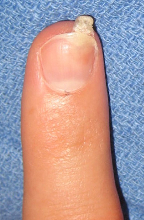
Subungual exostosis projecting dorsally toward the ulnar aspect of the right index finger distal phalanx.
Figure 2.

X-ray, anteroposterior view, of finger with arrow at subungual exostosis.
The patient underwent surgical excision under regional anesthesia. A portion of the native nail plate was lifted, showing the dorsal aspect of this lesion originating from the bony tuft below the sterile matrix. An incision was made down to the aberrant growth, and the elevator was used to dissect down to a normal rim of the tuft of distal phalanx. The lesion was removed entirely with a rongeur, flush to the normal tuft. Pathology examination of the growth confirmed fragments of cartilage, bone, aberrant nail bed, and nail plate suggestive of subungual exostosis (Fig. 3). The histological finding of an induced sterile matrix and nail plate has not previously been reported in the literature describing SE of the finger.
Figure 3.

Subungual exostosis (H&E at ×100 magnification). Arrow at fibrocartilaginous cap.
Discussion
Over the last 160 years, there have been scattered reports of subungual exostosis occurring in the foot and less commonly, in the hand. In 1857, Hutchinson was the first to describe an occurrence of SE on the thimble finger (the long finger) of a young woman. In 1966, Evison and Price reported on the occurrence of SE from the Bristol Bone Tumour Registry between 1947 and 1965; two patients among 20 had lesions in the fingers. The largest cases series to date came from Carroll et al. They reported 16 cases of SE of the finger in their patients seen at Columbia University from 1953 to 1989. Our case report adds to the scores of patients with subungual exostosis in the literature. However it is the first to describe the formation of aberrant induced nail plate growth.
Subungual exostosis is a benign fibrocartilaginous growth that is infrequently seen by physicians but should be included in the differential diagnosis of fingertip pathology. It is a tender lesion arising from the distal phalanx, often due to chronic traumatic stimulation. Early suspicion of subungual exostosis should lead one to verify the diagnosis by X-ray, and confirmation warrants prompt surgical excision.
References
- 1.Bennett RG, Grammer S. Painful callus of the thumb due to phalangeal exostosis. Arch. Dermatol. 1973;108:826–827. doi: 10.1001/archderm.108.6.826. [DOI] [PubMed] [Google Scholar]
- 2.Carroll RE, Chance JT, Inan Y. Subungual exostosis in the hand. J Hand Surg [Br] 1992;17B:569–574. doi: 10.1016/s0266-7681(05)80243-8. [DOI] [PubMed] [Google Scholar]
- 3.Cohen HJ, Frank SB, Minkin W, et al. Subungual exostoses. Arch Dermatol. 1973;107:431–432. doi: 10.1001/archderm.107.3.431. [DOI] [PubMed] [Google Scholar]
- 4.Dupuytren G. On exostosis on the upper surface of the ungual phalanx of the great toe. In: Gros C, editor. On injuries and diseases of bones. London: The Syndenham Society; 1847. pp. 408–415. [Google Scholar]
- 5.Evison G, Price CH. Subungual exostosis. Br J Radiol. 1966;39:451–455. doi: 10.1259/0007-1285-39-462-451. [DOI] [PubMed] [Google Scholar]
- 6.Guidetti MS, Stinchi C, Vezzani C, et al. Subungual exostosis of a finger resembling pterygium inversum unguis. Dermatology. 1996;193:354–355. doi: 10.1159/000246292. [DOI] [PubMed] [Google Scholar]
- 7.Hoehn JG, Coletta C. Subungual exostosis of the fingers. J Hand Surg. 1992;17:468–471. doi: 10.1016/0363-5023(92)90352-P. [DOI] [PubMed] [Google Scholar]
- 8.Hutchinson J. Subungual exostosis of the great toe: excision with success. Lancet. 1857;2:246–247. doi: 10.1016/S0140-6736(02)38683-5. [DOI] [Google Scholar]
- 9.Landon GC, Johnson KA, Dahlin DC. Subungual exostoses. J Bone Joint Surg [Am] 1979;61:256–259. [PubMed] [Google Scholar]
- 10.Lowenthal K. Subungual exostosis on a forefinger. N Y State J Med. 1964;64:2690–2695. [PubMed] [Google Scholar]
- 11.Matthewson MH. Subungual exostoses of the fingers. Are they really uncommon? Br J Dermatol. 1978;98:187–189. doi: 10.1111/j.1365-2133.1978.tb01620.x. [DOI] [PubMed] [Google Scholar]
- 12.Shapiro L, Baraf CS. Subungual epidermoid carcinoma and keratoacanthoma. Cancer. 1970;25:141–152. doi: 10.1002/1097-0142(197001)25:1<141::AID-CNCR2820250121>3.0.CO;2-H. [DOI] [PubMed] [Google Scholar]
- 13.Wissinger HA, McClain EJ, Boyes JH. Turret exostosis: ossifying hematoma of the phalanges. J Bone Joint Surg Am. 1966;48:105–110. [PubMed] [Google Scholar]