Abstract
Background
Approximately 20% of oocytes are classified as immature and discarded following intracytoplasmic sperm injection (ICSI) procedures. These oocytes are obtained from gonadotropin-stimulated patients, and are routinely removed from the cumulus cells which normally would mature the oocytes. Given the ready access to these human oocytes, they represent a potential resource for both clinical and basic science application. However culture conditions for the maturation of cumulus-free oocytes have not been optimized. We aimed to improve maturation conditions for cumulus-free oocytes via culture with ovarian paracrine/autocrine factors identified by single cell analysis.
Methodology/Principal Finding
Immature human oocytes were matured in vitro via supplementation with ovarian paracrine/autocrine factors that were selected based on expression of ligands in the cumulus cells and their corresponding receptors in oocytes. Matured oocytes were artificially activated to assess developmental competence. Gene expression profiles of parthenotes were compared to IVF/ICSI embryos at morula and blastocyst stages. Following incubation in medium supplemented with ovarian factors (BDNF, IGF-I, estradiol, GDNF, FGF2 and leptin), a greater percentage of oocytes demonstrated nuclear maturation and subsequently, underwent parthenogenesis relative to control. Similarly, cytoplasmic maturation was also improved as indicated by development to blastocyst stage. Parthenogenic blastocysts exhibited mRNA expression profiles similar to those of blastocysts obtained after IVF/ICSI with the exception for MKLP2 and PEG1.
Conclusions/Significance
Human cumulus-free oocytes from hormone-stimulated cycles are capable of developing to blastocysts when cultured with ovarian factor supplementation. Our improved IVM culture conditions may be used for obtaining mature oocytes for clinical purposes and/or for derivation of embryonic stem cells following parthenogenesis or nuclear transfer.
Introduction
Approximately 20% of oocytes routinely retrieved following hormone stimulation are classified as immature (termed germinal vesicle (GV) or metaphase I (MI) stage); these oocytes are discarded due to their reduced potential for embryo development under current culture conditions [1]. However, this cohort of oocytes is useful for studies aimed at elucidating the mechanisms of in vitro maturation of human oocytes and might ultimately contribute to the pool of embryos available for embryo transfer [2]. In addition, cumulus-free human oocytes may provide a platform for derivation of patient-specific embryonic stem cells (hESCs) including parthenogenic embryonic stem cells (pESCs) or somatic cell nuclear transfer-embryonic stem cells (SCNT-ESCs). Since the first report of in vitro human oocyte maturation in 1969 [3], several reports have documented blastocyst (BL) development or live birth achieved from oocytes matured in vitro [1], [4], [5]. However, nuclear and cytoplasmic maturation of cumulus-free oocytes remains suboptimal at best [2], [6], [7]. Moreover, ability of this cohort of oocytes to support the development of parthenogenic or nuclear transfer embryos to blastocyst stage has not been assessed.
The cumulus cells that surround oocytes within follicles modulate nuclear and cytoplasmic maturation by both physical cell-cell contact and the combined actions of paracrine factors [8]. However, during the process of intracytoplasmic sperm injection (ICSI), cumulus cells must be removed from oocytes. When these cumulus-free oocytes are directly exposed to commercially-available maturation media, they subsequently exhibit delayed oocyte maturation and abnormal embryo development [1], [2]. In the majority of studies of in vitro maturation (IVM), follicle stimulating hormone (FSH), luteinizing hormone (LH), estradiol and epidermal growth factor (EGF) are used as supplements. However, whether human oocytes express the receptors for FSH and LH is controversial [9], , and it is known that EGF affects oocyte maturation through the EGF receptor (EGFR), which is expressed by cumulus cells rather than the oocyte [11]. As outlined below, we used a microfluidic quantitative PCR (qPCR) system to elucidate the gene expression profiles of individual human oocytes and small numbers of cumulus cells using a combination of a large number of samples and targets [12], and then extended our studies via the use of parthenogenesis, in conjunction with gene expression profiling, as a functional assay of cytoplasmic maturation of oocytes.
Parthenogenesis in vitro is accomplished by artificial activation without sperm contribution [2]. In vivo, mammalian parthenotes are not able to develop to term due to aberrant, uniparental imprinting and consequent developmental abnormalities [13], [14]. Parthenogenesis, which can be induced in vitro by various electric, mechanical or chemical stimulations, is a valuable functional assay of oocyte developmental competence [15]. Also, pESC lines, that carry isogenic genome information from the oocyte donors, can be derived and may represent a potential alternative source of stem cells for basic scientific studies as well as novel therapeutic application. Although human pESCs have been derived from different oocyte sources such as donated mature or cryopreserved oocytes [16], [17], in vitro-matured oocytes present a potentially-abundant source for production of parthenogenic blastocysts.
Here, we report improved methods for in vitro maturation of cumulus-free human oocytes by supplemented culture media with ovarian paracrine/autocrine growth factors that were selected on the basis of gene expression profiles of single human oocytes and associated cumulus cells. Then we tested functionality of oocytes matured in vitro by assessing the ability to develop to the blastocyst stage, and by comparing the gene expression profiles of parthenogenic embryos and those obtained from IVF/ICSI.
Results
Expression of genes for paracrine/autocrine ligands and their cognate receptors in oocytes and cumulus cells
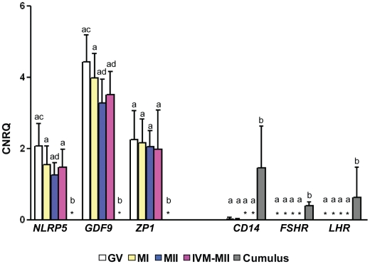
In order to improve culture conditions for maturation of cumulus-free oocytes in vitro, we began by assaying the expression of 15 growth factors (BDNF, IGF-I, estradiol, GDNF, leptin, FGF1, FGF2, GM-CSF, EGF, TGF-α, TGF-β1;2;3, and ET-1;2) and 27 of their cognate receptors that have been shown to regulate oocyte maturation. Selection was based on published literature deposited in two online databases (http://ovary.stanford.edu/ and http://receptome.stanford.edu/HPMR/) [18], [19]. Four different stages of oocytes, striped of their zona pellucida (ZP), were assayed: 1) GV (germinal vesicle), 2) MI (metaphase I), 3) MII (spontaneously matured metaphase II before IVM) and 4) IVM-MII (in vitro matured metaphase II). We confirmed the purity of ZP-free oocytes and cumulus cell samples based on the exclusive expression of known oocyte-specific and cumulus cell-specific genes. As expected, expression of the genes NLRP5, GDF9 and ZP1 was limited to oocytes, while expression of the genes CD14, FSHR and LHR, was limited to cumulus cells (Figure 1).
Figure 1. Expressions of oocyte-specific or cumulus cell-specific genes.
Values are the means of different stages of oocytes including GV (N = 10), MI (N = 8), MII (N = 9), IVM-MII (N = 10) and cumulus cells (N = 5). Error bars represent standard deviation (SD). CNRQ: calculated normalized relative quantity. *: Not detected in all samples. a,b or c,d: Values were significantly different (P<0.05).
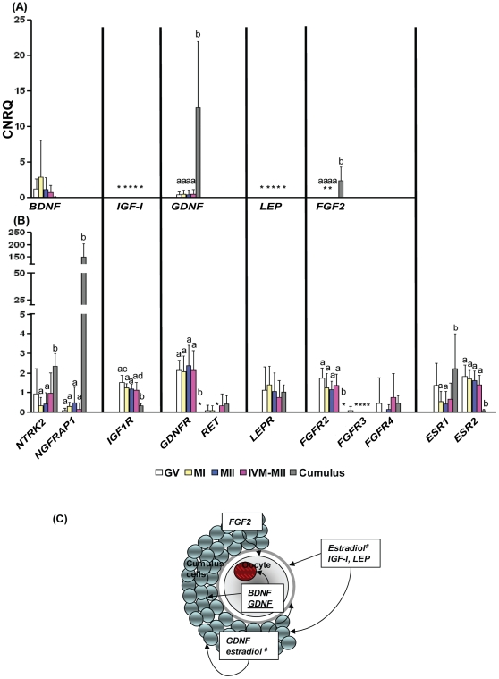
We examined expression of a set of candidate receptors and ligands in oocytes and cumulus cells, respectively (Figure 2A, 2B and S1). Among the growth factors, expression of mRNA of genes that encode for GDNF, FGF2, GM-CSF and TGF-β1 was significantly higher in cumulus cells than in oocytes. Conversely, the majority of receptors were detected at the mRNA level in oocytes with the exception for FGFR3, CSF2Rβ, EGFR and TGF-βR2. Among the receptors, expression of mRNA of genes that encode for NTRK2, NGFRAP1, ESR1 and EDNRA was higher in cumulus cells, while oocytes had significantly higher expression of mRNA of genes that encode for IGF1R, ESR2 and TGF-βR3. There were a few genes that were detected exclusively in only cumulus cells (CSF2Rβ and EGFR) or oocytes (GDNFR, FGF2R). We observed that even though the expression of the ligand, BDNF was low in cumulus cells compared to oocytes, its receptors, NTRK2 and NGFRAP1 were expressed in both oocytes and cumulus cells. In particular, NGFRAP1 was expressed more than 350 fold higher in cumulus cells than in oocytes. We also noted that mRNA of only two genes (IGF-I and leptin) was not expressed at detectable levels in either compartment, oocytes or cumulus cells. However, mRNA corresponding to their receptors, IGF1R and LEPR, was expressed in both oocytes and cumulus cells.
Figure 2. Expressions of ovarian paracrine/autocrine factors and their cognate receptors selected for supplementation in in vitro maturation media.
(A) Ovarian paracrine/autocrine ligands, (B) their cognate receptors, and (C) schematic representation of gene expressions. Values are the means of different stages of oocytes including GV (N = 10), MI (N = 8), MII (N = 9), IVM-MII (N = 10) and cumulus cells (N = 5). Error bars represent standard deviation (SD). CNRQ = calculated normalized relative quantity. *: No detected in all samples. a,b or c,d: Values were significantly different (P<0.05), underline: low expression level, #: adopted from Hillier et al. [46].
Six ovarian factors (BDNF, IGF-I, estradiol, GDNF, leptin and FGF2) were further studied based upon the expression of their receptors in oocytes and cumulus cells described above, as well as the availability of growth factor concentration information from previous animal model studies [1], [20], [21], [22], [23]. We derived a protocol for paracrine/autocrine supplementation for in vitro maturation of cumulus-free oocytes that includes supplementation with BDNF, IGF-I, GDNF, leptin, FGF2 and estradiol as shown in Figure 2C.
Effect of ovarian paracrine/autocrine supplement on oocyte nuclear maturation
To test whether culture supplementation to promote nuclear maturation, we assessed the polar body extrusion after culture of immature GV and MI oocytes in three different maturation conditions: 1) Commercial human oocyte maturation medium (Sage), 2) IVM-medium, and 3) IVM-medium supplemented with selected ovarian factors as described above and in Materials and Methods. As shown in Table 1, as expected, nuclear maturation of GV oocytes was less efficient than MI oocytes. Among the GV oocytes, 50–68% of oocytes extruded a first polar body within 48 h in the three different media. Significant differences were not observed with the GV oocyte nuclear maturation between any of the three media. In contrast, nuclear maturation of MI oocytes was much more efficient with values ranging from 75 to 94% in the three different media. The supplementation of MI oocytes in IVM-medium with ovarian factors demonstrated significantly higher rates of extruding a first polar body (94%) relative to media without supplementation (75%) (Table 1).
Table 1. Nuclear maturation of cumulus-free human oocytes in vitro from hormone-stimulated cycle.
| Oocytes | Culture media | No. of oocytes | Nuclear maturation rate (%) | |
| 24 h | 48 h | |||
| GV | Sage | 45 | 22 (48.9) | 31 (68.9) |
| IVM-medium | 46 | 19 (41.3) | 23 (50.0) | |
| Supplement | 98 | 45 (45.9) | 53 (54.1) | |
| MI | Sage | 42 | 37 (88.1) | 37 (88.1) |
| IVM-medium | 29 | 21 (72.4)a | 22 (75.9)a | |
| Supplement | 51 | 48 (94.1)b | 48 (94.1)b | |
Sage: commercial IVM medium supplemented with 10% SPS, FSH, hCG and estradiol.
IVM-medium: IVM-medium supplemented with 10% SPS.
Supplement: IVM-medium supplemented with 10% SPS, BDNF, estradiol, IGF-I, GDNF, FGF2, leptin.
GV: germinal vesicle stage.
MI: metaphase I stage.
Nuclear maturation: the presence of a first polar body.
Within columns, values with different superscripts were significantly different (P<0.05).
Functional analysis of ovarian paracrine/autocrine supplement via assessment of parthenote development
We used parthenogenic development as a functional assay of cytoplasmic maturation. Following chemical activation of oocytes matured in vitro, we monitored cleavage and development of the parthenotes daily. We observed that following activation, culture in both Sage and IVM-supplementation media demonstrated significantly higher cleavage rates (87.2% and 82.3%, respectively) compared to IVM-medium alone (55.6%). Most notably, however, parthenogenic blastocysts were only produced from embryos cultured with supplementation and not with Sage or IVM-medium alone (Table 2 and Figure S2).
Table 2. Embryo development after parthenogenetic activation of human oocytes matured in vitro.
| IVM media | Total oocytes | Activated (%)* | 2-cell (%)* | 8-cell (%)** | Blastocysts (%)** |
| Sage | 66 | 47 (71.1) | 41 (87.2)b | 4 (9.8) | - |
| IVM-medium | 44 | 36 (81.8) | 20 (55.6)a | 3 (15.0) | - |
| Supplement | 77 | 62 (80.5) | 51 (82.3)b | 7 (13.7) | 3 (5.9) |
Sage: commercial IVM medium supplemented with FSH, hCG and estradiol.
IVM-medium: IVM-medium supplemented with 10% SPS.
Supplement: IVM-medium supplemented with BDNF, estradiol, IGF-I, GDNF, FGF2, leptin.
*Percentage from activated oocytes.
**Percentage from cleaved embryos.
Within columns, values with different superscripts were significantly different (P<0.05).
Gene expression profiling of human parthenotes
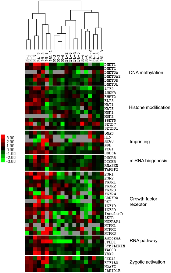
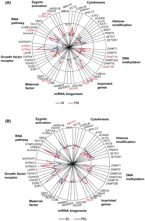
A total of 67 genes, from 10 functional categories related to embryo development were selected for analysis of parthenogenic and IVF/ICSI embryos. The categories included RNA pathway (5 genes), zygotic activation (4 genes), histone modification (10 genes), DNA methylation (6 genes), imprinting (6 genes), miRNA biogenesis (4 genes), pluripotency (3 genes), growth factor receptors (20 genes), maternal factors (4 genes) and cytokinesis (5 genes). These genes were selected for analysis of parthenogenic and IVF/ICSI embryos at the morula and blastocyst stage based primarily on published reports from non-human model systems (Figure 3, 4A, 4B and Table S2). In our data analysis, we observed 97% similarity in mRNA expression between parthenotes and IVF/ICSI embryos at the blastocyst stage (Figure 3 and 4B), with reduced similarity at the morula stage (83.6%, Figure 3 and 4A). More specifically, only the mRNAs of genes that encode for MKLP2 and PEG1 were expressed at lower levels in parthenogenic blastocysts in comparison to IVF/ICSI blastocysts (Figure 4B). Genes that encode for DNMT3A and MEG3 were expressed only in parthenotes but not in IVF/ICSI at the morula stage. Parthenogenic morula also had significantly higher expression levels of the mRNAs for DNMT3B, PEG1, DICER, NLRP5, NTRK2, NTRK3, CPEB1, CCNA1 and EIF1AX (Figure 4A).
Figure 3. Treeview display of cluster analysis of gene expression of all morulas and blastocysts from parthenogenesis and IVF/ICSI.
PM = parthenogenic morula, PBL = parthenogenic blastocyst.
Figure 4. Gene expression of parthenogenic embryos.
The average mRNA expression level of morulas (A) and blastocysts (B) from parthenogenesis and IVF/ICSI were displayed. Red color: Significantly differentially expressed. PM = parthenogenic morula, PBL = parthenogenic blastocyst.
Discussion
In this study, we first used a microfluidic quantitative PCR system to elucidate the gene expression profiles of single human oocytes and small groups of cumulus cells and subsequently improved upon in vitro maturation of cumulus-free oocytes to obtain parthenogenic blastocysts. We also observed that parthenotes from in vitro matured oocytes have similar mRNA expression levels of the majority of studied genes compared to IVF/ICSI embryos at the blastocyst stage.
Analysis of gene expression in single human oocytes and small groups of cumulus cells revealed notable similarities and differences from previous reports including human and model organisms. FSH, LH, estradiol and EGF are commonly used to promote maturation of cumulus-enclosed oocytes. According to the mRNA expression analysis in our study, there is negligible expression of the receptors for FSH, LH and EGF in human oocytes. This is consistent with a recent animal study [9] but contradictory to a previous report showing FSHR and LHR mRNA expression in mouse oocytes and pre-implantation embryos [10]. Furthermore, parthenote development in Sage medium with hormone supplementation did not improve late stage (further than 8-cells) embryo development compared to IVM-medium alone. Our gene expression and embryo development data suggest that FSH and LH may not affect oocyte nuclear and cytoplasmic maturation directly. In contrast, both ESR1 and ESR2, the receptors for estradiol, were detected in oocytes as well as cumulus cells in our study, suggesting a role for estradiol in oocyte maturation [24]. Therefore, the higher cleavage rate in Sage medium compared to IVM-medium alone is most likely due to the estradiol supplementation in Sage medium.
Both animal and human studies have demonstrated the important roles of neurotrophins (BDNF, NTF3 and NTF4) in oocyte maturation mediated by their receptors (NTRK1–3 and NGFRAP1) [20], [25]. We observed expression of BDNF mRNA and its receptors (NTRK2 and NGFRAP1) in both oocytes and cumulus cells. The higher expression of NGFRAP1 in cumulus cells suggests that BDNF may have a role in cumulus cell functions. It is important to note that the probe for NTRK2 in our study detected both the full length as well as the truncated forms of NTRK2. We suspect that the NTRK2 mRNA expression observed in the present study is the truncated form, since the full length form was not detected in either human oocytes or cumulus cells previously [26]. Interestingly, NGFRAP1 was detected in oocytes in our study but not in the previous study [26]. This may reflect the greater sensitivity of our microfluidic qPCR system over other traditional qPCR systems. Also, the previous study was conducted with cumulus-oocyte-complexes from small antral follicles without hormone stimulation, which may dramatically change the mRNA expression profiles compared to those undergoing hormone stimulation.
Expression of the mRNA encode for Glial cell line-Derived Neurotrophic Factor (GDNF) was detected in cumulus cells as shown previously [22] as well as oocytes in our study. GDNF is secreted by cumulus, granulosa, and theca cells as an ovarian factor stimulated by the preovulatory LH surge [22], and induces oocyte maturation and embryo developmental competence [27] by GDNFR and rearranged during transformation (RET). Based on the findings that GDNFR is only expressed in oocytes, and a low level of RET expression was detected in both oocytes and cumulus cells, our data suggest that GDNF might increase oocyte maturation by both a paracrine and autocrine manner in humans.
At the mRNA level, FGF2 was highly expressed in cumulus cells, while its receptor FGF2R was highly expressed in oocytes in our study. FGF2 and its receptors have been shown to be expressed at the protein level by both oocytes and granulosa cells up to secondary follicles [28], but the expression patterns in antral follicles have not been studied to date. Based on our gene expression data, FGF2 could affect oocyte maturation in a paracrine manner via the cumulus cells. This notion is supported by a previous study showing a higher concentration of FGF2 in follicular fluid compared to serum [29] and suggests that FGF2 production from the ovary has pivotal functions during follicle development and oocyte maturation.
Two parthenogenic blastocysts were produced from in vitro matured GV oocytes from hormone-stimulated cycles in our study. This is a proof of principle to demonstrate that immature oocytes are able to achieve the developmental competence with selected ovarian factors in vitro. In spite of ovarian factor supplementation, nuclear maturation of GV oocytes was not improved compared to MI stage oocytes. It may be due to the fact that the majority GV oocytes might be collected from pre-antral follicles which are not able to resume meiosis [30]. However, detailed protein expression of individual ovarian factors and their receptors in oocytes need to be further studied in order to elucidate the factors that may affect oocytes in specific maturation stages.
Once we had optimized the in vitro maturation of oocytes, we were able to obtain parthenogenic morulas and blastocysts in culture. It is noteworthy that this is the first study to show the gene expression profiles of human parthenogenic embryos at any stage. Overall, parthenotes have similar mRNA expression levels as IVF/ICSI embryos at the blastocyst stages (Figure 3 and 4B). The altered expression patterns of Cyclin A1 (CCNA1) in parthenotes changed from high (in morula) to low (in BLs) compared to IVF/ICSI embryos, which may due to it inefficient zygote genomic activation [31]. The altered expression of CCNA1 is similar to the expression of EIF1AX, another marker for zygotic genomic activation in our study as well as in a previous animal study [32]. However, we cannot suspect any abnormal effect on parthenotes due to the aberrant expression of CCNA1 since the protein is not expressed at the blastocyst stage [33], and its cell cycle regulation role is limited in meiotic cells [34]. Higher expression of cytoplasmic polyadenylation element-binding protein 1 (CPEB1) in parthenotes compared to normal embryos may increase cell cycle arrest and cellular senescence by inducing p53 translation [35]. This may cause lower cell number and higher apoptosis in parthenotes compared to normal embryos [14].
The mRNA expression pattern of growth factor receptors in parthenotes were different compared to normal embryos, suggesting that growth factors supplemented in oocyte/embryo culture media may alter the expression of their receptors [36]. Interestingly, BDNF improves pregnancy rates by increasing trophoblast cell growth and survival via its receptor NTRK2 expressed by the trophectoderm of blastocysts [37]. The effect of higher expression of NTRK2 and NTRK3 mRNA in parthenotes need to be further studied.
Aberrant imprinting is the major cause of embryo lethality of parthenogenic embryos. In our study, morula stage parthenotes exhibited high expression levels of MEG3 and PEG1. Maternally expressed MEG3 is regulated by a intergenic differentially methylated region in human gametes and embryos [38]. A possible explanation for this finding is that residual mRNA expression from oocytes was still present in morula stage parthenotes and/or chemical activation induced the aberrant expression of certain genes in parthenotes. Paternally expressed PEG1 exhibited significantly higher levels in parthenotes at the morula stage and dramatically decreased at the blastocyst stage. The high expression levels of two genes (DNMT3A and DNMT3B) related to DNA methylation in our study is also consistent with a mouse study showing the elevated methylation in parthenotes compared to IVF/ICSI embryos [39]. Analogous to CCNA1 and EIF1AX discussed above, the higher expression of de novo methyltransferase, DNMT3B, in parthenotes may be due to the altered zygotic activation since DNMT3B expression has been shown to be originated from embryo [40]. The regulation of epigenetic modifications in human pre-implantation embryos needs to be explored in further depth in order to understand the underlying molecular mechanisms behind early human development as well as the low efficiency of parthenogenesis and SCNT.
In our study, cleaved day 3 parthenotes were used to establish pESCs from single blastomeres using derivation methods as previously described [41]. Of the blastomeres isolated, approximately 21% (8 out of 38) divided once, but no blastocele could be detected in arrested parthenotes. Only blastomeres from non-arrested 8-cell stage parthenote formed blastoceles and proliferated (data not shown). From the 8 blastomeres isolated from an 8-cell parthenote, 3 blastomeres divided, which suggested that those 3 blastomeres are the most viable from the original 8-cell parthenote. This interpretation is supported by a previous study showing that the blastomeres from arrested human embryos still divided in vitro when isolated from the original embryos [42]. The results indicate the potential for individual blastomere growth and development from parthenotes obtained from in vitro maturation in spite of the fact that we did not derive a pESC line at this time. It would be useful to compare the potential of day 2 parthenotes which are more abundant since an ESC line was recently derived from 4-cell blastomeres [43].
In conclusion, cumulus-free human oocytes from hormone-stimulated cycles can be matured in vitro with the supplementation of ovarian paracrine/autocrine factors to achieve embryo developmental competence to blastocysts. Also, parthenogenic blatocysts from IVM oocytes exhibit similar gene expression profiles of selected genes compared to IVF/ICSI. Parthenogenesis is one of key steps towards successful human SCNT. Our improved IVM culture conditions may be used for supplying mature oocytes for regenerative medicine, including pESC and SCNT-ESC derivation.
Materials and Methods
Sample collection
Oocytes, cumulus cells and frozen embryos were collected from Stanford Fertility and Reproductive Medicine Center after approval by the Stanford Institutional Review Board (IRB); all samples were obtained with written informed consent from all participants involved in the study. Patients who had fewer than 50% mature oocytes were excluded from the oocyte donation. Protocols for controlled ovarian hyperstimulation and oocyte retrieval were as described previously [7]. Cumulus-free immature oocytes from consenting ICSI patients were obtained 6–8 h after oocyte retrieval and transferred to the research laboratory.
Gene expression profiling of oocytes and cumulus cells
For analyses of gene expression in oocytes and cumulus cells, the following samples were collected; 10 GV (germinal vesicle), 8 MI (metaphase I), 9 MII (spontaneously matured metaphase II before IVM), 10 IVM-MII (in vitro matured metaphase II) oocytes, and 5 cumulus aggregates containing 5–10 cells of corona radiata. Oocytes and cumulus cells were collected from 21 patients undergoing ICSI treatment. Two GV oocytes were collected from 2 PCOS (polycystic ovarian syndrome) patients. Removal of the zona pellucid (ZP) was accomplished by treatment with acidic tyrode (Millipore Co., Billerica, MA), and samples were stored at −80°C until use. The BioMark Dynamic Array microfluidic qPCR system (Fluidigm Corporation, San Francisco, CA) was used for gene expressions analysis of oocytes and cumulus cells. Individual oocytes or small groups of cumulus cells were pre-amplified according to the manufacturer's protocol (Fluidigm Co.) using 20× Taqman gene expression assays (Applied Biosystems, Foster City, CA) as listed in Table S1. Reaction mix contained 2.5 µl 2× Universal Master Mix (Applied Biosystems), 0.25 µl Sample Loading Buffer (Fluidigm Co.), and 2.25 µl pre-amplified cDNA for loading into the sample inlets of the 48 by 48 Dynamic Array (DA) (Fluidigm Co.). For probes, the reaction mix contained 2.5 µl 20× Taqman gene expression assay and 2.5 µl Assay Loading Buffer (Fluidigm Co.) for loading into the assay inlets on the DA. Each sample was assayed in duplicate, and CNRQ (calculated normalized relative quantity) values were calculated by classic delta-delta-Ct method and normalized to the multiple housekeeping genes, large, P0 (RPLPO) and glyceraldehydes-3-phosphate dehydrogenase (GAPDH) as a control using the qBasePlus 1.3 analysis software (http://www.biogazelle.com) [44].
In vitro maturation
Cumulus-free human oocytes from hormone stimulated ICSI patients were matured in three different conditions: 1) Sage (commercial oocyte maturation medium, Coopersurgical/Sage, Trumbull, CT) supplemented with 10% serum protein substitute (SPS), 0.075 IU/ml recombinant FSH (rFSH, Serono Laboratories, Randolph, MA), 0.025 µg/ml recombinant human chorionic gonadotrophin (rhCG, Serono Laboratories), 1 µg/ml estradiol (Sigma-Aldrich, St. Louis, MO) based on previous studies [1], [6], 2) IVM-medium based on previous report with minor modification of addition of 0.5 mg/l human transferrin supplemented with 10% SPS [1], [45], and 3) IVM-medium supplemented with 10% SPS and ovarian paracrine/autocrine factors [3 ng/ml of BDNF (PeptroTech Inc., Rocky Hill, NJ), 100 ng/ml of IGF-I (Sigma-Aldrich), 1 µg/ml of estradiol, 30 ng/ml of GDNF (R&D System Inc., Minneapolis, MN), 10 ng/ml of leptin (PeptroTech Inc.), and 0.5 ng/ml of FGF2 (R&D System Inc.)] based on results described above on expression of receptors in oocytes. A total of 189 GV and 122 MI cumulus-free oocytes from more than 90 patients were matured at 37°C in 6% CO2, 5% O2 and 89% N2. Nuclear maturation was characterized by the presence of a first polar body (metaphase II) at 24 and 48 h after IVM, and cytoplasmic maturation was determined by extent of early embryo development following chemical activation.
Functional assessment of maturation via parthenogenesis and embryo culture in vitro
Following in vitro maturation, oocytes at metaphase II with the presence of a first polar body were activated using 10 µM calcium ionophore A23187 (Sigma-Aldrich) for 5 min followed by 2 mM DMAP (Sigma-Aldrich) for 4 h at 37°C in 6% CO2, 5% O2 and 89% N2. Activated oocytes were subsequently cultured in biphasic culture medium, Quinn's Cleavage and Blastocyst media (Coopersurgical) supplemented with 10% SPS and growth factors [10 ng/ml of BDNF, 40 ng/ml of IGF-I, 5 ng/ml of EGF (R&D System Inc.), 2 ng/ml of GM-CSF (R&D System Inc.), 0.5 ng/ml FGF2, and 10 ng/ml of GDNF] at 37°C in 6% CO2, 5% O2 and 89% N2. Embryo development was monitored via microscopy daily for 7 days.
Gene expression profiling of parthenotes and IVF/ICSI embryos
For analyses of gene expression in embryos, cryopreserved day 3 embryos and day 6 blastocysts from 10 IVF/ICSI patients were thawed via an embryo thawing kit (Cooper Surgical) or a blastocyst thawing kit (Cooper Surgical), respectively, according to manufacturer's protocol. Eight morulas and seven blastocysts were harvested after culturing in Quinn's Blastocyst media supplemented with 10% SPS at 37°C in 6% CO2, 5% O2 and 89% N2. Two parthenogenic morulas and three parthenogenic blastocysts were also collected on day 5 and 6 of in vitro culture, respectively. The Taqman gene expression assays were as listed (Table S2). CNRQ values were calculated by classic delta-delta-Ct method and normalized to the multiple housekeeping genes, cadherin-associated protein, beta 1(CTNNB1) and GAPDH using the qBasePlus 1.3 analysis software. CNRQ values were converted into a log2 value, and imported into the clustering program, Cluster 3.0, and visualized by TreeView software (http://rana.lbl.gov).
Statistical analysis
Oocyte maturation and embryo development data were entered into a two-by-two contingency table, and Fisher's exact test was used to generate P-values in Prism version 5.02 for windows (GraphPad Software, Inc.). Gene expression data (CNRQ) of oocytes and cumulus cells were analyzed by one-way ANOVA with Bonferroni post test in Prism version 5.02. Gene expression data of embryos were analyzed by one-way ANOVA in JMP® software (SAS Institute, Cary, NC). Significant differences between experimental groups were noted when the comparison-wise P-value was less than 0.05.
Supporting Information
Expression data for ovarian paracrine/autocrine factors and their cognate receptors that were not subsequently used for supplementation in in vitro. (A) Paracrine/autocrine ligands, and (B) their cognate receptors. Values are the means of different stages of oocytes including GV (N = 10), MI (N = 8), MII (N = 9), IVM-MII (N = 10) and cumulus cells (N = 5). Error bars represent standard deviation (SD). CNRQ: calculated normalized relative quantity. *: Not detected in all samples. a,b or c,d: Values were significantly different (P<0.05).
(0.94 MB TIF)
Development of parthenogenic embryos. Embryos on day 1 (A) and day 3 (B,C); blastocysts on day 6 (D) and day 7 (E). Cells were stained with Hoechst 33342 (F). The images were taken either under 300× (A, B, E, F) or 200× (C, D) magnification.
(4.97 MB TIF)
Taqman probe assay numbers for genes tested in oocytes and cumulus cells.
(0.06 MB DOC)
Taqman probe assay numbers for genes tested in embryos.
(0.09 MB DOC)
Acknowledgments
We acknowledge all the study participants who kindly donated oocytes for research. We gratefully acknowledge Connie Wong and Vanessa Therese Angeles for experimental advice on gene expression study, Ha Nam Nyugen and Yimin Shu for their dedication to this project and technical support, and professor Ri-cheng Chian, McGill University, for the expert advice for the project. The authors also gratefully acknowledge Tasha Kalista Ladhardi who coordinates the Stanford RENEW (Regenerative medicine through the Ethical procurement of Nonviable or Excess cellular Waste) Biobank, and the embryologists in Stanford IVF clinics for their expertise, and members of the Reijo Pera laboratory for helpful comments and input.
Footnotes
Competing Interests: The authors have declared that no competing interests exist.
Funding: This work was supported by a postdoctoral fellowship to SLM from the California Institute for Regenerative Medicine (CIRM TG2-01159), a comprehensive research grant to RARP (CIRM RC1-00137) and matching funds from Stanford University. The funders had no role in study design, data collection and analysis, decision to publish, or preparation of the manuscript.
References
- 1.Chian RC, Tan SL. Maturational and developmental competence of cumulus-free immature human oocytes derived from stimulated and intracytoplasmic sperm injection cycles. Reprod Biomed Online. 2002;5:125–132. doi: 10.1016/s1472-6483(10)61614-8. [DOI] [PubMed] [Google Scholar]
- 2.McElroy SL, Kee K, Tran N, Menses J, Giudice LC, et al. Developmental competence of immature and failed/abnormally fertilized human oocytes in nuclear transfer. Reprod Biomed Online. 2008;16:684–693. doi: 10.1016/s1472-6483(10)60483-x. [DOI] [PubMed] [Google Scholar]
- 3.Edwards RG, Bavister BD, Steptoe PC. Early stages of fertilization in vitro of human oocytes matured in vitro. Nature. 1969;221:632–635. doi: 10.1038/221632a0. [DOI] [PubMed] [Google Scholar]
- 4.Liu J, Lu G, Qian Y, Mao Y, Ding W. Pregnancies and births achieved from in vitro matured oocytes retrieved from poor responders undergoing stimulation in in vitro fertilization cycles. Fertil Steril. 2003;80:447–449. doi: 10.1016/s0015-0282(03)00665-4. [DOI] [PubMed] [Google Scholar]
- 5.Friden B, Hreinsson J, Hovatta O. Birth of a healthy infant after in vitro oocyte maturation and ICSI in a woman with diminished ovarian response: case report. Hum Reprod. 2005;20:2556–2558. doi: 10.1093/humrep/dei109. [DOI] [PubMed] [Google Scholar]
- 6.Heindryckx B, De Sutter P, Gerris J, Dhont M, Van der Elst J. Embryo development after successful somatic cell nuclear transfer to in vitro matured human germinal vesicle oocytes. Hum Reprod. 2007;22:1982–1990. doi: 10.1093/humrep/dem106. [DOI] [PubMed] [Google Scholar]
- 7.Shu Y, Gebhardt J, Watt J, Lyon J, Dasig D, et al. Fertilization, embryo development, and clinical outcome of immature oocytes from stimulated intracytoplasmic sperm injection cycles. Fertil Steril. 2007;87:1022–1027. doi: 10.1016/j.fertnstert.2006.08.110. [DOI] [PubMed] [Google Scholar]
- 8.Kimura N, Hoshino Y, Totsukawa K, Sato E. Cellular and molecular events during oocyte maturation in mammals: molecules of cumulus-oocyte complex matrix and signalling pathways regulating meiotic progression. Soc Reprod Fertil. 2007;Suppl 63:327–342. [PubMed] [Google Scholar]
- 9.McNatty KP, Reader K, Smith P, Heath DA, Juengel JL. Control of ovarian follicular development to the gonadotrophin-dependent phase: a 2006 perspective. Soc Reprod Fertil. 2007;Suppl 64:55–68. doi: 10.5661/rdr-vi-55. [DOI] [PubMed] [Google Scholar]
- 10.Patsoula E, Loutradis D, Drakakis P, Kallianidis K, Bletsa R, et al. Expression of mRNA for the LH and FSH receptors in mouse oocytes and preimplantation embryos. Reproduction. 2001;121:455–461. doi: 10.1530/rep.0.1210455. [DOI] [PubMed] [Google Scholar]
- 11.Reeka N, Berg FD, Brucker C. Presence of transforming growth factor alpha and epidermal growth factor in human ovarian tissue and follicular fluid. Hum Reprod. 1998;13:2199–2205. doi: 10.1093/humrep/13.8.2199. [DOI] [PubMed] [Google Scholar]
- 12.Zimmermann BG, Grill S, Holzgreve W, Zhong XY, Jackson LG, et al. Digital PCR: a powerful new tool for noninvasive prenatal diagnosis? Prenat Diagn. 2008;28:1087–1093. doi: 10.1002/pd.2150. [DOI] [PubMed] [Google Scholar]
- 13.Surani MA, Barton SC, Norris ML. Development of reconstituted mouse eggs suggests imprinting of the genome during gametogenesis. Nature. 1984;308:548–550. doi: 10.1038/308548a0. [DOI] [PubMed] [Google Scholar]
- 14.Gomez E, Gutierrez-Adan A, Diez C, Bermejo-Alvarez P, Munoz M, et al. Biological differences between in vitro produced bovine embryos and parthenotes. Reproduction. 2009;137:285–295. doi: 10.1530/REP-08-0220. [DOI] [PubMed] [Google Scholar]
- 15.Rougier N, Werb Z. Minireview: Parthenogenesis in mammals. Mol Reprod Dev. 2001;59:468–474. doi: 10.1002/mrd.1054. [DOI] [PubMed] [Google Scholar]
- 16.Brevini TA, Gandolfi F. Parthenotes as a source of embryonic stem cells. Cell Prolif. 2008;41(Suppl 1):20–30. doi: 10.1111/j.1365-2184.2008.00485.x. [DOI] [PMC free article] [PubMed] [Google Scholar]
- 17.Cibelli JB, Cunniff K, Vrana KE. Embryonic stem cells from parthenotes. Methods Enzymol. 2006;418:117–135. doi: 10.1016/S0076-6879(06)18008-8. [DOI] [PubMed] [Google Scholar]
- 18.Ben-Shlomo I, Rauch R, Avsian-Kretchmer O, Hsueh AJ. Matching receptome genes with their ligands for surveying paracrine/autocrine signaling systems. Mol Endocrinol. 2007;21:2009–2014. doi: 10.1210/me.2007-0087. [DOI] [PubMed] [Google Scholar]
- 19.Leo CP, Vitt UA, Hsueh AJ. The Ovarian Kaleidoscope database: an online resource for the ovarian research community. Endocrinology. 2000;141:3052–3054. doi: 10.1210/endo.141.9.7679. [DOI] [PubMed] [Google Scholar]
- 20.Kawamura K, Kawamura N, Mulders SM, Sollewijn Gelpke MD, Hsueh AJ. Ovarian brain-derived neurotrophic factor (BDNF) promotes the development of oocytes into preimplantation embryos. Proc Natl Acad Sci U S A. 2005;102:9206–9211. doi: 10.1073/pnas.0502442102. [DOI] [PMC free article] [PubMed] [Google Scholar]
- 21.Wasielak M, Bogacki M. Apoptosis inhibition by insulin-like growth factor (IGF)-I during in vitro maturation of bovine oocytes. J Reprod Dev. 2007;53:419–426. doi: 10.1262/jrd.18076. [DOI] [PubMed] [Google Scholar]
- 22.Kawamura K, Ye Y, Kawamura N, Jing L, Groenen P, et al. Completion of Meiosis I of preovulatory oocytes and facilitation of preimplantation embryo development by glial cell line-derived neurotrophic factor. Dev Biol. 2008;315:189–202. doi: 10.1016/j.ydbio.2007.12.029. [DOI] [PubMed] [Google Scholar]
- 23.Boelhauve M, Sinowatz F, Wolf E, Paula-Lopes FF. Maturation of bovine oocytes in the presence of leptin improves development and reduces apoptosis of in vitro-produced blastocysts. Biol Reprod. 2005;73:737–744. doi: 10.1095/biolreprod.105.041103. [DOI] [PubMed] [Google Scholar]
- 24.Tesarik J, Mendoza C. Nongenomic effects of 17b-estradiol on maturation of human oocytes: relationship to oocyte developmental potential. J Clin Endocrinol Metab. 1995;80:1438–1443. doi: 10.1210/jcem.80.4.7714121. [DOI] [PubMed] [Google Scholar]
- 25.Martins da Silva SJ, Gardner JO, Taylor JE, Springbett A, De Sousa PA, et al. Brain-derived neurotrophic factor promotes bovine oocyte cytoplasmic competence for embryo development. Reproduction. 2005;129:423–434. doi: 10.1530/rep.1.00471. [DOI] [PubMed] [Google Scholar]
- 26.Anderson RA, Bayne RA, Gardner J, De Sousa PA. Brain-derived neurotrophic factor is a regulator of human oocyte maturation and early embryo development. Fertil Steril. 2010;93:1394–1406. doi: 10.1016/j.fertnstert.2009.04.007. [DOI] [PubMed] [Google Scholar]
- 27.Linher K, Wu D, Li J. Glial cell line-derived neurotrophic factor: an intraovarian factor that enhances oocyte developmental competence in vitro. Endocrinology. 2007;148:4292–4301. doi: 10.1210/en.2007-0021. [DOI] [PubMed] [Google Scholar]
- 28.Ben-Haroush A, Abir R, Ao A, Jin S, Kessler-Icekson G, et al. Expression of basic fibroblast growth factor and its receptors in human ovarian follicles from adults and fetuses. Fertil Steril. 2005;84(Suppl 2):1257–1268. doi: 10.1016/j.fertnstert.2005.05.018. [DOI] [PubMed] [Google Scholar]
- 29.Moncayo HE, Penz-Koza A, Marth C, Gastl G, Herold M, et al. Vascular endothelial growth factor in serum and in the follicular fluid of patients undergoing hormonal stimulation for in-vitro fertilization. Hum Reprod. 1998;13:3310–3314. doi: 10.1093/humrep/13.12.3310. [DOI] [PubMed] [Google Scholar]
- 30.Bachvarova R, De Leon V, Johnson A, Kaplan G, Paynton BV. Changes in total RNA, polyadenylated RNA, and actin mRNA during meiotic maturation of mouse oocytes. Dev Biol. 1985;108:325–331. doi: 10.1016/0012-1606(85)90036-3. [DOI] [PubMed] [Google Scholar]
- 31.Dobson A, Raja R, Abeyta M, Taylor T, Shen S, et al. The unique transcriptome through day 3 of human preimplantation development. Hum Mol Genet. 2004;13:1461–1470. doi: 10.1093/hmg/ddh157. [DOI] [PubMed] [Google Scholar]
- 32.Magnani L, Johnson CM, Cabot RA. Expression of eukaryotic elongation initiation factor 1A differentially marks zygotic genome activation in biparental and parthenogenetic porcine embryos and correlates with in vitro developmental potential. Reprod Fertil Dev. 2008;20:818–825. doi: 10.1071/rd08072. [DOI] [PubMed] [Google Scholar]
- 33.Fuchimoto D, Mizukoshi A, Schultz RM, Sakai S, Aoki F. Posttranscriptional regulation of cyclin A1 and cyclin A2 during mouse oocyte meiotic maturation and preimplantation development. Biol Reprod. 2001;65:986–993. doi: 10.1095/biolreprod65.4.986. [DOI] [PubMed] [Google Scholar]
- 34.Wolgemuth DJ, Laurion E, Lele KM. Regulation of the mitotic and meiotic cell cycles in the male germ line. Recent Prog Horm Res. 2002;57:75–101. doi: 10.1210/rp.57.1.75. [DOI] [PubMed] [Google Scholar]
- 35.Burns DM, Richter JD. CPEB regulation of human cellular senescence, energy metabolism, and p53 mRNA translation. Genes Dev. 2008;22:3449–3460. doi: 10.1101/gad.1697808. [DOI] [PMC free article] [PubMed] [Google Scholar]
- 36.Lee SH, Kim DY, Nam DH, Hyun SH, Lee GS, et al. Role of messenger RNA expression of platelet activating factor and its receptor in porcine in vitro-fertilized and cloned embryo development. Biol Reprod. 2004;71:919–925. doi: 10.1095/biolreprod.103.026138. [DOI] [PubMed] [Google Scholar]
- 37.Kawamura K, Kawamura N, Sato W, Fukuda J, Kumagai J, et al. Brain-derived neurotrophic factor promotes implantation and subsequent placental development by stimulating trophoblast cell growth and survival. Endocrinology. 2009;150:3774–37782. doi: 10.1210/en.2009-0213. [DOI] [PubMed] [Google Scholar]
- 38.Geuns E, De Temmerman N, Hilven P, Van Steirteghem A, Liebaers I, et al. Methylation analysis of the intergenic differentially methylated region of DLK1-GTL2 in human. Eur J Hum Genet. 2007;15:352–361. doi: 10.1038/sj.ejhg.5201759. [DOI] [PubMed] [Google Scholar]
- 39.Liu JH, Zhu JQ, Liang XW, Yin S, Ola SI, et al. Diploid parthenogenetic embryos adopt a maternal-type methylation pattern on both sets of maternal chromosomes. Genomics. 2008;91:121–128. doi: 10.1016/j.ygeno.2007.10.005. [DOI] [PubMed] [Google Scholar]
- 40.Hirasawa R, Chiba H, Kaneda M, Tajima S, Li E, et al. Maternal and zygotic Dnmt1 are necessary and sufficient for the maintenance of DNA methylation imprints during preimplantation development. Genes Dev. 2008;22:1607–1616. doi: 10.1101/gad.1667008. [DOI] [PMC free article] [PubMed] [Google Scholar]
- 41.Chung Y, Klimanskaya I, Becker S, Li T, Maserati M, et al. Human embryonic stem cell lines generated without embryo destruction. Cell Stem Cell. 2008;2:113–117. doi: 10.1016/j.stem.2007.12.013. [DOI] [PubMed] [Google Scholar]
- 42.Geber S, Winston RM, Handyside AH. Proliferation of blastomeres from biopsied cleavage stage human embryos in vitro: an alternative to blastocyst biopsy for preimplantation diagnosis. Hum Reprod. 1995;10:1492–1496. doi: 10.1093/humrep/10.6.1492. [DOI] [PubMed] [Google Scholar]
- 43.Geens M, Mateizel I, Sermon K, De Rycke M, Spits C, et al. Human embryonic stem cell lines derived from single blastomeres of two 4-cell stage embryos. Hum Reprod. 2009;24:2709–2717. doi: 10.1093/humrep/dep262. [DOI] [PMC free article] [PubMed] [Google Scholar]
- 44.Hellemans J, Mortier G, De Paepe A, Speleman F, Vandesompele J. qBase relative quantification framework and software for management and automated analysis of real-time quantitative PCR data. Genome Biol. 2007;8:R19. doi: 10.1186/gb-2007-8-2-r19. [DOI] [PMC free article] [PubMed] [Google Scholar]
- 45.Trounson A, Anderiesz C, Jones GM, Kausche A, Lolatgis N, et al. Oocyte maturation. Hum Reprod. 1998;13(Suppl 3):52–62; discussion 71-55. doi: 10.1093/humrep/13.suppl_3.52. [DOI] [PubMed] [Google Scholar]
- 46.Hillier SG, van den Boogaard AM, Reichert LE, Jr, van Hall EV. Intraovarian sex steroid hormone interactions and the regulation of follicular maturation: aromatization of androgens by human granulosa cells in vitro. J Clin Endocrinol Metab. 1980;50:640–647. doi: 10.1210/jcem-50-4-640. [DOI] [PubMed] [Google Scholar]
Associated Data
This section collects any data citations, data availability statements, or supplementary materials included in this article.
Supplementary Materials
Expression data for ovarian paracrine/autocrine factors and their cognate receptors that were not subsequently used for supplementation in in vitro. (A) Paracrine/autocrine ligands, and (B) their cognate receptors. Values are the means of different stages of oocytes including GV (N = 10), MI (N = 8), MII (N = 9), IVM-MII (N = 10) and cumulus cells (N = 5). Error bars represent standard deviation (SD). CNRQ: calculated normalized relative quantity. *: Not detected in all samples. a,b or c,d: Values were significantly different (P<0.05).
(0.94 MB TIF)
Development of parthenogenic embryos. Embryos on day 1 (A) and day 3 (B,C); blastocysts on day 6 (D) and day 7 (E). Cells were stained with Hoechst 33342 (F). The images were taken either under 300× (A, B, E, F) or 200× (C, D) magnification.
(4.97 MB TIF)
Taqman probe assay numbers for genes tested in oocytes and cumulus cells.
(0.06 MB DOC)
Taqman probe assay numbers for genes tested in embryos.
(0.09 MB DOC)


