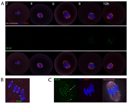
Fig. 1.
MCAK accumulates on centromeres, kinetochores and chromosome arms in mouse oocyte meiosis I. (A) Immunofluorescence of MCAK, CREST and microtubules (MTs) following IBMX washout. Note that MCAK accumulates on centromeres in mid-meiosis I. (B) Magnified image illustrating that MCAK overlaps CREST and chromosome ends, suggesting localisation at centromeres and kinetochores. (C) Magnified image of a late MI spindle highlighting the accumulation of MCAK on chromosome arms (arrow) and at the spindle pole (arrowhead).

