Abstract
A major issue in studies of amyloid formation is the difficulty of preparing the polypeptide of interest in an initially monomeric state under physiologically relevant conditions. This is particularly problematic for polypeptides which are natively unfolded in their unaggregated state, and perhaps the most challenging such system is islet amyloid polypeptide (Amylin), the causative agent of amyloid formation in type-2 diabetes. Preparation of islet amyloid polypeptide with the Ser-19 Ser-20 amide bond replaced by an ester circumvents these problems. The modified peptide is unstructured and monomeric at slightly acidic pH's as judged by analytical ultra centrifugation, gel-filtration, dynamic light scattering, and CD. A rapid pH jump leads to deprotonation of the Ser-20 amide group, and a subsequent rapid O to N acyl shifts regenerates normal human islet amyloid polypeptide. The half time, t1/2, for the conversion to normal islet amyloid polypeptide is 70 seconds at pH 7.4. The amyloid fibrils which are formed by the regenerated islet amyloid polypeptide are indistinguishable from those formed by the wild type polypeptide. The approach allows studies of amyloid formation by islet amyloid polypeptide to be carried out from a well defined, physiologically relevant starting state in the absence of denaturants or organic co-solvents.
Amyloid formation plays a role in at least twenty different human diseases and a broad range of proteins which do not form amyloid in vivo can be induced to do so in vitro.1 A major issue in studies of amyloid formation is the difficulty of preparing highly aggregation prone polypeptides in an initially monomeric state under physiological relevant conditions. This is particularly problematic for polypeptides which are unfolded in their monomeric state and perhaps the most challenging such system is islet amyloid polypeptide (IAPP or amylin). IAPP is responsible for the pancreatic amyloid associated with type-2 diabetes.2 Its role in islet amyloid deposition and its putative complicating role in islet cell transplantation have motivated mechanistic studies of amyloid formation by IAPP and the search for inhibitors of the process.2 Unfortunately, the extremely high propensity of the polypeptide to aggregate means that it is not possible to prepare IAPP in an initially monomeric state under physiologically relevant conditions. This has led to considerable variation in experimental measures of the kinetics of amyloid formation and has made it extraordinarily difficult to quantitatively compare studies which use different solubilization protocols. Here we demonstrate a simple, highly reproducible method for preparing monomeric IAPP which allows amyloid formation to be reliably triggered under defined, physiologically relevant conditions and avoids the use of organic co-solvents, denaturants, or dried films of peptide.
A wide range of methods have been used to prepare IAPP in an initial apparently unaggregated state but, all suffer from drawbacks.3 A common approach is to prepare the polypeptide in either neat DMSO or neat hexafluoroisopropanol (HFIP) and then to trigger amyloid formation by diluting the stock solution into buffer. Unfortunately, even trace amounts of residual DMSO or HFIP can have dramatic effects on the kinetics of amyloid formation by IAPP and the shape of the kinetic progress curves can vary depending upon the cosolvent used.3d In addition, trace amounts of organic cosolvents can complicate cell toxicity assays and even low levels of DMSO can interfere with spectroscopic studies. IAPP has also been prepared by dissolving in fluoroalcohols and preparing a dry film by removing the solvent. Amyloid formation is triggered by adding buffer to the dried film. A major complication with this method is that the initial state of the polypeptide is not well characterized and a range of aggregated species are almost certainty present at the earliest times. Notably, use of this method can result in significantly more rapid amyloid formation than observed with other protocols. The same concerns arise if the initial step of dissolving in fluoroalcohols is omitted.
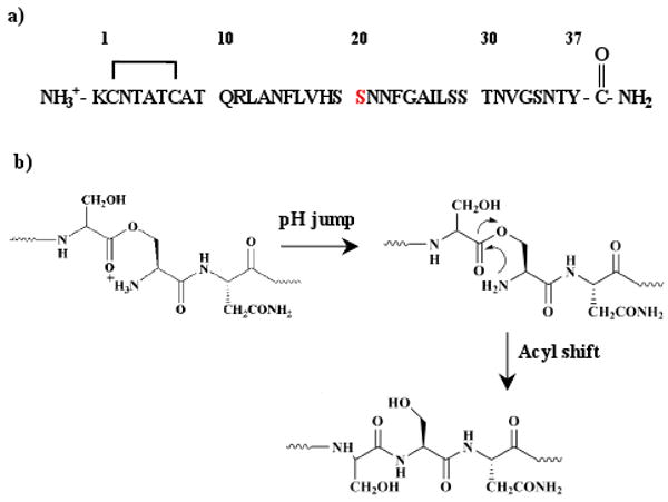
Our approach makes use of the recently described “switch peptide” concept in which an amide linkage is replaced by an ester linkage to a serine or threonine side chain (Figure 1).4 The ester to amide “switch peptide” approach was originally developed to aid in the synthesis of difficult peptides. Human IAPP contains five serines (Figure 1), two of which, Ser-19 and Ser-20, are located in a critical amyloidogenic region.5 IAPP was prepared via Fmoc chemistry and a Boc-Ser(Fmoc-Ser(tBu))-OH dipeptide derivative was utilized to replace the Ser19-Ser20 amide linkage with an ester linkage (Supporting Information). A pH jump leads to deprotonation of the Ser-20 amino group and a subsequent rapid O to N acyl shift leads to regeneration of normal IAPP.
Figure 1.

(a) Sequence of human IAPP with Ser-20 highlighted. The natural peptide has an amidated C-terminus and a Cys-2 Cys-7 disulfide bond. (b) The basis of the switch peptide approach.
IAPP contains a disulfide bond and this presents some potential complications since most protocols for disulfide bond formation involve incubation at neutral or slightly basic pH and the switch peptide is not stable under these conditions, owing to deprotonation of the Ser-20 amino group. Consequently the DMSO based method of disulfide formation was utilized which is effective even at acidic pH values.6
The ester linkage introduces a kink into the polypeptide chain within the critical amyloidogenic region, disrupts backbone hydrogen bonding, and introduces an additional charge due to the protonation of the amino group on Ser-20 (Figure 1). These factors significantly improve the solubility of IAPP. The ester linkage is stable provided the amino group of the serine is protonated which is readily achieved by keeping the peptide under mildly acidic conditions. An added benefit is that the single His residue in human IAPP is protonated under these conditions which is known to slow aggregation.3d, 7
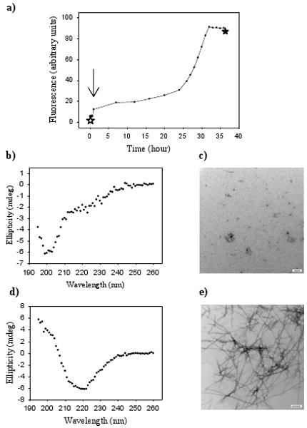
Figure 2 displays the results of kinetic measurements of IAPP amyloid formation. The curve is a fluorescence monitored thioflavin-T binding experiment. Thioflavin-T binds to IAPP amyloid fibrils, but not to pre-fibril intermediates or monomelic IAPP. A 16 μM sample of the switch peptide was incubated at pH 4.2 in buffer with no organic cosolvent. The CD spectrum collected of the freshly dissolved switch peptide indicates that it is unstructured (Figure 2b). Dynamic light scattering (DLS); gel filtration chromatography; analytical ultracentrifugation (AUC) and ultra filtration studies demonstrate that the switch peptide is monomeric (Supporting Information). The average diameter determined by DLS is consisted with a 37 residue monomeric peptide. The single species molecular weight deduced by AUC is within ±3% of the actual monomer molecular weight. The switch peptide has the same elution time from a superdex 75 column as rat IAPP, which is known to be monomeric. Transmission electron microscope (TEM) images of the switch peptide collected before the pH jump confirm that no fibrils were formed (Figure 2c). A pH jump (black arrow) to pH 7.4 results in rapid conversion of the switch peptide into normal IAPP. The regenerated IAPP exhibits the normal sigmoidial time course for fibril formation and TEM images reveal that dense mats of amyloid fibrils are formed at the end of the reaction (Figure 2). The CD spectrum recorded at this time point is typical of those measured for IAPP amyloid fibrils and is rich in β-structure (Figure 2d). As a control, a dried sample of normal wild type IAPP was dissolved in buffer at pH 7.4 at the same final peptide concentration. The sample aggregates rapidly. Thioflavin-T binding is observed, the CD spectrum indicates β-sheet structure and fibrils are detected by TEM after less than 1 minute (Supporting Information).
Figure 2.

The Ser-20-IAPP switch peptide does not aggregate but forms amyloid after a pH jump. (a) Thioflavin-T fluorescence vs time. The pH jump occurred at the point indicated by the arrow. (b) CD spectrum recorded at the time point indicated by the ✩ before the pH jump. (c) TEM image recorded before the pH jump (✩). (d) CD spectrum recorded at the time point indicated by the ★ after the pH jump and after amyloid formation is completed. (e) TEM image recorded after amyloid formation is completed (★). Scale bars in the TEM images represent 100 nanometers.
pH 4.2 samples of the switch peptide will eventually form amyloid-like aggregates if incubated for 7 days. However this is of no practical concern since there is no need to perform a lengthy incubation of the switch peptide before initiating the pH jump.
The switch peptide approach would be of little utility if the rate of conversion from the ester to the amide was comparable to the time for normal IAPP to form amyloid since the observed kinetic profiles would then be a convolution of the time course of amyloid formation with the time course of the O to N acyl shift. Fortunately, the acyl shift is considerably faster. We used an HPLC based assay to directly measure the rate for the conversion of the ester to the amide of the full length switch peptide (Supporting Information). The t1/2 for the reaction is 70 seconds at pH 7.4, 25°C, 20 mM Tris-HCl.
The methodology demonstrated here provides a convenient approach for preparing monomeric IAPP and for initiating amyloid formation in the absence of complications caused by standard solubilization protocols. The method can be used in cell culture based assays of cytotoxicity with the advantage that the potential toxic cosolvents are avoided, but the initial state of the peptide is well defined. The approach should be readily accessible, given that the necessary protected derivates required for the synthesis of the switch peptide are commercially available and are compatible with Fmoc based chemistry.
Supplementary Material
Acknowledgments
This work was supported by NIH Grant GM 078114. We thank members of the Raleigh group for helpful discussions.
Footnotes
Supporting Information Available: Experimental methods and additional figures. This material is available free of charge via the internet at http://pubs.acs.org.
References
- 1.a Westermark P. Febs J. 2005;272:5942–5949. doi: 10.1111/j.1742-4658.2005.05024.x. [DOI] [PubMed] [Google Scholar]; b Chiti F, Dobson CM. Annual Review of Biochemistry. 2006;75:333–366. doi: 10.1146/annurev.biochem.75.101304.123901. [DOI] [PubMed] [Google Scholar]
- 2.a Westermark P, Wernstedt C, Wilander E, Hayden DW, Obrien TD, Johnson KH. Proc Natl Acad Sci USA. 1987;84:3881–3885. doi: 10.1073/pnas.84.11.3881. [DOI] [PMC free article] [PubMed] [Google Scholar]; b Cooper GJS, Willis AC, Clark A, Turner RC, Sim RB, Reid KBM. Proc Natl Acad Sci USA. 1987;84:8628–8632. doi: 10.1073/pnas.84.23.8628. [DOI] [PMC free article] [PubMed] [Google Scholar]; c Kahn SE, Dalessio DA, Schwartz MW, Fujimoto WY, Ensinck JW, Taborsky GJ, Porte D. Diabetes. 1990;39:634–638. doi: 10.2337/diab.39.5.634. [DOI] [PubMed] [Google Scholar]; d Cooper GJS. Endocr Rev. 1994;15:163–201. doi: 10.1210/edrv-15-2-163. [DOI] [PubMed] [Google Scholar]; e Janson J, Ashley RH, Harrison D, McIntyre S, Butler PC. Diabetes. 1999;48:491–498. doi: 10.2337/diabetes.48.3.491. [DOI] [PubMed] [Google Scholar]
- 3.a Higham CE, Jaikaran ETAS, Fraser PE, Gross M, Clark A. Febs Lett. 2000;470:55–60. doi: 10.1016/s0014-5793(00)01287-4. [DOI] [PubMed] [Google Scholar]; b Konarkowska B, Aitken JF, Kistler J, Zhang SP, Cooper GJS. Febs J. 2006;273:3614–3624. doi: 10.1111/j.1742-4658.2006.05367.x. [DOI] [PubMed] [Google Scholar]; c Strasfeld DB, Ling YL, Shim SH, Zanni MT. J Am Chem Soc. 2008;130:6698. doi: 10.1021/ja801483n. [DOI] [PMC free article] [PubMed] [Google Scholar]; d Abedini A, Raleigh DP. Biochemistry. 2005;44:16284–16291. doi: 10.1021/bi051432v. [DOI] [PubMed] [Google Scholar]; e Padrick SB, Miranker AD. Biochemistry. 2002;41:4694–4703. doi: 10.1021/bi0160462. [DOI] [PubMed] [Google Scholar]; f Yonemoto IT, Kroon GJA, Dyson HJ, Balch WE, Kelly JW. Biochemistry. 2008;47:9900–9910. doi: 10.1021/bi800828u. [DOI] [PMC free article] [PubMed] [Google Scholar]
- 4.a Mutter M, Chandravarkar A, Boyat C, Lopez J, Dos Santos S, Mandal B, Mimna R, Murat K, Patiny L, Saucede L, Tuchscherer G. Angew Chem Int Edit. 2004;43:4172–4178. doi: 10.1002/anie.200454045. [DOI] [PubMed] [Google Scholar]; b Dos Santos S, Chandravarkar A, Mandal B, Mimna R, Murat K, Saucede L, Tella P, Tuchscherer G, Mutter M. J Am Chem Soc. 2005;127:11888–11889. doi: 10.1021/ja052083v. [DOI] [PubMed] [Google Scholar]; c Taniguchi A, Skwarczynski M, Sohma Y, Okada T, Ikeda K, Prakash H, Mukai H, Hayashi Y, Kimura T, Hirota S, Matsuzaki K, Kiso Y. Chembiochem. 2008;9:3055–3065. doi: 10.1002/cbic.200800503. [DOI] [PubMed] [Google Scholar]
- 5.a Westermark P, Engstrom U, Johnson KH, Westermark GT, Betsholtz C. Proc Natl Acad Sci USA. 1990;87:5036–5040. doi: 10.1073/pnas.87.13.5036. [DOI] [PMC free article] [PubMed] [Google Scholar]; b Shim SH, Gupta R, Ling YL, Strasfeld DB, Raleigh DP, Zanni MT. Proc Natl Acad Sci USA. 2009;106:6614–6619. doi: 10.1073/pnas.0805957106. [DOI] [PMC free article] [PubMed] [Google Scholar]
- 6.a Tam JP, Wu CR, Liu W, Zhang JW. J Am Chem Soc. 1991;113:6657–6662. [Google Scholar]; b Abedini A, Singh G, Raleigh DP. Anal Biochem. 2006;351:181–186. doi: 10.1016/j.ab.2005.11.029. [DOI] [PubMed] [Google Scholar]
- 7.Charge SBP, Dekoning EJP, Clark A. Biochemistry. 1995;34:14588–14593. doi: 10.1021/bi00044a038. [DOI] [PubMed] [Google Scholar]
Associated Data
This section collects any data citations, data availability statements, or supplementary materials included in this article.


