The crystallization of the Cry4Ba toxin from B. thuringiensis is described.
Keywords: Cry4Ba mosquito-larvicidal protein, Bacillus thuringiensis
Abstract
To obtain a complete structure of the Bacillus thuringiensis Cry4Ba mosquito-larvicidal protein, a 65 kDa functional form of the Cry4Ba-R203Q mutant toxin was generated for crystallization by eliminating the tryptic cleavage site at Arg203. The 65 kDa trypsin-resistant fragment was purified and crystallized using the sitting-drop vapour-diffusion method. The crystals belonged to the rhombohedral space group R32, with unit-cell parameters a = b = 184.62, c = 187.36 Å. Diffraction data were collected to at least 2.07 Å resolution using synchrotron radiation and gave a data set with an overall R merge of 9.1% and a completeness of 99.9%. Preliminary analysis indicated that the asymmetric unit contained one molecule of the active full-length mutant, with a V M coefficient and solvent content of 4.33 Å3 Da−1 and 71%, respectively.
1. Introduction
Bacillus thuringiensis is a rod-shaped aerobic spore-forming Gram-positive bacterium that is widespread in nature (Schnepf et al., 1998 ▶). Insecticidal proteins (known as δ-endotoxins) that are produced as cytoplasmic inclusions during sporulation have been classified via sequence similarities into two main families: the Cry (crystal) and the Cyt (cytolytic) toxins (Crickmore et al., 1998 ▶). The three-domain Cry toxins are of particular interest as they have been shown to be toxic towards a wide variety of insect larvae (Schnepf et al., 1998 ▶). For instance, the 130 kDa Cry4Ba toxin produced from B. thuringiensis subsp. israelensis is specifically toxic to mosquito larvae of the genera Aedes and Anopheles, which are the most important vectors of life-threatening human diseases such as dengue haemorrhagic fever and malaria (Federici et al., 2007 ▶).
B. thuringiensis Cry toxins are synthesized as inactive protoxins found within inclusions and are transformed into cytolytic pore-forming toxins via a multistep process (Gomez et al., 2007 ▶). Upon ingestion by susceptible insect larvae, the protoxins are solubilized in the midgut lumen and activated by larval gut proteases to yield ∼65 kDa toxic fragments comprising three domains: a bundle of α-helices, a three-β-sheet domain and a β-sandwich (Schnepf et al., 1998 ▶; Crickmore et al., 1998 ▶). The active toxins subsequently bind to specific receptors in the luminal plasma membrane, allowing the insertion of their pore-forming portion into the target cell membrane and leading to the eventual death of the larvae (Soberon et al., 2009 ▶). However, the actual underlying mechanisms of the toxic process are still not completely understood.
The protoxins of the 130 kDa mosquito-specific Cry4Aa and Cry4Ba toxins are proteolytically activated by trypsin in vitro to two protease-resistant fragments of ∼47 and ∼20 kDa that remain noncovalently associated, forming a 65 kDa active toxin (Angsuthanasombat et al., 2004 ▶). These two trypsin fragments are produced by cleavage at Arg235 and Arg203, which are located in the exposed loop connecting helices 5 and 6 within the pore-forming domain of Cry4Aa and Cry4Ba, respectively (Angsuthanasombat et al., 2004 ▶). As reported previously, glutamine substitution of Arg235, resulting in blockage of the tryptic cleavage site, in Cry4Aa has been shown to lead to retention of larvicidal activity (Boonserm, Poonwiroon et al., 2004 ▶) and the 65 kDa toxin has successfully been crystallized and modelled (Boonserm, Angsuthanasombat et al., 2004 ▶; Boonserm et al., 2006 ▶). An incomplete structure of the 65 kDa chymotrypsin-treated Cry4Ba toxin that lacked the N-terminal helices owing to proteolysis has also been reported (Boonserm et al., 2005 ▶). It is possible that chymotrypsin treatment produced heterogeneous protein fragments by cleavage at alternative N-terminal sites of the Cry4Ba native toxin. Thus, glutamine substitution of Arg203 in Cry4Ba, which results in resistance to further proteolytic digestion, should be of great use in determining the full-length three-dimensional structure. In this study, we therefore generated a full-length Cry4Ba-R203Q mutant that retains biological activity. The purification and crystallization of the 65 kDa trypsin-activated mutant protein are reported here.
2. Materials and methods
2.1. Construction of a trypsin-site elimination mutant
The pMU388 plasmid encoding the 130 kDa Cry4Ba protoxin under control of the lacZ promoter (Angsuthanasombat et al., 1987 ▶) was used as a template for generating a trypsin-site elimination mutant (R203Q) using a pair of mutagenic oligonucleotides (forward primer, 5′-GGTCTTTAGCACAGTC TGC A GGTGACCAACTATATAAC-3′; reverse primer, 5′-GTTATAGTTGGTCACC T GCA GACTGTGCTAAGACC-3′; changed nucleotides are indicated in bold and PstI restriction sites are indicated in italics). The trypsin site at Arg203 was deleted by substitution with glutamine via PCR-based site-directed mutagenesis following the procedure of the QuikChange Mutagenesis Kit (Stratagene).
2.2. Protein expression, tryptic activation and purification
Escherichia coli strain JM109 harbouring the Cry4Ba-R203Q plasmid was grown at 310 K in Luria–Bertani medium containing ampicillin (100 µg ml−1) and protein expression was induced with isopropyl β-d-1 thiogalactopyranoside (IPTG; 0.1 mM) for 4 h. E. coli cultures that overexpressed the mutant protein as cytoplasmic inclusions were harvested by centrifugation, resuspended in distilled water and disrupted using a French pressure cell (69 MPa). After centrifugation, the inclusion pellets were washed once with wash buffer consisting of 100 mM KH2PO4 pH 6.5, 0.8 M NaCl and 0.1% Triton X-100 and washed twice with cold distilled water by sonication. The partially purified inclusions (5–10 mg ml−1) were solubilized in 50 mM sodium carbonate pH 9.0 by incubation at 310 K for 1 h and insoluble materials were removed by centrifugation at 12 000g for 15 min.
The 130 kDa solubilized protoxin was activated with trypsin (l-1-tosylamide-2-phenylethyl chloromethyl ketone-treated, Sigma) at a 1:20(w:w) enzyme:toxin ratio in 50 mM sodium carbonate pH 9.0 for 16 h at 310 K. Proteolysis was stopped by the addition of 1 mM phenylmethanesulfonylfluoride. The trypsin-treated fraction was subjected to size-exclusion chromatography on Superdex 200 (GE Healthcare) equilibrated with 50 mM sodium carbonate pH 9.0 at a flow rate of 0.4 ml min−1. The eluted peak fractions containing the 65 kDa mutant protein were pooled and concentrated to ∼10 mg ml−1 by ultrafiltration using a Centriprep column (30 kDa cutoff, Amicon) prior to crystallization trials.
A mosquito-larvicidal assay of the toxin inclusions was performed using two-day-old Aedes aegypti larvae. The assay was carried out in 1 ml distilled water containing a serial dilution of toxin inclusions (0.2, 1.0, 2.5, 5, 10 and 25 µg ml−1) in a 48-well microtitre plate at room temperature (298–303 K) with ten larvae per well and a total of 100 larvae per toxin concentration. Mortality was recorded after 24 h incubation. LC50 values and 95% confidence were calculated using the Probit method of Finney analysis.
2.3. Protein crystallization
Crystals were grown by the vapour-diffusion method in sitting drops. 0.5 µl purified protein solution (∼10 mg ml−1) was mixed with an equal volume of reservoir solution consisting of 0.05 M ammonium acetate, 0.01 M MgCl2, 5–10%(v/v) (±)-2-methyl-2,4-pentanediol (MPD) in 0.05 M Tris–HCl buffer pH 7.0–8.0 (modified from Natrix screening kit condition No. 43, Hampton Research). The drop was then equilibrated against 100 µl reservoir solution at 293 K. Crystals appeared after a period of between 15 d and four weeks and grew to their maximum dimensions in three months.
2.4. Sequence analysis of Cry4Ba-R203Q by LC/MS/MS
Purified Cry4Ba-R203Q protein and dissolved Cry4Ba-R203Q crystals were subjected to SDS–PAGE (12% gel); they were subsequently eluted from the excised gel and digested with trypsin according to the standard protocol. Trypsin-generated peptide fragments were separated on a C18 column (0.18 × 100 mm, Thermo Electron, USA) and analyzed by liquid chromatography–tandem mass spectrometry (Finnigan LTQ Linear Ion Trap Mass Spectrometer) as performed by the Genome Institute, BIOTEC, Thailand.
2.5. Crystallographic data collection and processing
The crystal was transferred from the crystallization drop into a cryoprotectant solution (5 µl) consisting of 0.05 M ammonium acetate, 0.01 M MgCl2, 10%(v/v) MPD and 20%(v/v) glycerol in 0.05 M Tris buffer pH 7.5 for a few seconds, mounted on a synthetic nylon loop (0.1–0.2 mm, Hampton Research) and flash-cooled in liquid nitrogen. X-ray diffraction data were collected on beamline BL13B1 equipped with a CCD detector (Q315, ADSC) at the National Synchrotron Radiation Research Center (Hsinchu, Taiwan). For complete data collection, 90° of rotation was measured with 1.0° oscillation using an X-ray wavelength of 1.00 Å, an exposure duration of 10 s and a crystal-to-detector distance of 300 mm from a crystal cooled to 110 K in a nitrogen stream using an X-Stream cryosystem (Rigaku/MSC). All data were indexed, integrated and scaled using the HKL-2000 program suite (Otwinowski & Minor, 1997 ▶).
3. Results and discussion
3.1. Protein preparation of the 65 kDa Cry4Ba-R203Q mutant toxin
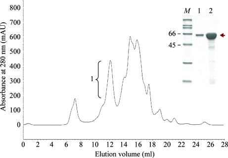
Upon IPTG induction, the 130 kDa Cry4Ba-R203Q mutant protoxin was predominantly produced as a crystalline inclusion that was partially purified from the E. coli lysate and solubilized in carbonate buffer pH 9.0; it had a solubility that was >70% of that of the wild-type inclusion under similar conditions. After activation by trypsin, the activated mutant toxin was successfully purified by gel filtration as a single peak at an elution volume corresponding to a 65 kDa monomer, which was obtained with a purity greater than 90% as analyzed by SDS–PAGE (Fig. 1 ▶).
Figure 1.
Protein purification of the 65 kDa trypsin-activated Cry4Ba-R203Q mutant using Superdex 200 size-exclusion chromatography. The inset shows SDS–PAGE analysis of the pooled (lane 1) and concentrated (10 mg ml−1; lane 2) fractions of peak 1 containing the 65 kDa mutant protein. Lane M contains molecular-mass standards (with values in kDa).
To assess the effect of the mutation on toxicity, the Cry4Ba-R203Q mutant toxin inclusion was tested for its relative biological activity against A. aegypti mosquito larvae. The results given in Table 1 ▶ show that the mutant toxin retained a high larvicidal activity that was comparable with that of the wild-type toxin. This indicated that the elimination of the tryptic cleavage site at Arg203 by glutamine substitution (R203Q) had no detrimental effect on the toxicity of Cry4Ba.
Table 1. Comparison of the toxicity of Cry4Ba and its mutant inclusions towards A. aegypti larvae.
| Toxin inclusion | LC50† (µg ml−1) |
|---|---|
| Wild-type Cry4Ba | 0.50 (0.36–0.71) |
| Cry4Ba-R203Q | 0.59 (0.37–0.93) |
LC50 represents the toxin inclusion concentration that leads to 50% mortality after 24 h exposure to a serial dilution of each toxin inclusion. 95% confidence limits are indicated in parentheses.
3.2. Crystallization of the trypsin-activated Cry4Ba-R203Q mutant toxin
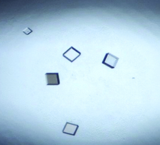
In analogy to our previous study of Cry4Aa (Boonserm, Angsuthanasombat et al., 2004 ▶), we successfully crystallized the 65 kDa trypsin-activated Cry4Ba-R203Q protein. Well formed crystals were obtained by using 0.05 M ammonium acetate, 0.01 M MgCl2 and 10%(v/v) MPD in 0.05 M Tris–HCl pH 7.5 (Fig. 2 ▶). It is noteworthy that when the dissolved crystals were verified by LC/MS/MS in order to confirm the presence of the intact 65 kDa Cry4Ba-R203Q protein, all parts of the trypsin-generated peptide sequences were found to perfectly match the corresponding Cry4Ba mutant sequence (residues Lys57–Ile633). This indicated that the crystals of the activated Cry4Ba-R203 toxin still contained the complete molecule containing the N-terminal fragment, in contrast to those of the 65 kDa chymotrypsin-treated native toxin, which lacked the N-terminal α1–α2b helices (Boonserm et al., 2005 ▶).
Figure 2.
Crystals of the 65 kDa trypsin-activated Cry4Ba-R203Q protein. The approximate dimensions of the largest crystal are 0.07 × 0.07 × 0.02 mm.
3.3. X-ray diffraction analysis of the Cry4Ba-R203Q activated toxin
Analysis of the X-ray diffraction data indicated that the crystals belonged to the rhombohedral space group R32, with unit-cell parameters a = b = 184.62, c = 187.36 Å. A complete data set was collected to a resolution of 2.07 Å and consisted of 74 138 unique reflections with an R merge of 9.1%. Assuming the presence of one activated toxin molecule (molecular weight of ∼65 000) in the asymmetric unit, the Matthews coefficient (V M) of the crystal (Matthews, 1968 ▶) was calculated to be 4.33 Å3 Da−1, corresponding to an estimated solvent content of 71%. The data statistics are summarized in Table 2 ▶. Structure determination of the Cry4Ba-R203Q mutant (602 residues) by the molecular-replacement method using the atomic coordinates of the truncated Cry4Ba structure (558 residues; PDB code 1w99; Boonserm et al., 2005 ▶) as a search model is in progress.
Table 2. Diffraction statistics for Cry4Ba-R203Q.
Values in parentheses are for the highest resolution shell.
| Wavelength (Å) | 1.00 |
| Temperature (K) | 110 |
| Resolution range (Å) | 30.0–2.07 (2.15–2.07) |
| Space group | R32 |
| Unit-cell parameters (Å) | a = b = 184.62, c = 187.36 |
| Unique reflections | 74138 (8174) |
| Completeness (%) | 99.9 (100) |
| 〈I/σ(I)〉 | 19.8 (4.3) |
| Average redundancy | 6.2 |
| Rmerge† (%) | 9.1 (40.4) |
| Mosaicity (°) | 0.25 |
| No. of molecules per asymmetric unit | 1 |
| Matthews coefficient (Å3 Da−1) | 4.33 |
| Solvent content (%) | 71.0 |
R
merge = 
 , where I
i(hkl) is the ith observation of reflection hkl and 〈I(hkl)〉 is the weighted average intensity for all observations i of reflection hkl.
Acknowledgments
This work was generously funded by the Thailand Research Fund (TRF) in cooperation with the Commission of Higher Education. The Royal Golden Jubilee PhD scholarship from TRF (to NT) is gratefully acknowledged. We thank Yuch-Cheng Jean and supporting staff for technical assistance at the synchrotron-radiation facility during data collection at BL13B1 of NSRRC in Taiwan. This study was also supported by the National Synchrotron Radiation Research Center (973RSB02 to C-JC).
References
- Angsuthanasombat, C., Chungjatupornchai, W., Kertbundit, S., Luxananil, P., Settasatian, C., Wilairat, P. & Panyim, S. (1987). Mol. Gen. Genet.208, 384–389. [DOI] [PubMed]
- Angsuthanasombat, C., Uawithya, P., Leetachewa, S., Pornwiroon, W., Ounjai, P., Kerdcharoen, T., Katzenmeier, G. R. & Panyim, S. (2004). J. Biochem. Mol. Biol.37, 304–313. [DOI] [PubMed]
- Boonserm, P., Angsuthanasombat, C. & Lescar, J. (2004). Acta Cryst. D60, 1315–1318. [DOI] [PubMed]
- Boonserm, P., Davis, P., Ellar, D. J. & Li, J. (2005). J. Mol. Biol.348, 363–382. [DOI] [PubMed]
- Boonserm, P., Mo, M., Angsuthanasombat, C. & Lescar, J. (2006). J. Bacteriol.188, 3391–3401. [DOI] [PMC free article] [PubMed]
- Boonserm, P., Pornwiroon, W., Katzenmeier, G., Panyim, S. & Angsuthanasombat, C. (2004). Protein Expr. Purif.35, 397–403. [DOI] [PubMed]
- Crickmore, N., Zeigler, D. R., Feitelson, J., Schnepf, E., Van Rie, J., Lereclus, D., Baum, J. & Dean, D. H. (1998). Microbiol. Mol. Biol. Rev.62, 807–813. [DOI] [PMC free article] [PubMed]
- Federici, B. A., Park, H. W., Bideshi, D. K., Wirth, M. C., Johnson, J. J., Sakano, Y. & Tang, M. (2007). J. Am. Mosq. Control Assoc.23, 164–175. [DOI] [PubMed]
- Gomez, I., Pardo-Lopez, L., Munoz-Garay, C., Fernandez, L. E., Perez, C., Sanchez, J., Soberon, M. & Bravo, A. (2007). Peptides, 28, 169–173. [DOI] [PubMed]
- Matthews, B. W. (1968). J. Mol. Biol.33, 491–497. [DOI] [PubMed]
- Otwinowski, Z. & Minor, W. (1997). Methods Enzymol.276, 307–326. [DOI] [PubMed]
- Schnepf, E., Crickmore, N., Van Rie, J., Lereclus, D., Baum, J., Feitelson, J., Zeigler, D. R. & Dean, D. H. (1998). Microbiol. Mol. Biol. Rev.62, 775–806. [DOI] [PMC free article] [PubMed]
- Soberon, M., Gill, S. S. & Bravo, A. (2009). Cell. Mol. Life Sci.66, 1337–1349. [DOI] [PMC free article] [PubMed]


