Abstract
We present a new image reconstruction algorithm for helical cone-beam computed tomography (CT). This algorithm is designed for data collected at or near maximum pitch, and provides a theoretically exact and stable reconstruction while beneficially using all measured data. The main operations involved are a differentiated backprojection and a finite-support Hilbert transform inversion. These operations are applied onto M-lines, and the beneficial use of all measured data is gained from averaging three volumes reconstructed each with a different choice of M-lines. The technique is overall similar to that presented by one of the authors in a previous publication, but operates volume-wise, instead of voxel-wise, which yields a significantly more efficient reconstruction procedure. The algorithm is presented in detail. Also, preliminary results from computer-simulated data are provided to demonstrate the numerical stability of the algorithm, the beneficial use of redundant data and the ability to process data collected with an angular flying focal spot.
1. Introduction
The ongoing increase in the number of detector rows has created a need for more and more sophisticated helical cone-beam (CB) reconstruction algorithms in x-ray computed tomography (CT). So far, for computational complexity reasons, preference has been given to analytical reconstruction algorithms. These can be divided into two classes: the approximate and the theoretically exact and stable (TES) algorithms. The approximate algorithms are currently the method of choice for commercial CT scanners; see, e.g., Feldkamp et al (1984), Stierstorfer et al (2002), Flohr et al (2003), Stierstorfer et al (2004), Heuscher et al (2004), Kudo et al (2004), Shechter et al (2004), Köhler et al (2006), Tang and Hsieh (2007), Zamyatin et al (2006), Katsevich et al (2007). They are attractive because they are able to use all measured data. But they result in reconstructions that are distorted by CB artifacts that can be severe for imaging with 64 detector rows or more.
In this paper, we focus on the class of TES algorithms, which due to their mathematically exact nature do not suffer from CB artifacts. We are particularly interested in the difficult problem of performing a TES reconstruction while beneficially using all measurements. Several TES algorithms have been published; we categorize them into 1-π and n-π methods.
The 1-π methods, such as Katsevich’s algorithm (Katsevich 2002) and the π-line two-step Hilbert algorithm (Zou and Pan 2004, Pack et al 2005, Ye et al 2005, Schöndube et al 2007a), essentially use a data angular coverage of 180° for each voxel, as viewed from the location of the voxel. That is, they perform reconstruction by backprojecting only filtered data defined inside the Tam–Danielsson (TD) window (Tam et al 1998, Danielsson et al 1997). For a standard helical CB data acquisition geometry, this means that even in the best case scenario only 73% of the measured data are effectively used in terms of noise. The best case scenario is data acquisition at maximum pitch, which is the pitch value such that the TD window just fits within the detector area. Therefore, the 1-π methods significantly waste dose when applied to standard helical CB data. This issue could be solved by adapting the x-ray beam to the TD window to prevent the patient from being exposed to unnecessary dose, but such an adaptation is mechanically challenging to build and could potentially remove flexibility in pitch selection. A preferable solution is to find a way to efficaciously include the data measured outside the TD window into a TES algorithm such that no dose is wasted. Including these data then serves as a way for reducing image noise while maintaining resolution.
The n-π methods, and in particular the 3-π methods, rely on the theoretical observations made in Proksa et al (2000). They extend the data set on which the reconstruction is based to the general case of a n-π detector window (Bontus et al 2003, Katsevich 2004, Bontus et al 2005, Katsevich 2006), where n is an odd integer. Thus, they essentially perform reconstruction at any given location using a relative data angular coverage of n times 180°. This allows a good increase in noise-effective data usage, namely to 86% for n = 3 and 89% for n = 5 (Proksa et al 2000), but does not allow full data utilization. Also, it requires fixing the pitch to relatively small values, e.g., for n = 3, the pitch needs to be fixed at about one third of the maximum pitch that was defined above for the 1-π methods. This results in a significant increase of the data acquisition time for a given axial coverage and thus induces a higher risk of motion artifacts, especially for imaging applications where a high pitch is typically required.
Up to now, it has not been clear if efficient TES reconstruction can be achieved while using all data acquired at or near maximum pitch with a standard rectangular (flat or curved) detector. It is also unclear if a TES algorithm can be built such that it uses most or all data on the detector for arbitrary pitch values. This paper focuses on the first of these questions. We start our investigation from an observation that was made in Pack et al (2005), namely that in some circumstances, the x-ray linear attenuation coefficient at a given location can be reconstructed in three different ways that involve each a different amount of data and yield together full data utilization. Moreover, a reduction of image noise can be achieved by averaging the three different reconstructions. Unfortunately, this observation was based on a computationally demanding procedure, as reconstruction at any location x̱ required a differentiated backprojection (DBP) and a subsequent inverse Hilbert transform (HT) on three lines that contain x̱.
We present here an efficient algorithm that allows TES reconstruction using all the data acquired at or near maximum pitch with a standard cylindrical detector. This algorithm is a volume-based implementation of the observation made in Pack et al (2005), which was found by extending our efficient π-line-based DBP–HT method (Schöndube et al 2007a, 2007b) to the general case of M-lines (i.e., lines that connect a source position with a point on the detector; see section 2.2) (‘M-line’ is an acronym for ‘measured line’). The extension provides a means to efficiently reconstruct the attenuation function on any desired stack of nutating slices that are made of M-lines and cover the volume of interest (VOI). We apply it to reconstruct the VOI three times, using stacks of nutating slices that require different amounts of data and that involve, altogether, all measurements. Averaging of the multiple VOI reconstructions then yields a result that effectively utilizes all measured data. Reconstruction on the different stacks of nutating slices can be performed in a sequential manner, or as a whole, within a single loop on the projections, which offers some advantages in terms of computational effort and data management, and is our preferred approach.
The paper is organized as follows. First, a mathematical description of the data acquisition geometry and of the DBP on M-lines is given in section 2. Then, in section 3, we explain how data outside the TD window can effectively be involved using the DBP, and describe our new algorithm in detail. Last, preliminary reconstruction results are shown in section 4, demonstrating high accuracy, the ability to make significant use of redundant data and the ability to reduce noise through this use. A discussion-and-conclusion section is given after these results.
2. Mathematical background
2.1. Data acquisition geometry
To describe the data acquisition geometry we use a system of Cartesian coordinates, x, y and z, that is attached to the object under examination. This system is laid out such that the z-axis corresponds to the direction in which the object is translated. A vector in this coordinate system is denoted as x̱ = [x, y, z]T, with [·]T denoting the transpose of a matrix or vector. The 3D distribution of x-ray attenuation coefficients to be reconstructed is f (x̱).
The motion of the vertex point (i.e., the radiation source) relative to the object is described as
| (1) |
where R0 is the helix radius, and 2πh is the helix pitch. The position of the vertex point is therefore defined by a single parameter, λ ∈ [λstart, λend], where λstart denotes the first and λend denotes the last position where measurements are taken. The vertex path is adjusted by λ0 and z0 such that at λ = 0 the source is located in the plane z = z0 at a polar angle λ0.
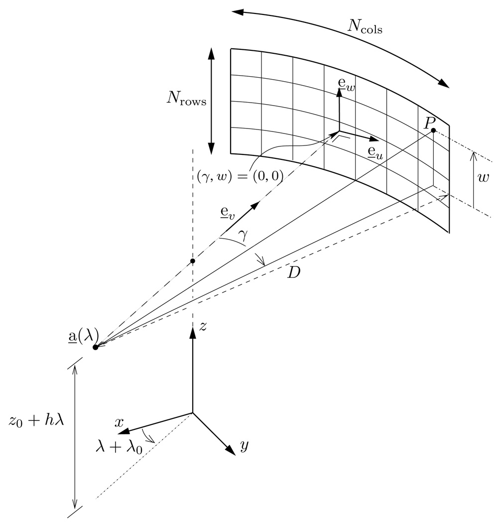
The algorithm is designed for a third generation CT scanner, where the x-ray data are measured using a curved-area detector; see figure 1. The curvature of the detector is such that the detector sits on the cylindrical surface of radius D that is centered on the line parallel to the z-axis through the vertex point. Any point P on the area detector can be specified using two coordinates, w and γ, which are defined relative to a midplane and a midline within this plane, as explained below. The area detector is pixelated into Nrows rows of Ncols pixels that are equidistantly spaced in w and γ; the sampling steps are denoted as Δw and Δγ, so that each pixel is of size (DΔγ) × Δw, with Δγ expressed in radians.
Figure 1.
Data acquisition geometry. Our algorithm is designed for the geometry of third generation CT scanners with a multi-row detector.
We call the plane that contains the vertex point and is orthogonal to the z-axis the midplane, and we call the line within the midplane that diverges from the vertex point and intersects the z-axis the midline. Coordinate w specifies the signed distance between P and the midplane; it is measured in the direction of z. Coordinate γ is the angle between the midline and the line that connects the vertex point with the orthogonal projection of P onto the midplane. This angle is measured positively in the clockwise direction, relative to the z-axis. By construction, (γ, w) = (0, 0) defines the point where the midline intersects the area detector.
The detector is centered on the origin, (γ, w) = (0, 0), except for a quarter-detector-pixel offset in γ, as in current CT scanners. The maximum fan angle is denoted as γmax, and implicitly defines the radius of the field of view (FOV) within which the object is contained. The first detector row is at position w = wmin and the last detector row is at position w = wmax, with wmin = −wmax and wmax = (Nrows − 1)Δw/2. Note that these definitions will be extensively used later.
To mathematically describe the data measured at a given (γ, w) location, we introduce three orthogonal unit vectors:
| (2) |
| (3) |
| (4) |
These vectors are such that e̱v(λ) is parallel to the midline, and e̱u(λ) = e̱v(λ) × e̱w. Thus, e̱u(λ), e̱v(λ) and e̱w form a 3D orthonormal basis rotating with the vertex point.
The line that connects the vertex point a̱(λ) to the (γ, w) location on the area detector is of direction
| (5) |
and the data measured along this line are
| (6) |
The TD window plays a significant role in our algorithm. It is defined as the region of the detector inside which the data are measured that are necessary (Tuy 1983, Danielsson et al 1997) and sufficient (Zou and Pan 2004) for TES reconstruction. When using only data inside this window, each voxel is reconstructed from a 180° range of data, as viewed from the voxel perspective. The upper and lower boundaries of the TD window are the CB projections of the helix turns above and below the current source position onto the detector; in our notation, they are given by (Noo et al 2003)
| (7) |
We call the pitch value such that the TD window just fits within the detector area maximum pitch. This value, denoted by 2π hmax, corresponds to a pitch factor, , that depends on γmax and the number of detector rows, Nrows, according to the formula
| (8) |
For a maximum fan angle of 26°, we have pmax ≃ 1.4.
Our algorithm is not applicable for arbitrary values of γmax and of the pitch factor, p. The following conditions must be met:
| (9) |
where γcrit is the smallest positive root of the equation (π/2 + δ) tan δ = 1, namely γcrit ≃ 26.24°, and
| (10) |
The condition on γmax is only introduced to guarantee pmin < pmax, whereas the constraint on p is derived from the algorithm, as will be discussed later.
2.2. Differentiated backprojection on M-lines
The backprojection part of the algorithm presented in this paper is essentially a generalization of the DBP-0 variant of our DBP–HT algorithm (Schöndube et al 2007a) from π-lines to ‘M-lines’. A π-line is a line that connects two source positions separated by less than one helix turn. In contrast, an M-line is any line that connects a source position with a point on the detector. By construction, any point within the helix cylinder belongs to a multitude of M-lines, and any π-line is an M-line, whereas the converse is not true.
In the same way as for the DBP on π-lines, the outcome of the DBP on M-lines is not the function f (x̱). It is the HT of f (x̱) along an M-line. To achieve the reconstruction of f (x̱), an inverse HT has to be applied after the DBP is performed. For this task, we use an implementation of the formula first presented by Söhngen (1937) (Noo et al 2004), as it requires the HT of f (x̱) only to be known over the support of f (x̱). A detailed description of how we implement Söhngen’s formula can be found in Schöndube et al (2007a equations (12) and (13)).
The link between the DBP and the HT along M-lines can be described as follows. Let (ℋf)(x̱, ω̱) be the HT of f at x̱ along the unit vector ω̱, i.e.,
| (11) |
where the integral has to be understood in the sense of a Cauchy principal value. Let ω̱(λ, x̱) denote the unit vector pointing from a̱(λ) toward x̱:
| (12) |
Furthermore, let 𝒟ℬ𝒫{λa, λb, x̱} be an operator that denotes the DBP of g(λ, γ, w) over the interval [λa, λb] at x̱. Using this notation we can formulate the relation that allows the reconstruction of f (x̱) along M-lines using the DBP, as introduced in Pack et al (2005), as
| (13) |
In this relation, λ1(x̱) and λ2(x̱) correspond to the source positions where the projection of x̱ just enters (respectively, leaves) the TD window on the detector, and λM specifies the direction of the HT, which is along the M-line that connects a̱(λM) to x̱, i.e., along ω̱(λM, x̱). Relation (13) is defined for any point x̱ inside the helix cylinder, and can be implemented whenever x̱ is illuminated with no interruptions over the intervals3 [λM, λ2(x̱)] and [λ1(x̱), λM]. A point x̱ is said to be illuminated over the interval [a, b] if the line connecting a̱(λ) to x̱ is covered by the measurements for all λ ∈ [a, b].
The value at a given point x̱ on the left-hand side of equation (13) depends on λM, as, for different choices of λM, the value of ω̱(λM, x̱) indicating the direction of the HT through x̱ will be different. It is only after performing the inverse HT that the reconstruction result at x̱ is mathematically independent from the choice of λM. It is also important to note that λM is not required to be within the interval [λ1(x̱), λ2(x̱)]. Let λi(x̱) and λo(x̱) be the first (respectively, last) source position at which the projection of x̱ just enters (respectively, leaves) the area detector. If there is no interrupted illumination over [λi(x̱), λo(x̱)], then λM can be selected anywhere between λi(x̱) and λo(x̱) (Pack et al 2005). Conditions (9) guarantee that there is no such interrupted illumination for all points x̱ within the FOV, as will be explained later. The possibility of choosing λM freely from a large number of values provides us with a degree of freedom in selecting the set of M-lines used for reconstruction. In the following section, we will discuss how this degree of freedom can be used to take redundant data measured outside the TD window into account.
The DBP can be implemented in various ways; see, e.g., Pack et al (2005), Zou and Pan (2004), Schöndube et al (2007a, 2007b), Yu et al (2007). In Schöndube et al (2007a, 2007b), Yu et al (2007), it was shown that using a rebinning step for the DBP significantly improves efficiency and noise properties. Using a rebinning step also provides an easy way of processing flying focal spot (FFS) data, since this feature affects then only the rebinning step, not the actual backprojection. In our implementation, we thus carry out the backprojection in a rebinned pseudo-parallel ‘wedge’ geometry (Flohr et al 2005, Heuscher et al 2004).
The rebinning is performed according to the equation
| (14) |
with
| (15) |
and w remaining untouched. In this rebinned geometry, the boundaries of the TD window are given by
| (16) |
and the DBP is expressed with an operator, 𝒟ℬ𝒫r{ϑa, ϑb, x̱}, which designates the DBP of gr(ϑ, sr,w) over the interval [ϑa, ϑb] at x̱.
The link between the wedge definition of the DBP and the expression used in equation (13) is
| (17) |
where
| (18) |
with γ*(λ, x̱) being the value of γ that corresponds to the line through a̱(λ) and x̱, i.e.,
| (19) |
Therefore, equation (13) can be rewritten in the form
| (20) |
where ϑ1(x̱) = ϑ*(λ1(x̱), x̱) and ϑ2(x̱) = ϑ*(λ2(x̱), x̱) denote the values of ϑ where the projection of x̱ just enters (respectively leaves) the TD window in the rebinned geometry.
The precise expression for 𝒟ℬ𝒫r{ϑa, ϑb, x̱} is
| (21) |
with
| (22) |
| (23) |
| (24) |
where ϑ0 = λ0 + π/2.
Inserting equation (21) into the right-hand side of equation (20) and combining the resulting terms together into a single weighted backprojection integral offers a more practical expression for implementing equation (20). This expression given below is used in our algorithm as described later:
| (25) |
where ϑmin and ϑmax are the minimum and maximum values among the three angles, ϑ1(x̱), ϑ2(x̱) and ϑ*(λM, x̱), and where
| (26) |
Note that α(ϑ, x̱) is zero for ϑ ∉ [ϑmin, ϑmax], so the integration bounds in equation (25) can be extended to the nearest view samples outside [ϑmin, ϑmax] to simplify the implementation.
3. DBP–HT with redundant data
3.1. Principle
As discussed in the previous section, λM can be chosen freely over a wide range of values when implementing equation (20) for a given point x̱. By construction, the selection of λM plays a significant role in the range of projections involved in the DBP. In order to illustrate this, figure 2 shows three different choices of λM with the corresponding M-lines. The part of the vertex path marked in bold indicates the source positions that contribute to the backprojection terms on the right-hand side of equation (13). The backprojection range spans over the interval [λ1(x̱), λ2(x̱)] for λ1(x̱) ≤ λM ≤ λ2(x̱), whereas it is extended to [λM, λ2(x̱)] if λM < λ1(x̱) and to [λ1(x̱), λM] if λM > λ2(x̱). The backprojection interval in the case λM ∈ [λ1(x̱), λ2(x̱)] is precisely the same as in the 1-π methods for TES reconstruction (Katsevich 2002, Noo et al 2003, Zou and Pan 2004, Schöndube et al 2007a). This interval is exactly covered by the TD window and thus does not allow using the redundant data. On the other hand, the backprojection range extends beyond the boundaries of the TD window when λM ∉ [λ1(x̱), λ2(x̱)]. Thus, in this case, redundant data are actually used for reconstruction at x̱. This interesting result was first noted in Pack et al (2005).
Figure 2.
M-lines and backprojection range. Reconstruction at x̱ can be achieved by computing and then inverting the HT on any M-line through x̱, but this procedure requires a different range of projections depending on the position of λM (the angle defining the M-line) relative to λ1 and λ2 (the angles defining the π-line through x̱). Three cases are possible: λM can be located before λ1 (left diagram), after λ2 (right diagram) or between λ1 and λ2 (middle diagram). In each case, the highlighted portion of the helix depicts the required backprojection range. Note also that the selection of λM is restricted to the interval [λi, λo] over which no interrupted illumination occurs.
Unfortunately, when evaluating their method, Pack et al (2005) found that the amount of noise in the reconstruction at x̱ increases for a choice of λM ∉ [λ1(x̱), λ2(x̱)], compared to λM ∈ [λ1(x̱), λ2(x̱)]. This somewhat surprising result can be understood by considering the simplified 2D case of h = 0 and w = 0. In this case, the relation gd(ϑ, sr, 0) = −gd(ϑ + π, −sr, 0) holds, which shows that, in contrast to the standard filtered backprojection (FBP) case, there is a minus sign between opposite filtered projections. If the differentiated backprojection is extended over more than a π-interval, the contributions from data that are 180° apart begin to cancel out each other, whereas they are averaged in the standard FBP. Because there is no noise correlation between measurements that are 180° apart, noise contributions add up regardless of whether we perform a DBP or an FBP reconstruction. Therefore, in the case of the DBP, extending the backprojection range yields an increase in the noise level. Pack et al (2005) concluded that a similar behavior can be assumed for the 3D case. In summary, when using the DBP–HT reconstruction method on M-lines it is not possible to reduce the noise level in reconstructed images just by naively extending the backprojection range over more than the π-interval.
Nonetheless, Pack et al (2005) found that noise can be reduced by averaging the three different reconstructions of f (x̱) that correspond to the following choices for λM: λM = λi(x̱), λM ∈ [λ1(x̱), λ2(x̱)] and λM = λo(x̱). Unfortunately, this procedure requires that a backprojection on all points along the intersections of three specific M-lines with the support of f must be performed to obtain the final outcome at any given point x̱; it is thus inefficient and not suitable for practical use. Specifically, the required number of operations is of order O(N5), which can be seen as follows. Let Nx = Ny = Nz = N be the number of voxels that is desired in x, y and z. Let NM ≃ N be the number of samples on one M-line. Let Np ≃ N be the number of source positions per half-turn of the helix. To reconstruct one voxel value, we need to backproject onto all NM samples of one M-line through the center of the voxel, so that the inverse HT can subsequently be applied. Moreover, to perform this backprojection, we need about one half helix turn of projections. Thus, the required number of operations is of order O(NM · Np) for one voxel value and one M-line. Since the procedure proposed in Pack et al (2005) requires three M-lines per voxel, and we wish for Nx · Ny · Nz = N3 voxels, we end up with a number of operations being O(NM · Np · 3 · N3), which is essentially O(N5).
In the remainder of this paper, the ideas from Pack et al (2005), Schöndube et al (2007a, 2007b) are combined to obtain a new TES reconstruction algorithm that effectively uses the redundant data while requiring a number of operations of order O (N4), instead of O (N5).
3.2. Our method
In contrast to the inefficient voxel-based method outlined in section 3.1, our approach performs a volume-based reconstruction similar to our π-line method presented in Schöndube et al (2007a). More precisely, we perform three volumetric reconstructions of the desired volume V, and then compute their average to obtain a result that beneficially involves the redundant data.
Each volumetric reconstruction is obtained by partitioning V into a stack of nutating surfaces of M-lines, performing reconstruction on those surfaces and then interpolating to a Cartesian grid. The partitioning of V is based on a single parameter, denoted by wsurf. This parameter specifies a w-coordinate on the detector toward which all M-lines of all surfaces are required to point to. The three partitionings of V that we consider correspond to the following choices for wsurf: wsurf = 0, wsurf = wmin and wsurf = wmax, with wmin and wmax being the w-coordinate for the first and the last detector row, respectively, as described in section 2.1. The reconstructions of V using these three values of wsurf are denoted as V*(0), V*(wmin) and V*(wmax), and their average as . These four volumes differ from V only due to data noise and discretization errors.
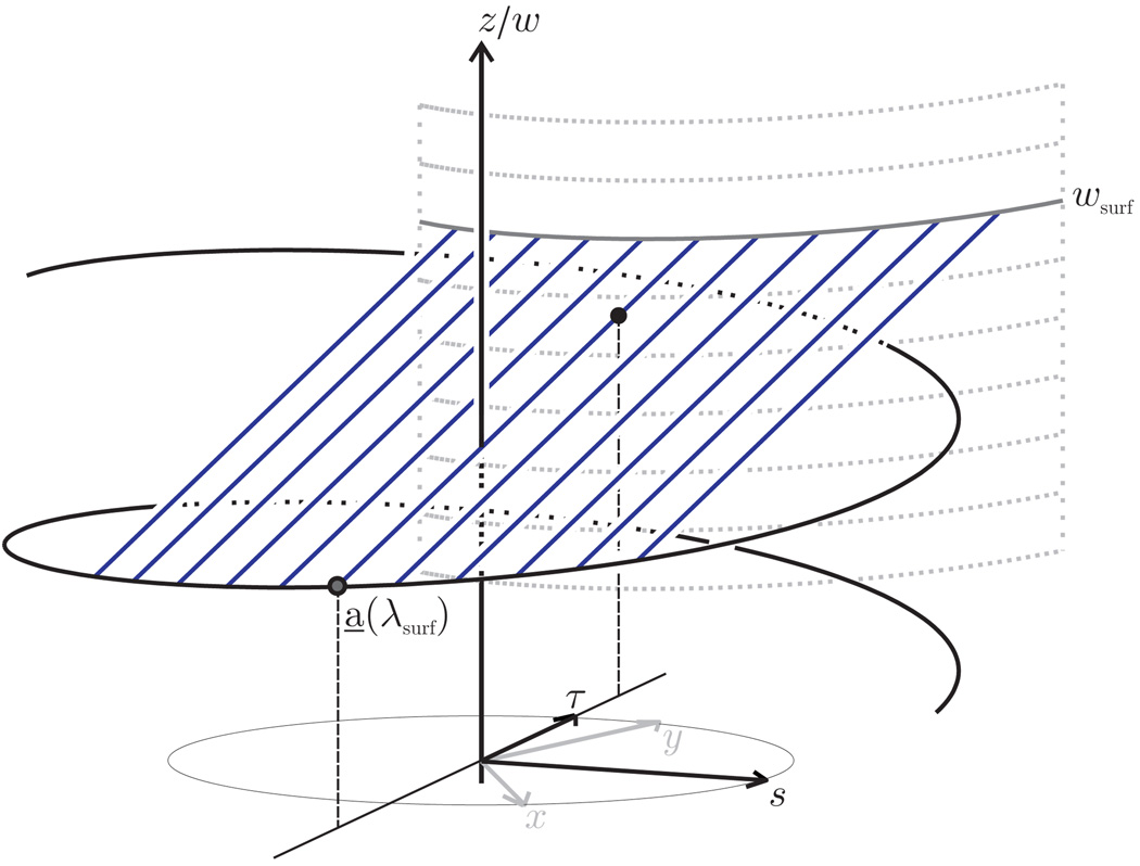
In addition to pointing toward w = wsurf, the M-lines forming each surface in any given partitioning of V are chosen to be equidistant and parallel to each other when projected onto the (x, y)-plane. There is thus one and only one M-line on each surface that intersects the z-axis. We use the angle that specifies the source position of this M-line to index the surfaces; this angle is called λsurf. Figure 3 depicts the surface of index λsurf for partitioning of V based on a given value of wsurf. We denote this surface as ℳ (λsurf;wsurf) with a semicolon between the two parameters, λsurf and wsurf, to emphasize that the first parameter is a variable, whereas the second is a constant for a given partitioning of V.
Figure 3.
Depiction of the M-line surface ℳ(λsurf; wsurf). Each M-line on ℳ(λsurf; wsurf) hits the detector at w = wsurf, i.e., on a fixed row of the virtual detector onto which the data are mapped by the wedge rebinning. By varying λsurf from −∞ to +∞ at fixed wsurf, we get a stack of nutating slices that covers the entire FOV.
To specify locations onto ℳ(λsurf; wsurf), we use two Cartesian coordinates, s and τ, that are defined within the (x, y)-plane, as shown in figure 3. These coordinates are measured along the following orthonormal axes, which rotate with the direction of the M-lines:
| (27) |
| (28) |
Vector e̱τ(λsurf) is along the projection of each M-line onto the (x, y)-plane and e̱s(λsurf) is defined such that e̱s(λsurf) × e̱τ (λsurf) = e̱z for any λsurf. Coordinate s identifies the M-lines on the surface, whereas τ is the positioning parameter on the M-line at the location s. The meaning of s is that of the signed distance from the origin to the projection of any M-line onto the (x, y)-plane. Location (s, τ) = (0, 0) corresponds to the intersection of ℳ(λsurf; wsurf) with the z-axis.
For any given wsurf, the variables s, τ and λsurf parameterize together the three-dimensional space within the helix cylinder. The Cartesian coordinates of the point, x̱(s, τ, λsurf; wsurf), specified by these three variables are
| (29) |
| (30) |
| (31) |
where is the value of λM that corresponds to the M-line at coordinate s on the surface ℳ(λsurf; wsurf), that is
| (32) |
Note that, by construction, the quantity based on equation (18) is the same for all (s, τ)-locations on ℳ(λsurf; wsurf). It is equal to
| (33) |
Note also that the z-coordinate of x̱(s, τ, λsurf; wsurf), given by equation (31), is a continuous function of λsurf that tends toward +∞(respectively, −∞)when λsurf tends to +∞(respectively, −∞). Hence, any point Q inside the FOV can be associated with at least one value of λsurf such that Q belongs to ℳ(λsurf; wsurf).
To accommodate the geometry of nutating slices, all reconstructions of V are performed with equal, equidistant sampling in x and y; s and τ are sampled using the same grid as that used for x and y. The number of samples in x, y, s and τ is denoted as Nlat and the sampling step as Δlat, where ‘lat’ stands for lateral. The main steps to compute V*(wsurf) on a Cartesian grid of Nlat × Nlat × Nz voxels of size (Δlat, Δlat, Δz) are as follows for any relevant value of wsurf, namely wsurf ∈ {0,wmin, wmax}:
Calculate the range of λsurf needed to fully cover V by the M-line surfaces corresponding to the fixed value of wsurf.
Create a stack of M-line surfaces, ℳ(λsurf; wsurf), spaced by Δλsurf = Δz/h over the calculated range of λsurf.
Rebin the data to the wedge geometry using the method described in Flohr et al (2005), which is applicable to data collected both without and with FFS.
- Differentiate the rebinned data in sr using a simple two-point scheme, i.e., for a generic function k(sr):
(34) Weight the data in w according to equation (22).
Compute the DBP onto the stack of M-line surfaces according to equation (25).
Apply a finite HT inversion on each M-line.
Linearly interpolate from the M-line surfaces to Cartesian coordinates. We first interpolate from (s, τ, λsurf) to an intermediate (x, y, λsurf) grid using equations (29) and (30) and then from these intermediate coordinates to (x, y, z) using equation (31).
Step 6 is most demanding in terms of managing computational effort and discretization errors. We provide further details on our implementation of this step in section 3.3. Before going into these details, two observations are, however, in order.
First, the steps outlined above are only applicable when there is no interrupted illumination for all points within the FOV. We show in the appendix that this condition is satisfied as long as the pitch factor, p, and the maximum fan angle, γmax, are selected as in conditions (9). Interestingly, the condition of no interrupted illumination is closely linked to the requirement that the M-line surfaces have no intersection with each other within the FOV when |wsurf| = wmax. In fact, under conditions (9), relation (31) with s and τ defined from equations (29) and (30) establishes a one-to-one relationship between z and λsurf for any (x, y) in the FOV and any |wsurf| ≤ wmax. This property facilitates the implementation of step 8 and is discussed in the appendix along with the issue of interrupted illumination.
Second, the three targeted reconstructions of V involve different data as they are based on different values of wsurf. On one hand, reconstruction V*(0) involves no data outside the TD window. On the other hand, reconstructions V*(wmin) and V*(wmax) involve together all measured data, with V*(wmin) using the data below and within the TD window, and V*(wmax) using the data within and above the TD window. This is illustrated in figure 4. Following our discussion in section 3.1, the noise level in V*(wmin) and in V*(wmax) may be expected to be higher than in V*(0), whereas the average may be expected to be significantly less noisy than any of these three reconstructions of V, as is demonstrated in the following section.
Figure 4.
Illustration of the three different kinds of M-line surfaces used in our approach. From left to right: wsurf = wmax, wsurf = 0 and wsurf = wmin. The curved black lines on the rebinned detector denote the boundaries of the TD window; the shaded detector area shows the amount of data needed for reconstruction on each surface.
3.3. Details on the backprojection step
Given that the backprojection step involves many arithmetic operations that do not depend on the value of wsurf, it is more efficient to perform the backprojection for the three volumes, V*(0), V*(wmin) and V*(wmax), altogether. We describe the steps for such a joint backprojection below. These steps compute a numerically stable version of equation (25) using the simple rectangular rule for numerical integration. The numerical stabilization amounts to replacing α(ϑ, x̱) in (25) by
| (35) |
where signa(·) is a smooth approximation of the signum function, namely,
| (36) |
Note that only the first appearance of the signum function is smoothed; the other two signum functions are left untouched. These two functions are sufficiently randomly sampled over ϑ, x, y and z that no smoothing is needed, whereas the first signum function is used on a fairly deterministic basis that requires numerical stabilization.
Before starting the backprojection, we compute a range of λsurf that allows covering V for all three values of wsurf. This range is defined as [λsurf,min, λsurf,max], where λsurf,min is the minimum value of λsurf for which ℳ(λsurf; wmax) just intersects V, and λsurf,max is the maximum value of λsurf for which ℳ(λsurf; wmin) just intersects V. Since the M-line surfaces are sampled with a step Δλsurf = Δz/h, the number of surfaces for each partition of V is then selected as
| (37) |
where ceil (·) is the operation that rounds any number to the nearest larger integer. By construction, Nsurf differs from Nz only by some overhead resulting from the obliqueness of the surfaces of M-line relative to the z-axis.
The backprojection is performed over the range ϑ ∈ [λsurf,min + π/2, λsurf,max + π/2] and fills the entries of three arrays of size Nlat × Nlat × Nsurf, where each array corresponds to one of the desired values of wsurf. The steps for any filtered projection of angle ϑ that is within the backprojection range are as follows:
-
For every λsurf ∈ [λsurf,min, λsurf,max]:
-
Test if the current projection contributes to samples that are within the FOV on ℳ(λsurf; wsurf) for any wsurf ∈ {0,wmin, wmax}. If not, increment the value of λsurf; otherwise, proceed with the steps below. The test is always (conservatively) positive when ϑ̂ = ϑ − ϑsurf satisfies the following condition, with ϑsurf = λsurf + π/2:
(38) This condition can be used to speed up this first step.
-
For every s on the grid perform the following:
Determine the range of τ which is inside the FOV for the current value of s.
-
For every τ within that interval:
Compute the value of for the current value of (s, τ) according to equation (23).
For this value of , compute wtop and wbottom, the upper and lower boundaries of the TD window, using linear interpolation through tabulated values of equation (16).
-
For each wsurf ∈ {0,wmin, wmax}:
Compute the value of w* for the current (s, τ, wsurf) triple according to equations (24) and (31), using again interpolation through tabulated values, this time of the functions arcsin(·) and .
- Compute αa(ϑ, x̱) of equation (35) using the formula
(39) Compute , with bilinear interpolation as the backprojection contribution to the location x̱ associated with the current values of wsurf, τ, s and λsurf.
-
The required number of operations of this algorithm is of order O(N4). Let N be the number of voxels that is desired in x, y and z. Thus, Nlat = N and Nz = N. Let Np = N be the number of source positions per half-turn of the helix. To reconstruct one M-line surface, we need to backproject about one half helix turn of projections onto Nlat · Nlat = N2 locations. This means that the required number of operations is of order O(N2 · Np). The procedure outlined above requires Nsurf M-line surfaces per volume, and we need three volumes for the combined reconstruction, thus we end up with a number of operations being O (3 · Nsurf · N2 · Np), which is essentially O(N4), since Nsurf ≈ Nz = N.
4. Algorithm validation
We present here preliminary results on the performance of our algorithm. These results were obtained from computer-simulated data defined using parameters that are representative of state-of-the-art commercial CT scanners; see table 1. Two configurations were considered for the data simulation, namely, without and with a lateral FFS. Moreover, the effect of the finite size of the focal spot and of the detector elements was consistently included, using 3 × 3 detector-lets and 2 × 2 source-lets at each source position, with exponential averaging of all line integrals connecting a source-let to a detector-let.
Table 1.
Data-simulation parameters.
| No of detector rows | 64 |
| No of detector columns | 736 |
| Detector pixel height | 0.10947 cm |
| Detector pixel width | 0.12858 cm |
| Source-to-detector distance (D) | 108.56 cm |
| Height of the focal spot | 0.135 cm |
| Width of the focal spot | 0.12 cm |
| Anode angle | 7.0° |
| Radius of the helix (R0) | 59.5 cm |
| No of projections per turn (no FFS) | 1152 |
| No of projections per turn (with FFS) | 2304 |
| Pitch factor | 1.35 |
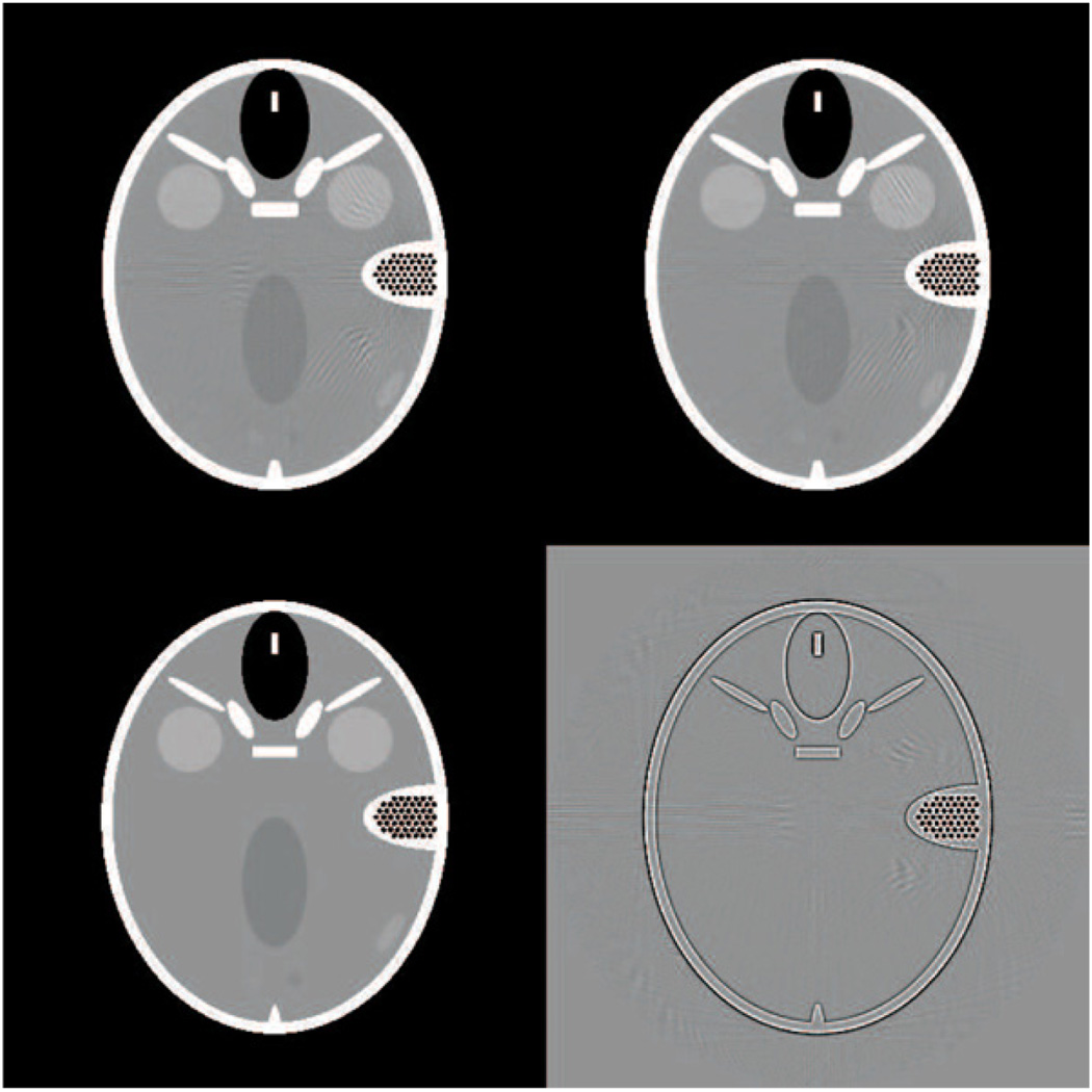
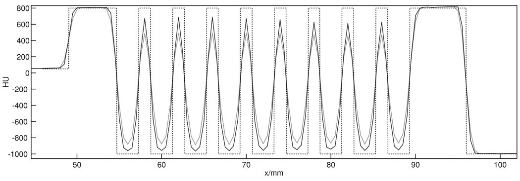
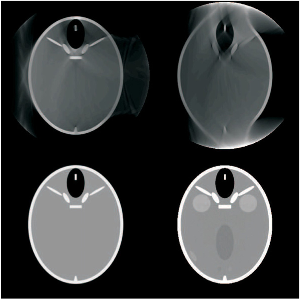
Three experiments were performed. In the first experiment, we used the FORBILD head phantom to visually assess robustness in terms of CB artifacts, discretization errors and effective handling of the lateral FFS feature. Figure 5 displays the outcome of this experiment for the slice z = 0 cm, as obtained on a grid of 401 × 401 square pixels of side 0.75 mm. The top row of this figure shows the results of our algorithm without (left) and with (right) lateral FFS, whereas the bottom row shows the ground truth and the difference image between the two images of the first row. These results are complemented by figure 6, where the profile through the ear at y = 0 cm is given for the reconstructions without and with FFS, and for the ground truth. It can be observed that the new algorithm yields accurate results, and that the FFS feature is effectively accounted for by the algorithm; in that it yields a clear improvement in resolution.
Figure 5.
Reconstructions of the FORBILD head phantom (with right ear only). Upper row: DBP–HT reconstructions from data simulated without (left) and with (right) angular FFS. Bottom row: ground truth (left) and difference image between the two reconstructions in the top row. The grayscale window for the ground truth and the reconstructions is [0, 100] HU. The window for the difference image is [−50, 50] HU.
Figure 6.
Profile at y = 0 cm through the ear of the reconstructions shown in figure 5; dotted line: ground truth, solid black line: DBP–HT from data simulated with angular FFS, solid gray line: DBP–HT from non-FFS data.
The second experiment aimed at demonstrating the effective use of the data outside the TD window, and at assessing the magnitude of the contribution of these data to the reconstruction. To achieve this goal, we performed two reconstructions using the FORBILD head phantom data of the first experiment. One reconstruction was obtained with a binary weight applied to the data that set all values inside the TD window to zero and left the other values unchanged. The other reconstruction was obtained with all detector values outside the TD window being set to zero. Naturally, none of these two reconstructions is theoretically exact, but their sum is due to the linearity of all operations in our algorithm. This data splitting technique allows singling out the impact of the data outside the TD window. The two reconstructions and their sum are displayed in figure 7. It can be observed that the data outside the TD window significantly impact the whole reconstruction process.
Figure 7.
Impact of redundant data in our reconstruction process. The top row shows the reconstruction of the FORBILD head phantom with no ears, as obtained when setting to zero either all data outside the TD window (left) or all data inside the TD window (right). The grayscale is [−1000, 1000] HU. Bottom row: sum of the two images in the top row, displayed with two different grayscales: (left) [−1000, 1000] HU, (right) [0, 100] HU.
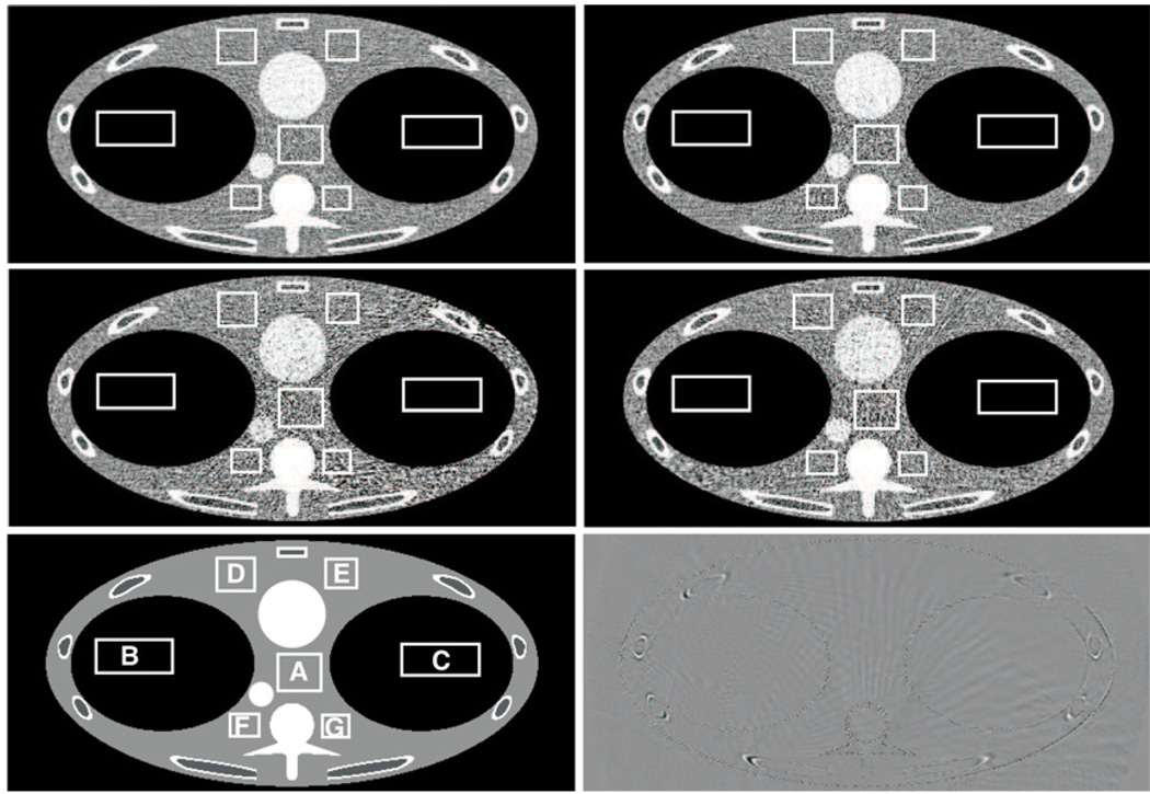
The third and last experiment aimed at verifying that noise performance behaves as expected. For this experiment, we used the FORBILD thorax phantom with addition of Poisson noise corresponding to an emission of 150 000 photons per ray. Figure 8 displays the reconstructions of slice z = 2.18 cm through each of the three sub-volumes, V*(0), V*(wmin) and V*(wmax), and also through the combined volume . These reconstructions were performed on a grid of 512 × 512 square pixels of side 0.9mm. We also included the difference image between the reconstructions of V*(0) and from noise-free data. This image shows that the difference in resolution between V*(0) and is very small, as the edges in the phantom are hardly visible, whereas the noise level is significantly lower in . Quantitative measurements of the standard deviation within seven regions of interest, labeled as A through G, are given in table 2 and support the visual assessment.
Figure 8.
Reconstruction of the FORBILD thorax phantom with Poisson noise added to the data, assuming an emission of 150 000 photons per ray. The standard deviation measurements reported in table 2 were evaluated in the rectangular areas. Top left: combined reconstruction . Top right: reconstruction V*(0), which is based only on the M-line surfaces defined with wsurf = 0. Middle left: reconstruction V*(wmax), which is based only on the surfaces defined with wsurf = wmax. Middle right: reconstruction V*(wmin), which is based only on the surfaces defined with wsurf = wmin. Bottom left: ground truth. Bottom right: difference image between and V*(0). The following grayscale window was used in all cases: [−50, 50] HU.
Table 2.
Standard deviation in HU within seven different regions of interest of the reconstructed images displayed in figure 8.
| V*(wmax) | V*(wmin) | V*(0) | |||
|---|---|---|---|---|---|
| A | 32.74 | 32.36 | 25.56 | 18.33 | |
| B | 12.29 | 13.57 | 11.94 | 8.56 | |
| C | 17.52 | 14.61 | 11.34 | 9.02 | |
| D | 20.45 | 21.00 | 14.44 | 13.62 | |
| E | 24.55 | 25.25 | 17.73 | 14.76 | |
| F | 29.84 | 25.81 | 28.29 | 18.34 | |
| G | 32.81 | 27.57 | 21.32 | 17.41 |
5. Discussion and conclusion
We have presented a new algorithm for helical CB CT that allows TES reconstruction while beneficially using all measured data. This algorithm was designed as an extension of the π-line DBP–HT algorithm presented in Schöndube et al (2007a) to the more general case of M-lines. The noise improvement that this algorithm offers is achieved using a principle that was first described in Pack et al (2005). Specifically, three different reconstructions of the VOI based on data sets that are only partially overlapping are averaged together to obtain a noise-improved image. Each individual reconstruction is obtained using a differentiated backprojection onto a specific type of M-lines. To ensure the usage of all redundant data on the detector, our proposed method uses one set of M-lines directed to the upper end of the detector, one set directed to the lower end of the detector and one set directed to the central detector row. Unlike the technique presented in Pack et al (2005) our algorithm performs the reconstruction volume-wise, instead of voxel-wise; this allows a significant decrease in computational effort, which we have shown to be of the order of the cubic root of the number of voxels.
Our algorithm only applies when there is no interrupted illumination for all points within the FOV, and we have proven that conditions (9) guarantee this to be the case. We did not discuss so far the optimality of these conditions. Regarding this issue, we have observed that the M-line surfaces defined with |wsurf| = wmax intersect each other non-tangentially within the FOV when p is decreased below pmin. Such intersections appear first near the edge of the FOV, and can be shown to imply the appearance of interrupted illumination. For this reason, we believe that conditions (9) are optimal.
Conditions (9) are not very restrictive for a small FOV, such as that corresponding to a head scan. But they are very stringent for thorax imaging, especially with wide patients. We are actively investigating new methodologies allowing the handling of interrupted illumination, so that lower pitch values may be considered for an arbitrary FOV size.
Preliminary results from computer-simulated data were provided to show effective use of redundant data and also of the angular FFS. Further quantification of gains in image quality, especially for real data, is important. Noise comparison with other approximate and TES algorithms is also of high interest. However, these two topics are beyond the scope of this paper, particularly because a careful resolution analysis needs to be performed at the same time. We will report on such evaluations in the near future.
An important issue when evaluating noise is whether uniform weighting of independent M-line results is optimal or not. Also, reconstruction using more than three sets of M-lines may be preferred to mitigate patient-motion artifacts along with noise. An early investigation of these issues was reported inKöhler et al (2008) at the 2008 IEEE Medical Imaging Conference, and yielded very encouraging results regarding patient-motion handling. No algorithm details were provided in Köhler et al (2008), but it is likely that a methodology that is very similar to ours was used. A first description of our algorithm was also given at the 2008 IEEE Medical Imaging Conference (Schöndube et al 2008).
Acknowledgments
This work was partially supported by NIH grant R01 EB007236 and by Siemens Healthcare. Its contents are solely the responsibility of the authors.
Appendix
In this appendix, we discuss how conditions (9) guarantee that there is no interrupted illumination for the points within the FOV. Our proof involves a close examination of intersections between the surfaces of M-lines defined with |wsurf| = wmax. More precisely, we demonstrate that: (i) under conditions (9) and the assumption that |wsurf| ≤ wmax, the surfaces ℳ(λsurf; wsurf) do not intersect each other within the FOV when varying λsurf, (ii) there is no interrupted illumination when the surfaces ℳ(λsurf; wsurf) do not intersect each other within the FOV for |wsurf| = wmax. Taken together, these two statements prove that there is no interrupted illumination under conditions (9).
To best understand our argument, recall that the condition p < pmax is required for any TES reconstruction, and is thus independent from our algorithm. Recall also that the object is always within the FOV, so the CB projection of a point that is within the FOV can only leave the detector through its top and bottom rows. Last, note that the rebinning step of equation (14) does not change the data properties with respect to interrupted illumination: if there is no interrupted illumination in the initial data geometry, there is also none after rebinning, and conversely.
To prove statement (i) above, we rewrite equation (31) into an expression that yields the z-coordinate of the point (x, y, z) that belongs to ℳ(λsurf; wsurf) at given x and y:
| (A.1) |
with
| (A.2) |
| (A.3) |
Then, we examine the behavior of ∂z/∂λsurf for any (x, y) within the FOV. Straightforward calculations yield
| (A.4) |
| (A.5) |
| (A.6) |
where r ≥ 0 and ϕ ∈ [0, 2π) are the polar coordinates of x and y, and where . It turns out that k(t) > 0 for all t when r < R0 because
for any t if r < R0,
limt→−∞ k(t) = 2, limt→+∞ k(t)=0.
Therefore, ∂z/∂λsurf > 0 whenever r < R0 and |wsurf|r < hD. However, under conditions (9), we have pmin < p, which can easily be rewritten in the form wmaxR0 sin γmax < hD from the definition of p, wmax and pmin. Given that R0 sin γmax is the radius of the FOV, we can conclude at this stage that ∂z/∂λsurf > 0 for any (x, y) within the FOV. This proves statement (i).
The proof of statement (ii) goes as follows. If there was interrupted illumination, there would exist a point within the FOV that is projected at least twice onto w = wmax (or w = wmin). But any point that is on the M-line surface ℳ(λsurf; wsurf) sits on an M-line that represents the course of a ray projecting it onto the detector row w = wsurf, with the direction ϑ = λsurf + π/2 in the rebinned geometry. Thus, if a point is projected onto the detector row ŵ = wmax (or ŵ = wmin) twice, say under the rebinned projection angles ϑ1 and ϑ2, it must also be located on two M-line surfaces, namely ℳ(ϑ1 − π/2; ŵ) and ℳ(ϑ2 − π/2; ŵ). This is only possible if those two M-line surfaces intersect. Hence, there is no interrupted illumination if the surfaces ℳ(λsurf; wsurf) do not intersect each other within the FOV for |wsurf| = wmax.
Footnotes
In this context, [a, b] means [b, a] when b < a.
References
- Bontus C, Köhler T, Proksa R. A quasiexact reconstruction algorithm for helical CT using a 3-pi acquisition. Med. Phys. 2003;30:2493–2502. doi: 10.1118/1.1601913. [DOI] [PubMed] [Google Scholar]
- Bontus C, Köhler T, Proksa R. EnPiT: filtered back-projection algorithm for helical CT using an n-pi acquisition. IEEE Trans. Med. Imaging. 2005;24:977–986. doi: 10.1109/TMI.2005.850545. [DOI] [PubMed] [Google Scholar]
- Danielsson PE, Edholm P, Eriksson J, Magnusson Seger M. Towards exact reconstruction for helical cone-beam scanning of long objects. a new detector arrangement and a new completeness condition. In: Townsend DW, Kinahan PE, editors. Proc. 1997 Meeting on Fully 3D Image Reconstruction in Radiology and Nuclear Medicine; Pittsburgh, PA. 1997. pp. 141–144. [Google Scholar]
- Feldkamp LA, Davis LC, Kress JW. Practical cone-beam algorithm. J. Opt. Soc. Am. A. 1984;1:612–619. [Google Scholar]
- Flohr T, Stierstorfer K, Bruder H, Simon J, Polacin A, Schaller S. Image reconstruction and image quality evaluation for a 16-slice CT scanner. Med. Phys. 2003;30:832–845. doi: 10.1118/1.1562168. [DOI] [PubMed] [Google Scholar]
- Flohr TG, Stierstorfer K, Ulzheimer S, Bruder H, Primak AN, McCollough CH. Image reconstruction and image quality evaluation for a 64-slice CT scanner with z-flying focal spot. Med. Phys. 2005;32:2536–2547. doi: 10.1118/1.1949787. [DOI] [PubMed] [Google Scholar]
- Heuscher D, Brown K, Noo F. Redundant data and exact helical cone-beam reconstruction. Phys. Med. Biol. 2004;49:2219–2238. doi: 10.1088/0031-9155/49/11/008. [DOI] [PubMed] [Google Scholar]
- Katsevich A. On two versions of a 3π algorithm for spiral CT. Phys. Med. Biol. 2004;49:2129–2143. doi: 10.1088/0031-9155/49/11/001. [DOI] [PubMed] [Google Scholar]
- Katsevich A. 3PI algorithms for helical computer tomography. Adv. Appl. Math. 2006;36:213–250. [Google Scholar]
- Katsevich A, Zamyatin AA, Silver MD. Optimized reconstruction algorithm for helical CT with fractional pitch between 1PI and 3PI. In: Beekman F, Kachelrieβ M, editors. Proc. 2007 Meeting on Fully 3D Image Reconstruction in Radiology and Nuclear Medicine; Lindau, Germany. 2007. pp. 333–336. [Google Scholar]
- Katsevich AI. Analysis of an exact inversion algorithm for spiral cone-beam CT. Phys. Med. Biol. 2002;47:2583–2597. doi: 10.1088/0031-9155/47/15/302. [DOI] [PubMed] [Google Scholar]
- Köhler T, Bontus C, Koken P. The radon-split method for helical cone-beam CT and its application to nongated reconstruction. IEEE Trans. Med. Imaging. 2006;25:882–897. doi: 10.1109/tmi.2006.876149. [DOI] [PubMed] [Google Scholar]
- Köhler T, Bontus C, Proksa R. BPF reconstruction for helical CT using all data; IEEE Nuclear Science Symp. Conf. Record NSS’08; 2008. pp. 4154–4156. [Google Scholar]
- Kudo H, Rodet T, Noo F, Defrise M. Exact and approximate algorithms for helical cone-beam CT. Phys. Med. Biol. 2004;49:2913–2931. doi: 10.1088/0031-9155/49/13/011. [DOI] [PubMed] [Google Scholar]
- Noo F, Clackdoyle R, Pack J. A two-step Hilbert transform method for 2D image reconstruction. Phys. Med. Biol. 2004;49:3903–3923. doi: 10.1088/0031-9155/49/17/006. [DOI] [PubMed] [Google Scholar]
- Noo F, Pack J, Heuscher D. Exact helical reconstruction using native cone-beam geometries. Phys. Med. Biol. 2003;48:3787–3818. doi: 10.1088/0031-9155/48/23/001. [DOI] [PubMed] [Google Scholar]
- Pack J, Noo F, Clackdoyle R. Cone-beam reconstruction using the backprojection of locally filtered projections. IEEE Trans. Med. Imaging. 2005;24:70–85. doi: 10.1109/tmi.2004.837794. [DOI] [PubMed] [Google Scholar]
- Proksa R, Köhler T, Grass M, Timmer J. The n-PI-method for helical cone-beam CT. IEEE Trans. Med. Imaging. 2000;19:848–863. doi: 10.1109/42.887834. [DOI] [PubMed] [Google Scholar]
- Schöndube H, Stierstorfer K, Dennerlein F, Noo F. Comparative evaluation of two analytical methods for helical cone-beam tomography; IEEE Nuclear Science Symp. Conf. Record NSS’07; 2007b. pp. 4467–4471. [Google Scholar]
- Schöndube H, Stierstorfer K, Dennerlein F, White TA, Noo F. Towards an efficient two-step Hilbert algorithm for helical cone-beam CT. In: Beekman F, Kachelrieβ M, editors. Proc. 2007 Meeting on Fully 3D Image Reconstruction in Radiology and Nuclear Medicine; Lindau, Germany. 2007a. pp. 120–123. [Google Scholar]
- Schöndube H, Stierstorfer K, Noo F. Accurate helical CT reconstruction with redundant data using nutating slices; IEEE Nuclear Science Symp. Conf. Record NSS’08; 2008. pp. 5158–5160. [Google Scholar]
- Shechter G, Köhler Th, Altman A, Proksa R. The frequency split method for helical cone-beam reconstruction. Med. Phys. 2004;31:2230–2236. doi: 10.1118/1.1773622. [DOI] [PubMed] [Google Scholar]
- Söhngen H. Die Lösungen der Integralgleichung und deren Anwendung in der Tragflügeltheorie. Math. Z. 1937;45:245–264. [Google Scholar]
- Stierstorfer K, Flohr T, Bruder H. Segmented multiple plane reconstruction: a novel approximate reconstruction scheme for multi-slice spiral CT. Phys. Med. Biol. 2002;47:2571–2581. doi: 10.1088/0031-9155/47/15/301. [DOI] [PubMed] [Google Scholar]
- Stierstorfer K, Rauscher A, Boese J, Bruder H, Schaller S, Flohr T. Weighted—a simple approximate 3D FBP algorithm for multislice spiral CT with good dose usage for arbitrary pitch. Phys. Med. Biol. 2004;49:2209–2218. doi: 10.1088/0031-9155/49/11/007. [DOI] [PubMed] [Google Scholar]
- Tam KC, Samarasekera S, Sauer F. Exact cone-beam CT with a spiral scan. Phys. Med. Biol. 1998;43:1015–1024. doi: 10.1088/0031-9155/43/4/028. [DOI] [PubMed] [Google Scholar]
- Tang X, Hsieh J. Handling data redundancy in helical cone beam reconstruction with a cone-angle-based window function and its asymptotic approximation. Med. Phys. 2007;34:1989–1998. doi: 10.1118/1.2736789. [DOI] [PubMed] [Google Scholar]
- Tuy HK. An inversion formula for cone-beam reconstruction. SIAM J. Appl. Math. 1983;43:546–552. [Google Scholar]
- Ye Y, Zhao S, Yu H, Wang G. A general exact reconstruction for cone-beam CT via backprojection-filtration. IEEE Trans. Med. Imaging. 2005;24:1190–1198. doi: 10.1109/TMI.2005.853626. [DOI] [PubMed] [Google Scholar]
- Yu L, Xia D, Zou Y, Sidky E, Bian J, Pan X. A rebinned backprojection-filtration algorithm for image reconstruction in helical cone-beam CT. Phys. Med. Biol. 2007;52:5497–5508. doi: 10.1088/0031-9155/52/18/003. [DOI] [PubMed] [Google Scholar]
- Zamyatin AA, Katsevich A, Silver MD, Nakanishi S. Helical CT reconstruction with large cone angle; IEEE Nuclear Science Symp. Conf. Record NSS’06; 2006. pp. 2264–2267. [Google Scholar]
- Zou Y, Pan X. Exact image reconstruction on PI-line from minimum data in helical cone-beam CT. Phys. Med. Biol. 2004;49:941–959. doi: 10.1088/0031-9155/49/6/006. [DOI] [PubMed] [Google Scholar]


