Abstract
Background
Fluoroscopy has been an integral part of modern interventional pain management. Yet fluoroscopy can be associated with risks for the patients and clinicians unless it is managed with appropriate understanding, skill and vigilance. Therefore, this study was designed to determine the amount of radiation received by a primary operator and an assistant during interventional pain procedures that involve the use of fluoroscopy
Methods
In order to examine the amount of radiation, the physicians were monitored by having them wear three thermoluminescent badges during each single procedure, with one under a lead apron, one under the apron collar and one on the leg during each single procedure. The data obtained from each thermoluminescent badge was reviewed from September 2008 to November 2008 and the annual radiation exposure was subsequently calculated.
Results
A total of 505 interventional procedures were performed with C-arm fluoroscopy during three months. The results of this study revealed that the annual radiation exposure was relatively low for both the operator and assistant.
Conclusions
With proper precautions, the use of fluoroscopy during interventional pain procedures is a safe practice.
Keywords: fluoroscopy, interventional pain management, radiation exposure
INTRODUCTION
Pain interventional procedures using the fluoroscopy are absolutely necessary in modern pain medical practice. As more and more physicians use fluoroscopy in their procedures, there has been heightened interest in radiation safety. Yet there are still many physicians who are not properly trained in radiation protection or radiation biology, and so they are often exposed to radiation. They are neither aware of the potential damage radiation can cause nor do they know simple methods that can decrease their exposure.
The number of reported cases of radiation-related complications has increased for both patients and the medical staff. The US FDA reported 26 burn complications due to fluoroscopy between 1992 and 1995 [1], and also anesthesiologists who had preformed a large number of nerve blocks reported burns of the hand [2]. The international Commission on Radiological Protection (ICRP) has set the annual maximum permissible radiation dose to reduce damages due to radiation exposure and the ICRP strongly suggest that radiation exposure be within these set limits. However, most doctors are not aware of their level of exposure to radiation.
Therefore, the authors of this report have aimed to measure the level of radiation exposure of physicians who are performing C-arm fluoroscopy-guided interventional pain procedures and we compared the data with the annual maximum permissible radiation dose.
MATERIALS AND METHODS
We conducted study on the radiation exposure of an operator and an assistant who performed C-arm fluoroscopy-guided pain interventional procedures from September to the end of November, 2008. The operator was a fellow and the assistant was a resident. Before the procedure started, the operator and the assistant wore a dosimeter (UD 802, Panasonic, Japan) inside their lead apron around the chest area, above their collar and on their legs where the lead apron does not cover (Fig. 1). Each dosimeter, which has its own serial number, was worn on the same area and the data was recorded. After the study period, the radiation exposure rates of the dosimeters were measured by the radiation safety unit of the author's hospital. The operator and the assistant always wore a 0.5 mm-thick lead apron and a thyroid protector. For the C-arm fluoroscopy, the X-ray tube was put under the patient and the image intensifier was placed above the patient. When taking lateral images, the operator stood on the side where the image intensifier was. For procedural convenience, the operator stood very close to the patient, and the assistant stood about 1 m away. Every day when a procedure was over, the cumulative radiation exposure time for the fluoroscopy was recorded. The C-arm fluoroscopy was a Philips BV 300 (Eindhoven, Nederland) with 70-100 kV and the ABC (automatic brightness control) was used at around 3-6 mA.
Fig. 1.
The positions of three dosimeters. The physician weares three thermoluminescent badges apron collar, under lead apron and leg.
RESULTS
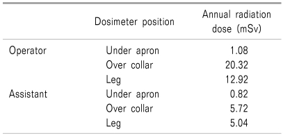
Five hundred five procedures were performed using C-arm fluoroscopy over a 3-month period. These procedures mainly included epidurograms, lumbar transforaminal epidural blocks, lumbar facet blocks, medial branch blocks, lumbar sympathetic nerve blocks, psoas compartment blocks, cervical nerve root blocks and cervical medial branch blocks. The cumulative exposure time reached the total of 676 minutes and 14 sec, with averaging about 80 sec of radiation exposure per each procedure. The level of radiation measured in the dosimeters placed in the 3 areas was calculated for one year (Table 1).
Table 1.
Predicted Annunal Radiation Dose Calculated From Dosimeter Measurements
DISCUSSION
In this study, the accumulated radiation exposure for the C-arm fluoroscopy-guided pain intervention procedures over a 3 month period was used to estimate the probable annual level of radiation exposure. The dosimeter worn inside the lead apron was used to measure the exposure level of the whole body. The dosimeter worn above the collar outside the lead apron was used to measure the radiation exposure at the level of the head and eyes. The dosimeter on the leg was used to measure the exposure level of the legs, which were not protected by the lead apron. The results showed that the radiation level in the dosimeter worn under the lead apron showing the exposure level of the whole body was not significantly different for the operator and the assistant. But there was a higher level of the operator's radiation exposure, as measured by the operator's dosimeters that were worn above the collar and on the leg, than that of the assistant. The whole body exposure levels of the operator and assistant were similar because they wore protective lead aprons, but the operator's exposure level was higher than the assistant's in the areas where the lead apron and the thyroid protector did not protect the body. We assume this difference is due to the fact that the assistant normally stands 1 m further behind the x-ray source than the operator. The level of exposure is inversely related to the square of the distance. Thus, if the distance doubles, the radiation exposure level drops 4 times [3]. In general, the scattered radiation level from the patients when standing 1 m apart is only about 0.1% of the patient's absorbed dose rate [4]. Thus, the study results show that standing 1 m away before obtaining an image can significantly reduce the staff member's level of radiation exposure.
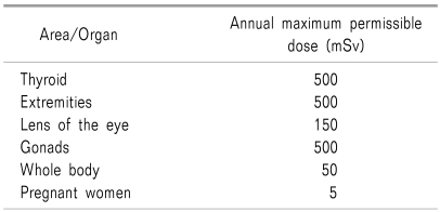
The annual maximum permissible radiation dose suggested by the ICRP is shown in Table 2 [4]. The three measurements of the dosimeters were all within the permissible range. Other studies have shown the radiation measurements for fluoroscopy-guided intervention pain procedures to be within the permissible range [4-6], but, the natural radiation exposure dose in daily life is 2.4 mSv. The radiation dose under the lead apron was below the natural radiation exposure dose. And although the areas which the lead apron did not protect were below the annual maximum permissible radiation dose, the operator was exposed to 6-10 times as much as the natural radiation exposure dose, and the assistant was exposed to twice as much.
Table 2.
Annual Maximum Target Area/Organ Permissible Radiation Doses [4]
The medical staff should be cautious of the scattered radiation of X-rays reflected from the patients' bodies, because the reflected dose is two to three times as great as the dose that enters the patients [7]. Therefore, it is suggested that C-arm fluoroscopy should be performed with the X-ray tube under the patient and the image intensifier above the patient. This way, the scattered radiation goes out underneath, which reduces the scattered radiation dose towards the medical staff more than that when the X-ray tube is placed above the patient [3]. Moreover, when the C-arm fluoroscopy is placed horizontally to obtain a lateral view, the operator should stand on the side of the image intensifier in order to be safe [8]. Also, placing the image intensifier as close as possible to the patient reduces the level of radiation exposure [3].
The authors anticipated that using the C-arm fluoroscopy as directed above will increase the scattered radiation going towards the lower limbs and this will increase the exposure level, and especially below the knees. However, the radiation level of the dosimeter worn on the leg was not very high. Appropriate protection and the correct placement of the C-arm fluoroscopy unit make intervention pain procedures relatively safe, as was found by the results of this study.
In this study, the radiation exposure time was on average 80 seconds. Botwin et al. [5,9,10] reported that when performing caudal blocks, the radiation exposure was 12.55 seconds, in transforaminal epidural blocks 15.16 seconds, in discography 57.24 seconds. Zhou et al. [6] noted that the exposure times for epidural blocks, facet joint blocks, sympathetic nerve blocks, sacroiliac joint blocks and discography were 46.6, 81.5, 64.4, 50.6 and 146.8 seconds, respectively. Botwin and Zhou et al. measured the length of each exposure time per procedure and they made comparisons, but the authors of those reports measured the total cumulative time without making comparisons of each procedure's exposure time, and this limited the depth of the study. Manchikanti et al. [4] stated that the radiation exposure time fluctuates with the level of experience the operator has. In this study, the operator was a fellow with relatively little experience, so the radiation exposure time may have been somwhat longer.
Radiation that causes damage to not only the patients but also the medical staff is increasing, and especially during C-arm fluoroscopy-guided needle placement. There are reported cases of operators whose hands were exposed to direct X-ray beams because they were not careful and they suffered radiation induced damage [2]. To reduce radiation-induced damage, it is suggested that operators wear 0.25 mm lead-rubber gloves [8]. However, even wearing lead-rubber gloves, the X-ray beams falling on the hand should be avoided. One should be especially careful when performing fluoroscopies with ABC, for if lead rubber gloves are seen in the image, then, there will automatically be more radiation exposure to the operator [3].
Even though the annual maximum cumulative dose is 50 mSv, wearing protective gear during procedures is highly recommended to reduce the dose of radiation exposure [3]. Obtaining images over several seconds should be avoided when placing a needle. It is better to instead to quickly obtain the images and to save the last image [7]. This way, one can plan the next movement of the needle from the final image and reduce the possible radiation exposure [4]. Also, people who are not needed for the procedure may step outside the procedure room while the image is being observed.
In conclusion, even though the radiation exposure time for C-arm fluoroscopy-guided intervention pain procedures in this study was higher than of other studies, the radiation dose fell in the range of the maximum allowable radiation dose, so it was possible to confirm that the medical staff was kept relatively safe.
References
- 1.Shope TB. Radiation-induced skin injuries from fluoroscopy. Radiographics. 1996;16:1195–1199. doi: 10.1148/radiographics.16.5.8888398. [DOI] [PubMed] [Google Scholar]
- 2.Valentin J. Avoidance of radiation injuries from medical interventional procedures. Ann ICRP. 2000;30:7–67. doi: 10.1016/S0146-6453(01)00004-5. [DOI] [PubMed] [Google Scholar]
- 3.Fishman SM, Smith H, Meleger A, Seibert JA. Radiation safety in pain medicine. Reg Anesth Pain Med. 2002;27:296–305. doi: 10.1053/rapm.2002.32578. [DOI] [PubMed] [Google Scholar]
- 4.Manchikanti L, Cash KA, Moss TL, Pampati V. Radiation exposure to the physician in interventional pain management. Pain Physician. 2002;5:385–393. [PubMed] [Google Scholar]
- 5.Botwin KP, Thomas S, Gruber RD, Torres FM, Bouchlas CC, Rittenberg JJ, et al. Radiation exposure of the spinal interventionalist performing fluoroscopically guided lumbar transforaminal epidural steroid injections. Arch Phys Med Rehabil. 2002;83:697–701. doi: 10.1053/apmr.2002.32439. [DOI] [PubMed] [Google Scholar]
- 6.Zhou Y, Singh N, Abdi S, Wu J, Crawford J, Furgang FA. Fluoroscopy radiation safety for spine interventional pain procedures in university teaching hospitals. Pain Physician. 2005;8:49–53. [PubMed] [Google Scholar]
- 7.Mahesh M. Fluoroscopy: patient radiation exposure issues. Radiographics. 2001;21:1033–1045. doi: 10.1148/radiographics.21.4.g01jl271033. [DOI] [PubMed] [Google Scholar]
- 8.Hororio TB. Fluoroscopy and radiation safety. In: Honorio TB, Srinivasa R, Robert EM, Spencer SL, Scott MF, editors. Essentials of pain medicine and regional anesthesia. 2nd ed. Philadelphia: Churchill Livingstone Publishers; 2005. pp. 516–524. [Google Scholar]
- 9.Botwin KP, Freeman ED, Gruber RD, Torres-Rames FM, Bouchtas CG, Sanelli JT, et al. Radiation exposure to the physician performing fluoroscopically guided caudal epidural steroid injections. Pain Physician. 2001;4:343–348. [PubMed] [Google Scholar]
- 10.Botwin KP, Fuoco GS, Torres FM, Gruber RD, Bouchlas CC, Castellanos R, et al. Radiation exposure to the spinal interventionalist performing lumbar discography. Pain Physician. 2003;6:295–300. [PubMed] [Google Scholar]


